* Study carried out in the Departments of Radiology of the Universidade Federal Fluminense – UFF, Fluminense Federal University – and of the Universidade Federal do Rio de Janeiro – UFRJ, Federal University of Rio de Janeiro – Rio de Janeiro (RJ) Brazil.
1. Full Professor and Head of the Department of Radiology. Universidade Federal Fluminense – UFF, Fluminense Federal University – Rio de Janeiro (RJ) Brazil. 2. Masters student in Radiology. Universidade Federal do Rio de Janeiro – UFRJ, Federal University of Rio de Janeiro – Rio de Janeiro (RJ) Brazil.
3. Adjunct Professor of Radiology. Faculdade de Medicina de São José do Rio Preto – FAMERP, São José do Rio Preto School of Medicine – São José do Rio Preto (SP) Brazil.
4. Adjunct Professor of Radiology. Universidade Federal do Paraná – UFPR, Federal University of Paraná – Curitiba (PR) Brazil.
5. Consultant Thoracic Radiologist. The Cardiothoracic Centre NHS Trust and The Royal Liverpool and Broadgreen University Hospitals, Liverpool, United Kingdom.
6. Assistant Professor of Radiology. Universidade Federal da Bahia – UFBA, Federal University of Bahia – Salvador (BA) Brazil.
7. Assistant Professor of Clinical Surgery. Faculdade de Medicina de Petrópolis – FMP, Petrópolis School of Medicine – Petrópolis (RJ) Brazil. 8. Assistant Professor. University of Ottawa, Ottawa, ON, Canada.
9. Professor of Clinical Medicine. Faculdade de Medicina de Petrópolis – FMP, Petrópolis School of Medicine – Petrópolis (RJ) Brazil. Correspondence to: Edson Marchiori. Rua Thomaz Cameron, 438, Valparaíso, CEP 25685-120, Petrópolis, RJ, Brasil.
Tel 55 21 2629-9076/55 24 2249-2777. E-mail: edmarchiori@gmail.com Submitted: 23 March 2007. Accepted, after review: 1 June 2007.
Diffuse abnormalities of the trachea: computed tomography findings*
Edson Marchiori1, Aline Serfaty Pozes2, Arthur Soares Souza Junior3,
Dante Luiz Escuissato4, Klaus Loureiro Irion5, César de Araujo Neto6,
Jorge Luiz Barillo7, Carolina Althoff Souza8, Gláucia Zanetti9
Abstract
The aim of this pictorial essay was to present the main computed tomography findings seen in diffuse diseases of the trachea. The diseases studied included amyloidosis, tracheobronchopathia osteochondroplastica, tracheobronchomegaly, laryngotracheobronchial papillomatosis, lymphoma, neurofibromatosis, relapsing polychondritis, Wegener’s granulomatosis, tuberculosis, paracoccidioidomycosis, and tracheobronchomalacia. The most common computed tomography finding was thickening of the walls of the trachea, with or without nodules, parietal calcifications, or involvement of the posterior wall. Although computed tomography allows the detection and characterization of diseases of the central airways, and the correlation with clinical data reduces the diagnostic possibilities, bronchoscopy with biopsy remains the most useful procedure for the diagnosis of diffuse lesions of the trachea.
such as the heart, the kidneys, and the gastroin-testinal tract.(3-5) Primary respiratory amyloidosis
presents three characteristic forms: nodular, diffuse parenchymal, and tracheobronchial.(3-6) The most
common is the tracheobronchial form. The most common symptoms are cough, dyspnea, hemoptysis, and hoarseness.(4) The predominant radiographic
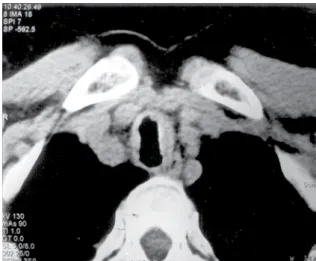
finding is nodular, irregular narrowing of the trachea. Computed tomography scans can reveal nodules protruding into the tracheal lumen or, more often, diffuse parietal thickening, with multifocal submucosal plaques(5,7) (Figure 1). In contrast to
what is seen in tracheobronchopathia osteochon-droplastica, the posterior wall of the trachea can be affected. The thickened wall can present calci-fications.(6,7) Parenchymal abnormalities secondary
to obstruction caused by bronchial collapse can be seen, as can recurrent infectious consolidations, bronchiectasis, and obstructive hyperinflation.(3,7)
Tracheobronchopathia
osteochondroplastica
Tracheobronchopathia osteochondroplastica is a benign disease of the tracheobronchial tree, of unknown etiology, characterized by the pres-ence of multiple submucosal osteocartilaginous nodules, which are primarily located in the anterior and lateral walls of the trachea and in the main bronchi. The posterior wall of the trachea is rarely Tracheal diseases are uncommon and can be
classified as focal, when they are located in only one region of the trachea, or as diffuse, when there is involvement of various tracheal segments. The main causes of focal involvement are primary tracheal neoplasms, lesions of traumatic origin, and some infectious diseases. Diffuse involvement, however, is caused by a wide array of conditions, including amyloidosis, tracheobronchopathia osteochondroplastica, relapsing polychondritis, laryngotracheobronchial papillomatosis, tracheo-bronchomegaly, neurofibromatosis, tuberculosis and other granulomatous infections, such as Wegener’s granulomatosis, as well as lymphomas.(1-3)
Abnormalities of the trachea are rare in daily clinical practice and, although some patients present significant symptoms, abnormalities of the central airways are frequently unapparent or are not seen on chest X-rays. In cases of clinical or radiological suspicion of tracheobronchial alteration, further evaluation using computed tomography is of great importance.(3) The aspect/location of the lesions,
the presence/location of the calcifications, and the association with abnormalities of the pulmonary parenchyma, as well as the correlation with the clinical data, can allow the differential diagnosis to be made among the diffuse abnormalities of the trachea.
The spiral acquisition makes it possible to perform multiplane two- or three-dimensional reconstruc-tions, which are especially useful for the evaluation of the extent of the disease and for surgical planning. Virtual bronchoscopy does not provide information additional to that provided by images obtained at the axial plane, nor do other three-dimensional reconstructions. However, it is useful for surgeons and pulmonologists who are already familiar with bronchoscopic images. In addition, it is less invasive and can be performed without general anesthesia. Furthermore, it allows the evaluation of the tracheal segments distal to the stenosis.(2,3) Nevertheless,
bronchoscopy with histopathological study remains the method of choice for the definitive diagnosis of diffuse abnormalities of the trachea.(3)
Tracheobronchial amyloidosis
Amyloidosis is characterized by local or systemic deposition of abnormal amyloid material in extra-cellular tissues and can involve multiple organs,
airways during expiration.(2,14) With the impairment
of the cough reflex and of the mucociliary defense mechanism, the airways are extremely widened and weakened, which causes mucus accumulation, recurrent pneumonia, and fibrosis.(2,13,15) Therefore,
the symptoms are less than specific and are related to recurrent infections, including excessive expec-toration, hemoptysis, and dyspnea.(2) Computed
tomography scans show an increase in the diam-eter of the upper airways (Figure 3). A decrease in the thickness of the tracheal wall, together with bronchiectasis and diverticula, which usually origi-nate from the posterior lateral wall, can also be observed.(2,13-15)
Laryngotracheobronchial papillomatosis
Laryngotracheobronchial papillomatosis is an infection of the airways, caused by the human papillomavirus, and is capable of presenting malig-nant degeneration.(2,3) The larynx is usually affected,
whereas tracheal involvement is rare, occurring in only approximately 5% of patients with laryngeal papillomatosis.(2,3,16) Papillomas can be single or
multiple.(3) The most common initial symptom is
hoarseness caused by the impairment of the true vocal cords. With the dissemination of the disease, the patient can present varying degrees of airway obstruction, with wheezing, atelectasis, recurrent pneumonia, and bronchiectasis. Hemoptysis occurs frequently, and the infection is commonly confused with active tuberculosis.(16) The radiographic
find-ings include polypoid lesions, either sessile or pedunculated, located in the trachea and in the main bronchi. When there is distal involvement of the airways, computed tomography scans show nodules with a centrilobular distribution, often cavitated, predominantly located in the basal and posterior halves of the lungs (Figure 4). When other infections are superimposed, air-fluid levels within the cavities, areas of consolidations, and atelectasis can be seen.(2,3,16,17)
Tracheal lymphoma
Lymphomas are primary neoplasms of the lymphoreticular system and are classified as Hodgkin’s disease or non-Hodgkin’s lymphoma. The primary tracheal form is extremely rare, and, when it occurs, it seems to be related to the mucosa-associated lymphoid tissue.(18) The most common symptoms
affected.(1,8-12) Many patients are asymptomatic, and
the disease, in such cases, is diagnosed through routine tests.(3) The most common symptoms are
cough, dyspnea, wheezing, and, occasionally, hemoptysis, which is caused by the friction between nodules.(9) Patients frequently present recurrent
respiratory infections.(8,9) Although
tracheobroncho-pathia osteochondroplastica is a benign disease, it can evolve to severe tracheal stenosis.(1,9) Computed
tomography is very sensitive in the identification of nodule calcification, in the definition of the extent and distribution of the tracheobronchial stenosis, and in the characterization of complications, such as atelectasis, bronchiectasis, and postobstruc-tive pneumonia.(10) As can be seen in Figure 2, the
most common tomographic finding is the pres-ence of multiple submucosal nodules, calcified or not, located in the anterior and lateral walls of the trachea.(1,3,9-12)
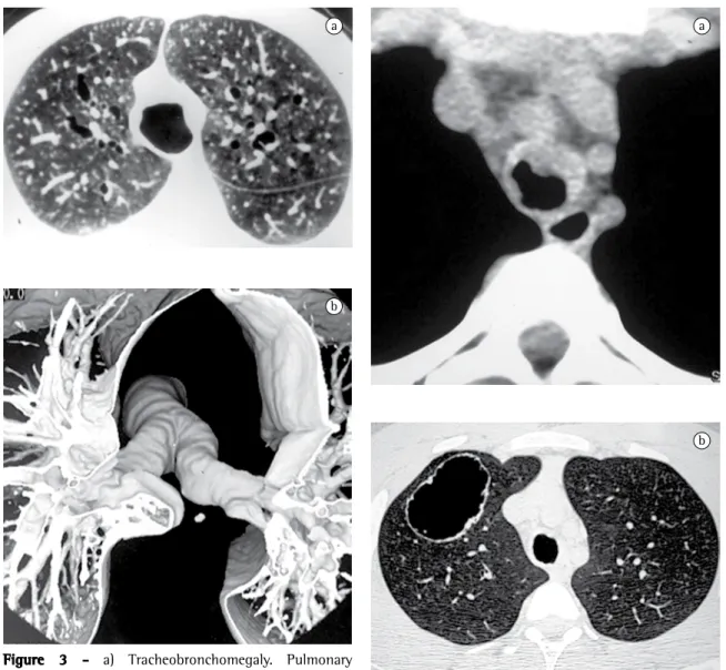
Tracheobronchomegaly
Tracheobronchomegaly, also known as Mounier-Kuhn syndrome, is characterized by marked dilatation of the trachea and main bronchi due to atrophy or to the absence of the longitudinal elastic fibers and the smooth muscles that form its walls.(13,14) The
main abnormality observed in the respiratory physi-ology of such patients is the total collapse of the
walls due to the diffuse infiltration of the submu-cosa.(18,19)
Tracheal neurofibromatosis
Neurofibromatosis is the most common phacoma-tosis and presents thoracic manifestations in 10 to of airway lymphoma are related to obstruction, and
they often delay the initiation of treatment due to the diagnostic confusion with bronchial asthma.(18)
Among such symptoms are dyspnea, nonproductive cough, and wheezing. Hemoptysis can also occur, although it is not common, since, in most cases, the bronchial mucosa is intact.(18,19) Computed
tomog-raphy scans, such as the one displayed in Figure 5, can reveal focal tracheal narrowing and a solitary mass or polypoid thickening of the tracheobronchial
a
b
Figure 3 - a) Tracheobronchomegaly. Pulmonary parenchyma window slice showing irregular and diffuse dilation of the trachea. Note also the presence of bronchiectasis; and b) Tracheobronchomegaly. Three-dimensional reconstruction revealing the marked dilatation of the trachea and of the main bronchi.
a
b
present airway involvement, and the larynx and the upper portion of the trachea are the most common sites. The clinical criteria for the diagnosis include the presence of three or more of the following abnormalities: bilateral auricular chondritis, seron-egative inflammatory polyarthritis, nasal chondritis, ocular inflammation, respiratory tract chondritis, and auditory-vestibular disorder. Confirmation 20% of the cases. As can be seen in Figure 6, upper
airway involvement is revealed by the presence of intraluminal or extraluminal nodular lesions.(20-22)
The typical clinical profile consists of numerous circumscribed areas of skin hyperpigmentation accompanied by dermal and neural tumors of various types. Additional diagnostic criteria include freckles in the axillary region, iris hamartomas, bone dysplasia, multiple tumors of the central nervous system, such as optic nerve gliomas, and having a first-degree relative affected by the disease.(21) Upper airway
obstruction occurs due to the presence of intralu-minal or extraluintralu-minal lesions that cause extrinsic compression of the tracheobronchial tree.(20-22)
Relapsing polychondritis
Relapsing polychondritis is a rare systemic disease characterized by recurrent episodes of cartilage inflammation involving the cartilage of the nose, ear, peripheral articulations, larynx, and tracheo-bronchial tree. In the early phase of the disease, laryngotracheobronchial involvement is present in approximately 10% of cases and indicates bad prognosis. Over the course of the disease, approxi-mately half of all patients with polychondritis
Figure 5 - Lymphoma. Computed tomography scan revealing nodulation and plaques in the tracheal lumen, which cause an irregular reduction in its caliber. Note also bronchiectasis and areas of parenchymal consolidations caused by an accompanying infectious process.
Figure 6 - Neurofibromatosis. Three-dimensional (volume rendering) reconstruction of the trachea and main bronchi in which one can identify, by transparency, three nodular polypoid images, with regular borders, located in the carina and in each of the main bronchi.
Wegener’s granulomatosis
Wegener’s granulomatosis is a systemic autoim-mune disease that primarily affects the kidneys, the upper airways, and the lower airways. It occurs more frequently in males and in middle-aged indi-viduals. The most common symptoms are cough, hemoptysis, chest pain, and dyspnea. The principal histopathological abnormality is necrotizing granu-lomatous vasculitis of the small blood vessels.(3,24,25)
The principal computed tomography findings include subglottic stenosis, and circumferential mucosal thickening, as well as irregularity and ulceration of the tracheobronchial walls. Although involvement of the cartilage rings is less common, it can also result in deformity and narrowing of the trachea. As shown in Figure 7, Wegener’s granulomatosis is often accompanied by pulmonary nodules, which can be cavitated, and by alveolar infiltration.(3,24,25)
Tracheal tuberculosis and other
granulomatous infections
The main finding in central airway tuberculosis is the narrowing of the lumen. Tuberculosis involving the central airways occasionally results in diffuse stenosis, which can lead to respiratory failure in the acute phase. On computed tomography scans, active tracheobronchial tuberculosis presents as irregular circumferential luminal narrowing and can be accompanied by mediastinitis. In the fibrotic is made by immunohistochemistry and
histo-logical analysis of the cartilage samples analyzed. Computed tomography findings include thickening of and calcifications in the tracheal wall, which can evolve to fibrotic scarring and destruction of the cartilage rings, causing deformity and narrowing of the trachea and bronchi. The posterior wall of the trachea is usually spared. The images obtained during expiration can reveal airway collapse.(3,23)
Figure 8 - Paracoccidioidomycosis. Pulmonary parenchyma window slice showing irregularities in the tracheal wall, in addition to consolidations and fibrotic lesions in the upper lobes, with emphysematous bullae and probable bronchiectasis.
a b
presenting as an isolated nodule in the right upper lobe bronchus with upper lobe collapse. J Thorac Cardiovasc Surg. 2005;130(3):901-2.
9. Khan AM, Shim C, Simmons N, Chung V, Alterman DD, Haramati LB, et al. Tracheobronchopathia osteochondroplastica: a rare cause of tracheal stenosis--”TPO stenosis”. J Thorac Cardiovasc Surg. 2006;132(3):714-6.
10. Sá JM, Almeida J, Amado J, Fernandes B, Caminha J, Ferraz JM. Traqueobroncopatia osteocondroplástica. Experiência de uma Unidade de Broncologia. Rev Port Pneumol. 2002;VIII(4): 329-39.
11. Tadjeddein A, Khorgami Z, Akhlaghi H. Tracheobronchopathia osteoplastica: cause of difficult tracheal intubation. Ann Thorac Surg. 2006;81(4):1480-2.
12. White BD, Kong A, Khoo E, Southcott AM. Computed tomography diagnosis of tracheobronchopathia osteochondroplastica. Australas Radiol. 2005;49(4):319-21. 13. Ghanei M, Peyman M, Aslani J, Zamel N. Mounier-Kuhn
syndrome: a rare cause of severe bronchial dilatation with normal pulmonary function test: a case report. Respir Med. 2007;101(8):1836-9.
14. Webb EM, Elicker BM, Webb WR. Using CT to diagnose nonneoplastic tracheal abnormalities: appearance of the tracheal wall. AJR Am J Roentgenol. 2000;174(5):1315-21. 15. Fortuna FP, Irion K, Wink C, Boemo JL. Mounier-Kuhn
syndrome. J Bras Pneumol. 2006;32(2):180-183.
16. Gruden JF, Webb WR, Sides DM. Adult-onset disseminated tracheobronchial papillomatosis: CT features. J Comput Assist Tomogr. 1994;18(4):640-2.
17. Neto CA, Campos RM, Bastos ML. Papilomatose respiratória recorrente com disseminação pulmonar - relato de dois casos. Radiol Bras. 2002;35(2):117-20.
18. Fidias P, Wright C, Harris NL, Urba W, Grossbard ML. Primary tracheal non-Hodgkin’s lymphoma. A case report and review of the literature. Cancer. 1996;77(11):2332-8.
19. Gollub MJ, Castellino RA. Diffuse endobronchial non-Hodgkin’s lymphoma: CT demonstration. AJR Am J Roentgenol. 1995;164(5):1093-4.
20. Cranshaw JH, Morgan C, Knowles G, Nicholson AG, Goldstraw P. Intramural neurofibroma of the trachea treated by multiple stents. Thorax. 2001;56(7):583-4.
21. Fortman BJ, Kuszyk BS, Urban BA, Fishman EK. Neurofibromatosis type 1: a diagnostic mimicker at CT. Radiographics. 2001;21(3):601-12.
22. Moorthy SS, Radpour S, Weisberger EC. Anesthetic management of a patient with tracheal neurofibroma. J Clin Anesth. 2005;17(4):290-2.
23. Luckey P, Kemper J, Niehues T, Schroten H, Fürst G. Diagnostic role of inspiration and expiration CT in a child with relapsing polychondritis. AJR Am J Roentgenol. 2001;176(1):61-2.
24. Summers RM, Aggarwal NR, Sneller MC, Cowan MJ, Wood BJ, Langford CA, et al. CT virtual bronchoscopy of the central airways in patients with Wegener’s granulomatosis. Chest. 2002;121(1):242-50.
25. Mayberry JP, Primack SL, Müller NL. Thoracic manifestations of systemic autoimmune diseases: radiographic and high-resolution CT findings. Radiographics. 2000;20(6):1623-35. 26. Moon WK, Im JG, Yeon KM, Han MC. Tuberculosis of the
central airways: CT findings of active and fibrotic disease. AJR Am J Roentgenol. 1997;169(3):649-53.
form of the disease, the lumen is smoother, and the wall is not thickened. Lymph node enlarge-ment is usually associated with the active form of the disease.(26) The disease can affect a large portion
of the trachea, also affecting the bronchi,(26) or can
involve only a small segment of the trachea or of one bronchus.(2,27) Other granulomatous infections,
such as histoplasmosis and paracoccidioidomycosis (Figure 8), can cause similar lesions.(2,28)
Tracheobronchomalacia
Tracheobronchomalacia is characterized by flac-cidity of the walls of the airways, which causes excessive collapse of the trachea and bronchi during expiration. The disease can be congenital or can be acquired secondary to a series of factors, such as selective intubation, trauma, infection, chronic extrinsic compression, and chronic obstruc-tive pulmonary disease.(29,30) Clinically, patients can
present dry cough, dyspnea, and recurrent infec-tions. The result of the tomographic examination obtained during the inspiratory phase is normal. As can be seen in Figure 9, the tomographic diagnosis is usually made based on slices obtained during expi-ration, or, in dynamic studies, when the inner area of the tracheal or bronchial lumen, on axial slices, suffers a greater than 50% reduction in comparison with the inspiratory phase.(29,30)
References
1. Leske V, Lazor R, Coetmeur D, Crestani B, Chatté G, Cordier JF, et al. Tracheobronchopathia osteochondroplastica: a study of 41 patients. Medicine (Baltimore). 2001;80(6):378-90. 2. Marom EM, Goodman PC, McAdams HP. Diffuse
abnormalities of the trachea and main bronchi. AJR Am J Roentgenol. 2001;176(3):713-7.
3. Prince JS, Duhamel DR, Levin DL, Harrell JH, Friedman PJ. Nonneoplastic lesions of the tracheobronchial wall: radiologic findings with bronchoscopic correlation. Radiographics. 2002;22 Spec No:S215-30. Erratum in: Radiographics. 2003 Jan-Feb;23(1):191.
4. Daniels JT, Cury JD, Diaz J. An unusual cause of postobstructive pneumonia. Chest. 2007;131(3):930-3. 5. Geusens EA, Verschakelen JA, Bogaert JG. Primary pulmonary
amyloidosis as a cause of interlobular septal thickening. AJR Am J Roentgenol. 1997;168(4):1116-7.
6. Chaoui AS, Barish MA, Fenlon HM. Thoracic case of the day. Primary diffuse tracheobronchial amyloidosis. AJR Am J Roentgenol. 1999;173(3):823, 827-8.
7. Marchiori E, Souza Jr AS, Ferreira A, Azevedo KC, Fialho SM, Crespo SJ. Amiloidose pulmonar: aspectos na tomografia computadorizada. Radiol Bras. 2003;36(2):89-94.
29. Baroni RH, Feller-Kopman D, Nishino M, Hatabu H, Loring SH, Ernst A, et al. Tracheobronchomalacia: comparison between end-expiratory and dynamic expiratory CT for evaluation of central airway collapse. Radiology. 2005;235(2):635-41. 30. Loring SH, O’Donnell CR, Feller-Kopman DJ, Ernst A.
Central airway mechanics and flow limitation in acquired tracheobronchomalacia. Chest. 2007;131(4):1118-24. 27. Kim Y, Lee KS, Yoon JH, Chung MP, Kim H, Kwon OJ, et al.
Tuberculosis of the trachea and main bronchi: CT findings in 17 patients. AJR Am J Roentgenol. 1997;168(4):1051-6. 28. Campos EP, Torchio LN, Cataneo AJ, Perotti EA.