J Bras Pneumol. 2008;34(9):749-752
749
Case Report
together with the fact that the diagnosis has only a limited impact on the long-term treatment plan, have motivated the development of new diagnostic tools that involve less invasive procedures, such as the combination of bronchoal-veolar lavage and computed tomography of the chest.
Case report
A 58-year-old, previously healthy, female patient present with a 4-year history of dyspnea on great exertion and cyanosis of the extremities. The symptoms progressed
Introduction
Pulmonary hypertension is a complex clinical condi-tion that can result from any one of a number of distinct processes; extensive investigation is therefore required in order to make an accurate diagnosis and institute the correct treatment.(1) Among the various forms of pulmonary
hypertension is pulmonary veno-occlusive disease (PVOD),(2)
which is characterized by the fact that it primarily affects the venous compartment of the pulmonary vascular system. The definitive diagnosis of PVOD is made through surgical lung biopsy.(3) However, the morbidity and mortality
associ-ated with performing biopsies in this population of patients,
Pulmonary veno-occlusive disease: diagnostic and therapeutic alternatives*
Doença veno-oclusiva pulmonar: alternativas diagnósticas e terapêuticas
Carlos Eduardo Galvão Barboza1, Carlos Viana Poyares Jardim2,
André Luís Dressler Hovnanian3, Bruno Arantes Dias3, Rogério Souza4
Abstract
Pulmonary veno-occlusive disease (PVOD) is a rare cause of pulmonary hypertension. Surgical biopsy has typically been required for diagnostic confirmation. However, the morbidity, mortality and limited benefit of this procedure have generated discussion regarding noninvasive diagnostic techniques. We present the case of a female patient with progressive dyspnea, hypoxemia and pulmonary hypertension, the last diagnosed via catheterization. Computed tomography revealed septal thickening and diffuse micronodules. Bronchoalveolar lavage revealed occult alveolar hemorrhage. Treatment with an endothelin antagonist was started, resulting in symptomatic and functional improvement. Occult alveolar hemorrhage differentiates PVOD from idiopathic pulmonary hypertension. We believe that this finding, in combination with characteristic tomographic findings, is sufficient to establish a diagnosis of PVOD.
Keywords: Hypertension, pulmonary; Pulmonary veno-occlusive disease; Bronchoalveolar lavage; Receptors, endothelin/antagonists & inhibitors.
Resumo
A doença veno-oclusiva pulmonar (DVOP) é uma causa rara de hipertensão pulmonar. A biópsia cirúrgica era usualmente necessária para seu diagnóstico; entretanto, sua morbidade, mortalidade e seu impacto limitado levantou a discussão sobre o diagnóstico não-invasivo. Apresen-tamos um caso de uma paciente com dispnéia progressiva, hipoxemia e hipertensão pulmonar no cateterismo. A tomografia computadorizada revelou espessamento septal e micronódulos difusos. O lavado broncoalveolar revelou hemorragia alveolar oculta. Iniciou-se tratamento com antagonista da endotelina, que resultou em melhora clínica e funcional. A hemorragia alveolar oculta é uma característica da DVOP capaz de diferenciá-la da hipertensão pulmonar idiopática. Acreditamos que sua presença, associada à tomografia característica, seja suficiente para o diagnóstico de DVOP.
Descritores: Hipertensão pulmonar; Pneumopatia veno-oclusiva; Lavagem broncoalveolar; Receptores de endotelina/antagonistas e inibidores.
* Study conducted in the Pulmonology Department of the University of São Paulo School of Medicine, São Paulo, Brazil. 1. Resident in the Pulmonology Department of the University of São Paulo School of Medicine, São Paulo, Brazil.
2. Physician in Charge of the Pulmonary Circulation Outpatient Clinic of the Department of Pulmonology. Instituto do Coração – InCor, Heart Institute – of the Hospital das Clínicas da Faculdade de Medicina da Universidade de São Paulo – HC-FMUSP, University of São Paulo School of Medicine Hospital das Clínicas – São Paulo, Brazil.
3. Physician in the Pulmonary Hypertension Group of the Pulmonology Department of the University of São Paulo School of Medicine, São Paulo, Brazil. 4. Head of the Pulmonary Circulation Group of the Department of Pulmonology. Instituto do Coração – InCor, Heart Institute – of the Hospital das Clínicas da Faculdade de Medicina da Universidade de São Paulo – HC-FMUSP, University of São Paulo School of Medicine Hospital das Clínicas – São Paulo, Brazil. Correspondence to: Rogério Souza. Disciplina de Pneumologia, Faculdade de Medicina da Universidade de São Paulo, Av. Dr. Enéas de Carvalho Aguiar, 255, sala 7079, CEP 05403-900, São Paulo, SP, Brasil.
Tel 55 11 3069-5695. E-mail: rogerio.souza@incor.usp.br Financial support: None.
750 Barboza CEG, Jardim CVP, Hovnanian ALD, Dias BA, Souza R
J Bras Pneumol. 2008;34(9):749-752
Discussion
Pulmonary hypertension is rarely caused by PVOD, which accounts for only 10% of all cases initially considered idiopathic.(3,4) Prior to 2003,
PVOD was classified as venous pulmonary hyper-tension. However, it is currently included in the arterial hypertension category, since it presents clinical characteristics similar to those of idiopathic pulmonary arterial hypertension and has the same risk factors.(2) Although PVOD is most often seen in
children and young adults, it can appear as early as the first year and as late as the seventh decade of life; among adult patients, it occurs predominantly in males (at a ratio of approximately 2:1).(1,5)
within the preceding 18 months, and, within the last 6 months, the patient had experienced dyspnea on minimal exertion (functional class III). She stated that she had not presented upper-limb edema, chest pain or syncope. By her own admission, she was a former smoker, with a 24 pack-year history, although she had quit smoking 26 years prior. The physical examination revealed cyanosis, regular cardiac rhythm with hyperphonesis of the second heart sound, normal breath sounds and peripheral oxygen saturation (SpO2) at 84% on room air.
A chest X-ray revealed prominence of the medial pulmonary artery, and a computed tomography scan of the chest showed dilation of the arterial branches, with an artery-bronchus ratio of > 1, septal thick-ening and diffuse micronodules (Figure 1). The pulmonary function tests results showed that airflow and lung volumes were normal, but that there was a significant reduction in the diffusing capacity of the lung (30% of predicted). An echocardiogram revealed dilated right heart chambers and pulmo-nary artery systolic pressure estimated at 84 mmHg. The patient was submitted to the six-minute walk test (6MWT), and the distance covered was 375 m. Tests for connective tissue diseases, HIV infection, portal hypertension and chronic pulmonary embo-lism were all negative.
Right heart catheterization was performed, and the findings confirmed the diagnosis of pulmonary hypertension: mean pulmonary artery pressure, 48 mmHg; right atrial pressure, 6 mmHg; pulmo-nary wedge pressure, 8 mmHg; cardiac output, 2.6 L/min; and pulmonary vascular resistance, 15.3 Woods units (unresponsive to nitric oxide).
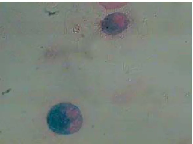
Based on the tomographic findings, we decided to investigate PVOD using bronchoscopy and bron-choalveolar lavage (BAL). The cytological analysis revealed a predominance of macrophages (89% of the total number of cells), 50% of which were hemo-siderin-laden (Figure 2). Therefore, the presumptive diagnosis was PVOD.
Full anticoagulation with warfarin was started, together with specific treatment of the pulmo-nary hypertension using the endothelin receptor antagonist bosentan, at 125 mg/day for four weeks and at 250 mg/day thereafter. After 6 months of follow-up, the patient presented significant func-tional improvement, evolving to funcfunc-tional class II, with an SpO2 of 90% on room air and covering
500 m on the 6MWT.
Figure 2 - Photomicrograph of the bronchoalveolar lavage showing heavily hemosiderin-laden macrophages (magnification, ×1,000).
Pulmonary veno-occlusive disease: diagnostic and therapeutic alternatives
J Bras Pneumol. 2008;34(9):749-752
751
Since such patients present functional limitation, and since surgical lung biopsy implies considerable morbidity and mortality, a presumptive diagnosis of PVOD should be made based on the combination of suggestive tomographic findings and BAL-proven occult alveolar hemorrhage.
There have been no controlled studies of treat-ments for PVOD. There have been only a few, isolated reports of clinical and functional improve-ment brought about through the use of medications, such as nifedipine and prostacyclin.(9,11) However,
prostacyclin and its derivatives should be used with extreme caution due to the risk of pulmonary edema resulting from the preferentially arterial vasodilation. Lung transplantation is currently considered the only treatment capable of significantly increasing survival.(3) Without treatment, the prognosis is poor,
and the majority of patients die within two years after diagnosis.(4)
In the case presented here, we opted to treat the patient with an endothelin receptor antagonist, since its antiremodeling effect tends to overpower its vasodilator effect,(14-16) thereby reducing the risk
of hemodynamic complication as well as slowing the progression of the disease. We believe this to be the first case of PVOD to present a favorable clinical response to this class of drug.
The case presented here highlights the possibility of making a presumptive diagnosis of PVOD using a noninvasive technique (bronchoscopy), as well as the potential benefit of endothelin receptor antago-nists in treating PVOD. This combination can have advantages for patients not only in that it offers a therapeutic option but also in that it can obviate the need for surgical lung biopsy.
References
1. Sociedade Brasileira de Pneumologia e Tisiologia. Diretrizes Brasileiras para Manejo da Hipertensão Pulmonar. J Bras Pneumol 2005;31(supl. 2):S1-S31.
2. Simonneau G, Galiè N, Rubin LJ, Langleben D, Seeger W, Domenighetti G, et al. Clinical classification of pulmonary hypertension. J Am Coll Cardiol. 2004;43(12 Suppl S):S5-S12.
3. Mandel J, Mark EJ, Hales CA. Pulmonary veno-occlusive disease. Am J Respir Crit Care Med. 2000;162(5):1964-73. 4. Pietra GG, Edwards WD, Kay JM, Rich S, Kernis J, Schloo B,
et al. Histopathology of primary pulmonary hypertension. A qualitative and quantitative study of pulmonary blood vessels from 58 patients in the National Heart, Lung, and Blood Institute, Primary Pulmonary Hypertension Registry. Circulation. 1989;80(5):1198-206.
The cause of PVOD is unknown. It might repre-sent an idiopathic process or a common pathological response to various injurious stimuli.(3) Among the
potential etiologies are genetic factors (familial cases of PVOD related to mutation of the bone morphogenetic protein receptor II gene having been reported),(6) autoimmune diseases and infections,(3)
It remains unknown whether smoking plays a role. From an anatomopathological point of view, PVOD is characterized by the following(3): intimal fibrosis
and medial hypertrophy, together with thrombi, in the pulmonary veins and venules; medial hyper-trophy, albeit without plexiform lesions, in the arteries; and interstitial edema, accompanied by hemosiderosis, in the parenchyma.
In PVOD, the clinical profile resembles that of pulmonary hypertension resulting from other causes, including progressive dyspnea on exertion, with or without signs of cor pulmonale.(7) The diagnosis
should be based on the clinical presentation and on any radiological alterations suggestive of pulmonary edema, such as septal lines, basal reticular opacities and pleural effusion.(3,5) Specifically, characteristic
findings on tomography scans of the chest include smooth septal thickening, diffuse micronodules and ground-glass opacities.(8) Pulmonary function tests
typically show a reduction in the diffusing capacity of the lung.(5)
In catheterized patients, an increase in pulmo-nary artery pressure can be seen, although pulmopulmo-nary wedge pressure remains normal.(9-11) Typically, PVOD
patients do not respond to vasodilators and can even develop acute pulmonary edema when treated with prostacyclin, a reaction considered strongly suggestive of a diagnosis of PVOD.(12) Nevertheless,
in the largest case-series study of PVOD to date, 10 PVOD patients received vasodilators, and 8 of those patients presented a favorable response, whereas only one developed pulmonary edema. Despite such suggestive clinical, radiological and hemodynamic characteristics, diagnostic confirmation can only be obtained through surgical lung biopsy.(3)
Dou to the predominance of the postcapil-lary component, occult alveolar hemorrhage, as evidenced by hemosiderosis, is considered another characteristic of PVOD.(3) It has recently been
752 Barboza CEG, Jardim CVP, Hovnanian ALD, Dias BA, Souza R
J Bras Pneumol. 2008;34(9):749-752
in a patient with pulmonary veno-occlusive disease. Chest. 2002;122(3):1096-8.
12. Palmer SM, Robinson LJ, Wang A, Gossage JR, Bashore T, Tapson VF. Massive pulmonary edema and death after prostacyclin infusion in a patient with pulmonary veno-occlusive disease. Chest. 1998;113(1):237-40.
13. Rabiller A, Jaïs X, Hamid A, Resten A, Parent F, Haque R, et al. Occult alveolar haemorrhage in pulmonary veno-occlusive disease. Eur Respir J. 2006;27(1):108-13.
14. Shi-Wen X, Chen Y, Denton CP, Eastwood M, Renzoni EA, Bou-Gharios G, et al. Endothelin-1 promotes myofibroblast induction through the ETA receptor via a rac/phosphoinositide 3-kinase/Akt-dependent pathway and is essential for the enhanced contractile phenotype of fibrotic fibroblasts. Mol Biol Cell. 2004;15(6):2707-19.
15. Kunichika N, Landsberg JW, Yu Y, Kunichika H, Thistlethwaite PA, Rubin LJ, et al. Bosentan inhibits transient receptor potential channel expression in pulmonary vascular myocytes. Am J Respir Crit Care Med. 2004;170(10):1101-7.
16. Michel RP, Langleben D, Dupuis J. The endothelin system in pulmonary hypertension. Can J Physiol Pharmacol. 2003;81(6):542-54.
5. Holcomb BW Jr, Loyd JE, Ely EW, Johnson J, Robbins IM. Pulmonary veno-occlusive disease: a case series and new observations. Chest. 2000;118(6):1671-9.
6. Runo JR, Vnencak-Jones CL, Prince M, Loyd JE, Wheeler L, Robbins IM, et al. Pulmonary veno-occlusive disease caused by an inherited mutation in bone morphogenetic protein receptor II. Am J Respir Crit Care Med. 2003;167(6):889-94. 7. Barst RJ, McGoon M, Torbicki A, Sitbon O, Krowka MJ, Olschewski H, et al. Diagnosis and differential assessment of pulmonary arterial hypertension. J Am Coll Cardiol. 2004;43(12 Suppl S):S40-S47.
8. Resten A, Maitre S, Humbert M, Rabiller A, Sitbon O, Capron F, Simonneau G, Musset D. Pulmonary hypertension: CT of the chest in pulmonary venoocclusive disease. AJR Am J Roentgenol. 2004;183(1):65-70.
9. Salzman GA, Rosa UW. Prolonged survival in pulmonary veno-occlusive disease treated with nifedipine. Chest. 1989;95(5):1154-6.
10. Davis LL, deBoisblanc BP, Glynn CE, Ramirez C, Summer WR. Effect of prostacyclin on microvascular pressures in a patient with pulmonary veno-occlusive disease. Chest. 1995;108(6):1754-6.