Evaluation of the exhaled carbon
monoxide levels in smokers with COPD*
Avaliação da concentração de monóxido de carbono no ar exalado em tabagistas com DPOC
Gustavo Chatkin, José Miguel Chatkin, Gabriel Aued,
Guilherme Oliveira Petersen, Edna Thais Jeremias, Flávia Valladão Thiesen
Abstract
Objective: To measure exhaled carbon monoxide (COex) levels in smokers with and without COPD. Methods: Smokers treated at outpatient clinics of São Lucas Hospital in the city of Porto Alegre, Brazil, between September of 2007 and March of 2009 were invited to participate in this study. The participants completed a questionnaire regarding demographic and epidemiologic characteristics and were submitted to spirometry, as well as to determination of COex and urinary cotinine levels. The participants were divided into two groups: those with COPD and those without COPD. Results: The study involved 294 smokers, of whom 174 (59.18%) had been diagnosed with COPD. All of the participants presented with urinary cotinine levels > 50 ng/mL. Smokers with COPD presented significantly higher median values for age and pack-years than did those without COPD (p < 0.001 and p = 0.026, respectively). No other statistically significant differences were found. When adjusted for gender, age at smoking onset, number of cigarettes/day and urinary cotinine level, the mean values of COex were higher, but not statistically so, in the COPD group than in the non-COPD group (17.8 ± 0.6 ppm and 16.6 ± 0.7 ppm, respectively; p = 0.200). The differences remained nonsignificant when plotted logarithmically. A wide dispersion of COex values was found when the participants were classified by FEV1 level (r = −0.06; p = 0.53) or by Global Initiative for Chronic Obstructive Lung
Disease classification (r = 0.08; p = 0.34). The proportions of false-negative results for smoking were 18.4% and 6.7%, respectively, in the COPD and non-COPD groups (p = 0.007). Conclusions: Since COex values did not differ significantly between smokers with COPD and those without, there seem to be no major contraindications to their use in smokers with COPD.
Keywords: Carbon monoxide; Smoking cessation; Pulmonary disease, chronic obstructive.
Resumo
Objetivo: Medir os níveis de monóxido de carbono no ar exalado (COex) em tabagistas com e sem DPOC.
Métodos: Tabagistas frequentadores dos ambulatórios do Hospital São Lucas em Porto Alegre (RS) entre setembro de 2007 e março de 2009 foram convidados a participar do estudo. Os participantes responderam a um questionário com características demográficas e epidemiológicas e realizaram espirometria, medição de cotinina urinária e de COex. Os participantes foram agrupados conforme a presença de DPOC. Resultados: Foram incluídos 294 tabagistas, 174 (59,18%) diagnosticados com DPOC. Todos os participantes apresentavam níveis de cotinina urinária > 50 ng/mL. Os fumantes com DPOC apresentaram medianas significativamente superiores as do grupo sem DPOC para as variáveis idade e maços-ano (p < 0,001 e p = 0,026, respectivamente). Não houve diferença significativa nas demais variáveis. Quando ajustados para sexo, início do tabagismo, cigarros/dia e cotinina urinária, os valores médios de COex foram mais altos no grupo DPOC que no grupo sem DPOC, mas sem significância estatística (17,8 ± 0,6 ppm e 16,6 ± 0,7 ppm, respectivamente; p = 0,200). As diferenças permaneceram não significativas quando o método de base logarítmica foi usado. Uma ampla dispersão dos valores de COex foi encontrada quando os participantes foram classificados conforme os valores de VEF1 (r = −0,06; p = 0,53) ou o sistema de classificação de Global Initiative for Chronic Obstructive Lung Disease (r = 0,08; p = 0,34). As proporções de resultados falso-negativos para tabagismo foram de 18,4% e 6,7%, respectivamente, nos grupos com e sem DPOC (p = 0,007). Conclusões: Esse estudo mostrou que os valores de COex não apresentaram diferenças significativas em fumantes com ou sem DPOC. Desse modo, parece não haver nenhuma restrição relevante para a sua aplicabilidade em fumantes com DPOC.
Descritores: Monóxido de carbono; Abandono do hábito de fumar; Doença pulmonar obstrutiva crônica.
* Study carried out at the Pontifícia Universidade Católica do Rio Grande do Sul – PUCRS, Pontifical Catholic University of Rio Grande do Sul – São Lucas Hospital, Porto Alegre, Brazil.
Correspondence to: José Miguel Chatkin. Hospital São Lucas da PUCRS, Avenida Ipiranga, 6690, sala 315, Pós-Graduação, CEP 90610-000, Porto Alegre, RS, Brasil.
Tel 55 51 3336-5043. E-mail: jmchatkin@pucrs.br
Financial support: Gabriel Aued is a recipient of a Bolsa de Pesquisa para Alunos de Graduação from the Pontifícia Universidade Católica do Rio Grande do Sul (BPA/PUCRS, Research Grant for Undergraduate Students/Pontifical Catholic University of Rio Grande do Sul). Guilherme Pettersen is a recipient of a Young Investigator Grant from the Fundação de Amparo à Pesquisa do Rio Grande do Sul (FAPERGS, Foundation for the Support of Research in the state of Rio Grande do Sul).
have raised doubts, and COPD is believed to be a potentially confounding factor in defining the cut-off point to distinguish smokers from nonsmokers. Airway inflammation, oxidative stress, structural remodeling, airflow obstruction and changes in gas exchange are factors that might affect the interpretation of the results obtained by the use of COex levels.(13-15)
Even in the most recent guidelines on addiction treatment and follow-up of smoking status, there are discrepancies in the definition of the cut-off points for the various biochemical markers in some situations and the COex cut-off point remains undefined for patients with COPD.(3,4,16) Some authors have even suggested an
increase in the cut-off point for COPD patients, but no consensus has been reached.(14,15,17)
In view of the harmful health effects of
nicotine dependence, there is a growing demand for methods that allow an accurate evaluation of continued exposure to smoking, especially in patients undergoing smoking cessation treatment. Since COPD is a common disease among smokers, determining whether the COex cut-off point adopted for smokers without COPD can also be used for those with COPD, as well as studying the potential intervening factors, is essential for improving the treatment for nicotine dependence and the maintenance of smoking abstinence. Therefore, the objective of this study was to measure COex levels in smokers with and without COPD.
Methods
Smokers treated at outpatient clinics of the São Lucas Hospital in the city of Porto Alegre, Brazil, were invited to participate in this controlled cross-sectional study.
Active smokers were defined as those having smoked at least 100 cigarettes over their lifetime and currently smoking daily or occasionally.(4)
Biochemical confirmation of the smoking status (urinary cotinine level ≥ 50 ng/mL) was also a criterion.(18)
The volunteers were divided into two groups (with and without COPD) on the basis of the clinical and spirometric diagnosis of COPD,
as defined by the Global Initiative for Chronic Obstructive Lung Disease (GOLD).(18) Data were
collected between September of 2007 and March of 2009.
Introduction
Although, in recent decades, the hazardous effects of smoking have been widely disseminated, more than one third of the world population over 15 years of age, corresponding to 1.2 billion people, continue to use some form of tobacco.(1,2)
Smoking cessation treatment is recognized as one of the effective measures to prevent smoking-related diseases and is founded on two cornerstones: cognitive-behavioral therapy, which targets the psycho-emotional component of nicotine dependence, and pharmacological treatment, which targets the biochemical
component. In order to achieve permanent
abstinence, these treatment modalities should be applied jointly, although they should be individually tailored to the needs of each patient.(3,4)
Biochemical confirmation of the smoking status of the individual is central to the reliable categorization of current and former (abstinent) smokers, so that the smoking cessation process can be monitored.(5) Cases in which patients
report abstinence without actually abstaining are common—in isolation, questions regarding smoking behavior tend to underestimate the true prevalence of smoking.(5-7) Therefore, although
there is little doubt about the reliability of the responses on questionnaires administered in the initial visit of a patient who is seeking smoking cessation treatment, many smokers misrepresent their real situation during the follow-up period.(8)
Among the biochemical markers that are most widely used in order to verify smoking status are exhaled carbon monoxide (COex) levels and the determination of cotinine levels in serum, urine or saliva, salivary cotinine level being considered the gold standard due to its specificity and sensitivity.(9) Since the determination of COex
levels is noninvasive, is inexpensive and yields immediate results, it is the method that has been the most widely used in research and in clinical practice.(10,11)
The clinical follow-up of the process of smoking cessation requires that patients, even patients with established pulmonary diseases, such as COPD, be monitored for recurrence of the habit.(12)
inhalation maneuver followed by breath-holding for 20 s, which is the period required for alveolar CO balance to occur. Subsequently, with their lips around the mouthpiece of the device, the individuals performed a slow and complete exhalation. The COex value, in ppm, was displayed on the analyzer screen after the end of this maneuver. The method has previously been validated.(21)
The determination of urinary cotinine levels was performed by HPLC at the Pontifícia
Universidade Católica do Rio Grande do Sul
(PUCRS, Pontifical Catholic University of Rio
Grande do Sul) Institute of Toxicology. Urinary
cotinine levels were calculated based on their absorbance at 260 nm, considering the peak area and the calibration curve (r > 0.99), by a technique previously standardized by one of the authors.(20)
Data were obtained through a standardized questionnaire, administered during medical visits and identified only by a number in order to ensure anonymity, and were entered into
a database. In addition, medical charts were
reviewed for any additional information and for confirmation of the information provided on the questionnaires.
Quantitative data with normal distribution are expressed as mean and standard deviation, whereas quantitative data without normal distribution are expressed as median and interquartile range (25th-75th percentile). Categorical data are expressed as number and percentage. Student’s t-test for independent All smoking volunteers, regardless of age
or gender, were eligible for inclusion. However, those with a history of atopy (asthma or rhinitis) were excluded, as were those who had suffered a respiratory infection or had used corticosteroids, including inhaled corticosteroids, in the last four weeks, those with severe systemic disease, such as an active tumor or neoplasia that had been treated recently (in the last six months), those with an autoimmune disease and those with a psychiatric disorder.
After having been duly informed of the objectives of the study and the procedures related thereto, all of the volunteers gave written informed consent. Subsequently, they completed a questionnaire regarding demographic data, comorbidities, use of medications and additional information on smoking (number of cigarettes smoked and total smoking time), together with the Fagerström test for nicotine dependence.
(19) The participants were then submitted to
determination of COex levels, collection of urine samples (for the determination of urinary cotinine levels) and spirometry, which was performed in accordance with a standardized protocol.(20) The
time elapsed since the last cigarette smoked was standardized at ≤ 60 min, and all procedures were performed in the early morning in order to minimize possible differences in tobacco intake.
The determination of COex levels was performed with a portable MicroCO device (Micro Medical Ltd; Rochester, Kent, United Kingdom), which has an electrochemical sensor. The volunteers learned how to perform a deep
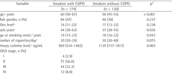
Table 1 - Demographic and smoking-related variables in smokers with and without COPD (n = 294).
Variable Smokers with COPD Smokers without COPD p*
(n = 174) (n = 120)
Age,a years 60 (50-67) 50 (45-55) < 0.001
Male gender, n (%) 84 (47) 46 (38) 0.237
COex levela 16 (11-22) 17 (13-22) 0.238
Pack-yearsa 44 (30-62) 37 (28-55) 0.026
Age at smoking onset,a years 19 (15-23) 18 (16-22) 0.943
Number of cigarettes/daya 20 (20-35) 20 (20-40) 0.075
Urinary cotinine level,a ng/mL 969 (534-1482) 1139 (737-1817) 0.005
GOLD stage, n (%)
I 4 (2.9)
II 77 (56.6)
III 44 (32.2)
IV 12 (8.8)
The PUCRS Scientific Committee and Research Ethics Committee approved the project.
Results
A total of 294 smokers, of whom 174 (59.18%) had been diagnosed with COPD, were considered eligible for inclusion in the study. For all of the volunteers, smoking status was confirmed by urinary cotinine levels ≥ 50 ng/mL. Those with urinary cotinine levels < 50 ng/mL were excluded, even if they classified themselves as smokers.
The demographic characteristics of the volunteers with and without COPD are described in Table 1. Median age and median pack-years were significantly higher among the patients with COPD than among those without (p < 0.001 and p = 0.026, respectively). No other statistically significant differences were found between the two groups.
As can be seen from Table 2, the mean COex value in patients with COPD was 17.1 ± 0.6 ppm, compared with 17.6 ± 0.6 ppm in those without COPD (p = 0.559). When the data were adjusted by ANCOVA for gender, age, age at smoking onset, number of cigarettes/day and urinary cotinine level, those values became 17.8 ± 0.6 ppm and 16.6 ± 0.7 ppm in the patients with and without COPD, respectively (p = 0.200). The lack of statistical significance remained when the data were plotted logarithmically (univariate ANOVA: p = 0.133; ANCOVA: p = 0.724).
A wide dispersion of COex values was found for each FEV1 level and for each GOLD stage.
No statistically significant relationships were identified. Analyzing the relationship between FEV1 and COex values by Spearman’s test, we
found values of r = −0.06 and p = 0.53. We also
found no statistically significant relationship
between the GOLD stage and COex values
(r = 0.08 and p = 0.34).
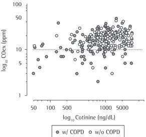
Figure 1 presents a scatter plot of urinary cotinine values, all ≥ 50 ng/ml, confirming the samples was used in order to compare the
groups in terms of quantitative data, whereas the chi-square test was used in order to compare the groups in terms of categorical data. For quantitative data without normal distribution, the nonparametric Mann-Whitney test was
used. In addition, the Levene test was used in
order to compare, specifically, the dispersion (variability) of COex values. Spearman’s correlation coefficient was used in order to evaluate the correlations with quantitative variables. To control for potential confounding factors and to evaluate their impact on COex levels, analysis of covariance (ANCOVA) was used. Logarithmic transformation of data related to COex and cotinine levels was performed in order to determine the reproducibility of the findings of the ANCOVA and for the presentation of the scatter plots. The level of significance was set at
α = 0.05. Data were processed with the Statistical Package for the Social Sciences, version 11.5
(SPSS Inc., Chicago, IL, USA).
Table 2 - Exhaled carbon monoxide levels in smokers with and without COPD (n = 294).a
Variable Smokers with COPD Smokers without COPD p p’
(n = 174) (n = 120)
Exhaled CO, ppm 17.1 ± 0.6 17.6 ± 0.6 0.559* 0.133
Adjusted exhaled CO, ppm 17.8 ± 0.6 16.6 ± 0.7 0.200** 0.724
aData presented as mean ± standard deviation. *Student’s t-test. **Analysis of covariance adjusted for gender, age, age at smoking onset, number of cigarettes/day and urinary cotinine level. p’: statistical analysis using the same tests, but with logCOex.
results suggests that the traditional criteria can be maintained.
The discrepancies among studies are attributed to several factors, such as the type of study design and the type of patients included in the study, as well as the presence of airway inflammation in varying degrees of severity, the different levels of change in lung architecture and the different degrees of airflow obstruction, all of which have frequently been cited.(14,15,17)
This has led some authors(15) to suggest
that the COex cut-off point for these patients be changed to 11 ppm. However, the fact that, in the present sample, similar COex values were found for smokers with COPD and for those without COPD suggests that the cut-off point can be maintained at 10 ppm.
Although there is evidence to support the use of a COex cut-off point between 6 and 8 ppm to differentiate smokers form nonsmokers,(4,11,15,22)
we chose to maintain the cut-off point of 10 ppm in the present study.
The use of this value is related to the situation of the patients included, who were under smoking cessation follow-up treatment at a specialized outpatient clinic, which necessitated greater specificity.
In studies investigating the prevalence of
current smoking in certain populations, sensitivity needs to be greater, and it would therefore be preferable to reduce the COex cut-off point.(7)
As previously mentioned, the results presented here are related to the cross-sectional analysis of a group of smokers upon their arrival at our outpatient clinic.
Although the smoking status of all of the patients was confirmed biochemically by the determination of urinary cotinine levels, COex levels < 10 ppm were found in some volunteers in the two groups studied. Possibly, these low values detected are related to the time elapsed since the last cigarette smoked, which might have been longer than predicted, despite the fact that the patients were emphatically instructed to respect the time frame, which was standardized at ≤ 60 min. The inverse (smokers with urinary cotinine levels < 50 ng/mL) was not detected, since this was an exclusion criterion.
Some patient characteristics presented here
merit consideration. In both of the groups
studied, females predominated, a trend similar to that detected in studies focusing on smoking smoking status of the volunteers. Nevertheless,
40 patients (14.0%) presented COex values below 10 ppm, which is the cut-off point used in order to classify an individual as a smoker
by this method. In this subgroup, 32 had COPD
and 8 did not. Therefore, among the patients with COPD, the rate of false-negative results was 18.4% (classified as smokers based on the urinary cotinine level but classified as nonsmokers based on the COex level), compared with 6.7% among the patients without COPD (p = 0.007).
Figure 2 shows the distribution of COex values in the patients with and without COPD. The equivalence between the groups is clear, as is the intersection between them, which accounts for the lack of a statistical difference in the distribution of the data.
Discussion
The present study shows that the evaluation of the smoking status in smokers with COPD based on the determination of COex levels can be performed using the same criteria applied to smokers without COPD.
Although proposals to change the COex cut-off points for categorizing individuals with COPD as smokers or nonsmokers can be found in the literature,(14,15) the analysis of the present
and their morphophysiological consequences for the COex cut-off points were not studied.
In addition, a wide dispersion of COex values was found for each GOLD stage, without a
statistically significant relationship (r = 0.08 and p = 0.34).
The need to use COex levels as a marker of smoking abstinence at smoking cessation clinics is growing, especially because it is easily applied and yields immediate results.(10,11) This
was confirmed in the present study, in which the patients had no difficulty in performing the necessary maneuvers and the results were obtained immediately.
The external validity of our results and conclusions needs to be corroborated by other studies. The high frequency of patients with COPD in smoking cessation clinics makes it imperative that the issues addressed here be definitively clarified.
References
1. Vollset SE, Tverdal A, Gjessing HK. Smoking and deaths
between 40 and 70 years of age in women and men.
Ann Intern Med. 2006;144(6):381-9.
2. World Health Organization. WHO Framework Convention
on Tobacco Control. Geneva: World Health Organization;
2005.
3. Fiore M, Jaén CR, Baker TB, Bailey WC, Bennett
G, Benowitz NL, et al. Treating tobacco use and dependence: 2008 Update. Clinical Practice Guideline.
Rockville: Public Health Service; 2008.
4. Reichert J, Araújo AJ, Gonçalves CM, Godoy I, Chatkin
JM, Sales MP, et al. Smoking cessation guidelines--2008. J Bras Pneumol. 2008;34(10):845-80.
5. Middleton ET, Morice AH. Breath carbon monoxide as an indication of smoking habit. Chest. 2000;117(3):758-63.
6. Pearce MS, Hayes L; Newcastle Heart Project; Newcastle Thousand Families Study. Self-reported smoking status and exhaled carbon monoxide: results from two population-based epidemiologic studies in the North of England. Chest. 2005;128(3):1233-8.
7. Rebagliato M. Validation of self reported smoking. J Epidemiol Community Health. 2002;56(3):163-4. 8. Barrueco M, Jiménez Ruiz C, Palomo L, Torrecilla M,
Romero P, Riesco JA. Veracity of smokers’ reports of abstinence at smoking cessation clinics [Article in Spanish]. Arch Bronconeumol. 2005;41(3):135-40. 9. Jatlow P, Toll BA, Leary V, Krishnan-Sarin S, O’Malley
SS. Comparison of expired carbon monoxide and plasma cotinine as markers of cigarette abstinence. Drug Alcohol Depend. 2008;98(3):203-9.
10. Javors MA, Hatch JP, Lamb RJ. Cut-off levels for breath carbon monoxide as a marker for cigarette smoking. Addiction. 2005;100(2):159-67.
11. Santos UP, Gannam S, Abe JM, Esteves PB, Freitas
FM, Wakassa TB, et al. Emprego da determinação de cessation clinics, especially in developing
countries.(23,24) This preponderance of females
can be explained by a possible selection bias, since it has been shown that women seek medical treatment for smoking cessation more frequently than do men.(21,25-27)
The mean age was higher in the COPD group than in the non-COPD group, which is consistent with the natural history of the disease.(28) In
addition, only 3% of the patients with COPD had had more than 8 years of schooling, a finding that is also in accordance with those reported in the literature, which indicate that the frequency of COPD is inversely proportional to the level of education of the smoker.
The internal validity of the present study is strengthened by aspects such as the adequate size of the sample, in number of participants with and without COPD, as well as the completeness of the demographic information compiled in the database. Although the design of the study required that all of the procedures be performed within the same period, no patients were lost to follow-up. The possibility of using the determination of urinary cotinine levels as the gold standard, which is feasible in few centers in the country, also conferred greater reliability upon the information obtained here.
Possible technical difficulties that could affect the results obtained by the use of urinary cotinine levels were overcome, and the method was only used after the validation process had been completed.(20) We were also careful not
to include individuals who were taking any medication or who had a clinical condition that could affect the results.
The present study has limitations. One is that the results obtained were not compared with data from nonsmokers with COPD, most of whom are possibly better classified as former smokers. With this comparison, it would have been possible to determine with certainty whether the acute changes caused by the components of cigarette smoke affect the COex cut-off point. Another acknowledged limitation is that, although the volunteers were categorized by functional severity of COPD, it was not possible to conduct a thorough evaluation of its possible influence on the results obtained by the use of COex levels in view of the small
de cotinina em urina por cromatografia líquida de alta eficiência. Rev Bras Toxicol. 2006;19(1):25-31. 21. Chatkin JM, Abreu CM, Haggstram FM, Blanco DC,
Rodini V, Martins D, et al. Exhaled CO: is 10 ppm a reliable threshold value to confirm current smoking? Am J Respir Crit Care Med. 2002;165:19s.
22. Oliveira MV, Oliveira TR, Pereira CA, Bonfim AV, Leitão Filho FS, Voss LR. Smoking among hospitalized patients in a general hospital. J Bras Pneumol. 2008;34(11):936-41.
23. Instituto Nacional de Câncer – INCA [homepage on the Internet]. Rio de Janeiro: Ministério da Saúde [cited
2009 Apr 20]. Tabagismo: dados e números. Available from: http://www.inca.gov.br/releases/press_release_
view.asp?ID=1856
24. Ministério da Saúde. Instituto Nacional de Câncer.
Coordenação de Prevenção e Vigilância. Prevalência do tabagismo no Brasil: dados de inquéritos epidemiológicos em capitais brasileiras. Rio de Janeiro: Coordenação de
Prevenção e Vigilância/INCA/MS; 2004.
25. Chatkin JM, Mariante de Abreu C, Haggsträm FM, Wagner MB, Fritscher CC. Abstinence rates and predictors of outcome for smoking cessation: do Brazilian smokers need special strategies? Addiction. 2004;99(6):778-84. 26. Chatkin JM, Abreu CM, Blanco DC, Tonietto R, Scaglia N,
Wagner MB, et al. No gender difference in effectiveness of smoking cessation treatment in a Brazilian real-life
setting. Int J Tuberc Lung Dis. 2006;10(5):499-503. 27. Santos SR, Gonçalves MS, Leitão Filho FS, Jardim JR.
Profile of smokers seeking a smoking cessation program. J Bras Pneumol. 2008;34(9):695-701.
28. Menezes AM, Perez-Padilla R, Jardim JR, Muiño A, Lopez
MV, Valdivia G, et al. Chronic obstructive pulmonary disease in five Latin American cities (the PLATINO study):
a prevalence study. Lancet. 2005;366(9500):1875-81. monóxido de carbono no ar exalado para a detecção do
consumo de tabaco. J Pneumol. 2001;27(5):231-6.
12. Tønnesen P, Carrozzi L, Fagerström KO, Gratziou
C, Jimenez-Ruiz C, Nardini S, et al. Smoking cessation in patients with respiratory diseases: a high priority, integral component of therapy. Eur Respir J. 2007;29(2):390-417.
13. Chung KF, Adcock IM. Multifaceted mechanisms in
COPD: inflammation, immunity, and tissue repair and destruction. Eur Respir J. 2008;31(6):1334-56. 14. Montuschi P, Kharitonov SA, Barnes PJ. Exhaled
carbon monoxide and nitric oxide in COPD. Chest. 2001;120(2):496-501.
15. Sato S, Nishimura K, Koyama H, Tsukino M, Oga T, Hajiro T, et al. Optimal cutoff level of breath carbon monoxide for assessing smoking status in patients with asthma and COPD. Chest. 2003;124(5):1749-54.
16. Raw M, Anderson P, Batra A, Dubois G, Harrington
P, Hirsch A, et al. WHO Europe evidence based recommendations on the treatment of tobacco dependence. Tob Control. 2002;11(1):44-6.
17. van Beurden WJ, Dekhuijzen PN, Smeenk FW. Exhaled biomarkers in COPD: their potential role in diagnosis, treatment and prognosis. Monaldi Arch Chest Dis. 2002;57(5-6):258-67.
18. Global Initiative for Chronic Obstructive Lung Disease.
Pocket guide to COPD diagnosis, management and
prevention - A Guide for Health Care Professionals – update 2008. Bethesda: Global Initiative for Chronic
Obstructive Lung Disease; 2008.
19. Heatherton TF, Kozlowski LT, Frecker RC, Fagerström KO. The Fagerström Test for Nicotine Dependence: a revision of the Fagerström Tolerance Questionnaire. Br J Addict. 1991;86(9):1119-27.
20. Cattaneo R, Alegretti AP, Sagebin FR, Abreu CM, Petersen
GO, Chatkin JM. Validação do método para determinação
About the authors
Gustavo Chatkin
Physician. Department of Pulmonology, Pontifícia Universidade Católica do Rio Grande do Sul – PUCRS, Pontifical Catholic University of Rio Grande do Sul – São Lucas Hospital, Porto Alegre, Brazil.
José Miguel Chatkin
Full Professor of Internal Medicine and Pulmonology. Pontifícia Universidade Católica do Rio Grande do Sul – PUCRS, Pontifical Catholic University of Rio Grande do Sul – School of Medicine, Porto Alegre, Brazil.
Gabriel Aued
Medical Student. Pontifícia Universidade Católica do Rio Grande do Sul – PUCRS, Pontifical Catholic University of Rio Grande do Sul – School of Medicine, Porto Alegre, Brazil.
Guilherme Oliveira Petersen
Pharmacy Student. Pontifícia Universidade Católica do Rio Grande do Sul – PUCRS, Pontifical Catholic University of Rio Grande do Sul – School of Pharmacy, Porto Alegre, Brazil.
Edna Thais Jeremias
Nurse. Department of Pulmonology, Pontifícia Universidade Católica do Rio Grande do Sul – PUCRS, Pontifical Catholic University of Rio Grande do Sul – São Lucas Hospital, Porto Alegre, Brazil.
Flávia Valladão Thiesen