426
DOI:
10.1590/0004-282X20160049
IMAGES IN NEUROLOGY
Hypertrophic olivary degeneration: unveiling
the triangle of Guillain-Mollaret
Degeneração olivar hipertrófica: descobrindo o triângulo de Guillain-Mollaret
William Alves Martins
1, Luiz Carlos Porcello Marrone
1,3, Ricardo Bernardi Soder
1,3, Jaderson Costa da Costa
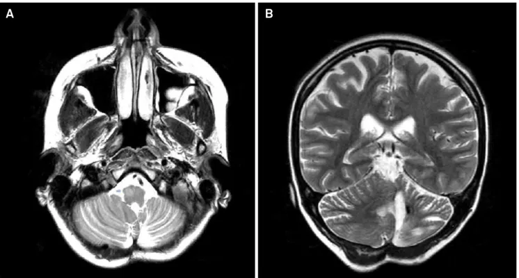
1,2,3A 28-year-old woman presented with hemi-dystonic
movements on the left. Four years earlier, a ruptured
vas-cular malformation caused a hemorrhagic stroke on the
left cerebellar hemisphere (Figure 1B), evacuated by
sur-gery. MRI revealed right
hypertrophic olivary
degenera-tion (HOD) (Figure 1A) and reducdegenera-tion of cerebellar-rubro
pathways (Figure 2).
HOD is a trans-synaptic degeneration afecting the
dentate-rubro-olivary pathway or Guillain-Mollaret’s
trian-gle
1. A lesion in any part of this synaptic pathway may lead to
HOD (Figure 3), including stroke, cranioencephalic trauma,
neurodegenerative diseases, inlammatory illnesses, among
others
2,3. his unique synaptic collapse is associated to pala
-tal myoclonus, parkinsonian features and dystonia
1,2.
1 Pontifícia Universidade Católica do Rio Grande do Sul, Hospital São Lucas, Departamento de Neurologia, Porto Alegre RS, Brasil; 2 Pontifícia Universidade Católica do Rio Grande do Sul, Faculdade de Medicina, Departamento de Medicina Interna, Porto Alegre RS, Brasil; 3 Pontifícia Universidade Católica do Rio Grande do Sul, Instituto do Cérebro, Porto Alegre RS, Brasil.
Correspondence: William Alves Martins; Departmento de Neurologia, Hospital São Lucas – PUCRS; Avenida Ipiranga 6690/220; 00610-000 Porto Alegre RS, Brasil; Email: [email protected]
Conflict of interest: There is no conflicts of interest to declare.
Received 22 August 2015; Received in final form 29 November 2015; Accepted 09 December 2015.
Figure 1.
(A) Axial T2-weighted image showing enlargement and hyperintensity of the
right
olivary nuclei, compatible to hypertrophic
olivary degeneration; (B) Coronal T2-weighted image displaying reduced
left
cerebellar hemisphere associated to hemosiderin.
427
William Alves Martins et al. Hyperthophic olivary degeneration
References
1. Guillain G, Mollaret P. Deux-cas de myoclonies synchrones et rhythmes velopharyngo-laringo-oculo-diaphragmatiques. Rev Neurol. 1931;2:545-66.
2. Martins WA, Schilling LCP, Neto FK, Becker J. Hypertrophic olivary degeneration secondary to Neuro-Behçet’s disease.
Clin Neuroradiol. 2015 Mar 24 [Epub ahead of print]. doi:10.1007/s00062-015-0384-0
3. Siebert E, Harms L, Herbst M. Posttraumatic bilateral hypertrophic olivary degeneration. Neurol Sci. 2013;34(10):1829-30.
doi:10.1007/s10072-013-1309-9