Clinical, radiological, and laboratory characteristics in
pulmonary tuberculosis patients: comparative study of
HIV‑positive and HIV‑negative inpatients at a referral hospital*
Manifestações clínicas, radiológicas e laboratoriais em indivíduos com tuberculose pulmonar: estudo comparativo entre indivíduos HIV positivos
e HIV negativos internados em um hospital de referência
Aline Besen, Guilherme Jönck Staub, Rosemeri Maurici da Silva
Abstract
Objective: To compare clinical, radiological, and laboratory characteristics of individuals with pulmonary tuberculosis co-infected or not with HIV. Methods: A cross-sectional study, in which signs and symptoms were assessed by anamnesis and physical examination in patients hospitalized with pulmonary tuberculosis. The results of sputum smear microscopy and culture for Mycobacterium tuberculosis, as well as hemoglobin levels and CD4+
T-cell counts, were obtained from medical records, and chest X-ray reports were consulted. Results: We included 50 pulmonary tuberculosis patients, who were divided into two groups (HIV-positive and HIV-negative; n = 25 per group). The mean age of the participants was 38.4 ± 10.5 years; 46 (92%) were males; and 27 (54%) were White. Expectoration was presented by 21 (84%) and 13 (52%) of the patients in the HIV-negative and HIV-positive groups, respectively (p = 0.016). Radiological findings of cavitation were present in 10 (43%) and 2 (10%) of the patients in the HIV-negative and HIV-positive groups, respectively (p = 0.016), whereas an interstitial pattern was observed in 18 (78%) and 8 (40%), respectively (p = 0.012). The mean hemoglobin level was 11.1 ± 2.9 g/ dL and 9.3 ± 2.2 g/dL in the HIV-negative and HIV-positive groups, respectively (p = 0.015). Conclusions: In our sample of tuberculosis patients, expectoration was less prevalent, hemoglobin levels were lower, and cavitation was less common, as was an interstitial pattern, among those co-infected with HIV than among those without HIV co-infection.
Keywords: HIV; Tuberculosis; Acquired immunodeficiency syndrome.
Resumo
Objetivo: Comparar as manifestações clínicas, radiológicas e laboratoriais de indivíduos com tuberculose pulmonar coinfectados com HIV com aqueles sem a coinfecção. Métodos: Estudo transversal, no qual sinais e sintomas foram analisados por meio de anamnese e exame físico em pacientes internados com tuberculose pulmonar. A baciloscopia, a cultura para Mycobacterium tuberculosis, a dosagem de hemoglobina e a contagem de células T CD4+ foram
obtidas de registros dos prontuários, assim como os laudos das radiografias de tórax. Resultados: Foram incluídos 50 pacientes com tuberculose pulmonar, que foram divididos em dois grupos (HIV positivo e HIV negativo; n = 25 por grupo). A média de idade dos participantes foi de 38,4 ± 10,5 anos, 46 (92%) eram do sexo masculino, e 27 (54%) eram caucasianos. Apresentaram expectoração 21 (84%) e 13 (52%) dos pacientes nos grupos HIV negativo e HIV positivo, respectivamente (p = 0,016). Achados radiológicos de cavitação estavam presentes em 10 (43%) e 2 (10%) dos pacientes nos grupos HIV negativo e HIV positivo, respectivamente (p = 0,016), ao passo que padrão intersticial estava presente em 18 (78%) e 8 (40%) dos pacientes nesses grupos (p = 0,012). O nível médio de hemoglobina foi de 11,1 ± 2,9 g/dL e 9,3 ± 2,2 g/dL nos grupos HIV negativo e HIV positivo, respectivamente (p = 0,015). Conclusões: Entre pacientes coinfectados com tuberculose e HIV desta amostra, houve menor prevalência de expectoração, foram menos frequentes os achados radiológicos de cavitação e de padrão intersticial, e os níveis de hemoglobina foram mais baixos do que naqueles sem essa coinfecção.
Descritores: HIV; Tuberculose; Síndrome de imunodeficiência adquirida.
* Study carried out at the University of Southern Santa Catarina and the Federal University of Santa Catarina, Florianópolis, Brazil. Correspondence to: Rosemeri Maurici da Silva. Rodovia Virgílio Várzea, 2236, Residencial Villa Vernazza, apto. 601, bloco A, Saco Grande II, CEP 88032-001, Florianópolis, SC, Brasil.
Tel. 55 48 3621-3363. E-mail: [email protected] Financial support: None.
to seven days before the initiation of treatment with antituberculosis drugs.
The participants underwent focused history taking, during which they were asked about the presence of the following symptoms: cough; wheezing; fever; sweating; weight loss; anorexia; asthenia; chest pain; dyspnea; and irritability (sudden mood changes, impatience, or both). The participants who confirmed the presence of cough were asked about the presence of hemoptysis and expectoration. Those who reported sweating were asked whether they had day sweats, night sweats, or both. Those who reported weight loss were asked how much weight they had lost. The duration of each symptom was also investigated.
Physical examination included palpation of the head and neck lymph nodes, including cervical and supraclavicular lymph nodes. Lymph node enlargement was defined as the presence of lymph nodes larger than 1 cm. Arthritis was defined as the presence of signs of inflammation of any of the joints. Ascites was defined as the presence of a fluid thrill or shifting dullness to percussion of the abdomen. Liver size was estimated by percussion and palpation of the liver, hepatomegaly being defined as a liver larger than 14 cm. The diagnosis of splenomegaly was established by percussion of Traube’s space and palpation of the spleen with the patient in the right lateral decubitus position, with the left arm raised, the forearm resting on the top of the head, and the left leg slightly flexed at hip and knee. Splenomegaly was defined as dullness to Traube’s space percussion or a palpable splenic tip. Digital clubbing was defined as a digital index ≥ 1.
History taking and physical examination were performed by two observers, and each participant was examined by one of the observers, chosen in a random manner.
The information regarding the anteroposterior and lateral chest X-ray findings was obtained from the chest X-ray reports found in the medical records of the patients, and the findings were classified as follows: alveolar consolidation; interstitial infiltrate; pleural effusion; cavitation; mediastinal or hilar lymph node enlargement (or both); mass; nodule; or any combination thereof.
The results of the laboratory tests (CD4+ T-cell count, determination of hemoglobin
Introduction
Infection with HIV is one of the major risk factors for tuberculosis. Among immunocompetent individuals with latent tuberculosis, there is a 10% chance that the infection will progress to active disease over the course of their lifetime; among HIV-positive individuals with latent tuberculosis, that chance is 8-10% per year.(1)
With the advent of AIDS, the manifestations of tuberculosis in HIV-positive individuals have been found to differ from those observed in HIV-negative individuals.(2) In most individuals with a CD4+ T-cell count below 200 cells/mm3, the radiological presentation of tuberculosis is atypical. In addition, there is usually no granuloma formation.(3,4)
Immunological changes have an impact on tissue manifestations, on radiological manifestations, and possibly on clinical manifestations. The objective of the present study was to compare pulmonary tuberculosis patients with and without HIV co-infection in terms of clinical, radiological, and biochemical characteristics.
Methods
mean CD4+ T-cell count was 174.6 ± 158.0 cells/mm3, the lowest being 25 cells/mm3.
The mean age of the patients in the HIV-positive group was 37.2 ± 10.6 years, compared with 39.6 ± 10.6 years for the HIV-negative group (p = 0.442). Of the 25 patients in the HIV-negative group, 13 (52%) were White, compared with 11 (44%) of those in the HIV-positive group (p = 0.500). In the HIV-negative group, 23 (92%) of the patients were male, as were 23 (92%) of those in the HIV-positive group (p = 0.695).
When asked about previous treatment for tuberculosis, 33 (66%) of the participants reported that they had never undergone tuberculosis treatment, and there was no significant difference between the two groups (p = 0.500).
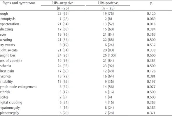
Of the sample as a whole, 42 (84%) reported cough, the mean duration of the symptom being 3.0 ± 4.5 months. In addition, 9 (18%) reported hemoptysis, and 34 (68%) reported expectoration. As can be seen in Table 1, expectoration was more common among the individuals in the HIV-negative group (p = 0.016).
levels, sputum smear microscopy for AFB, and culture for M. tuberculosis) were obtained from the medical records of the participants.
All of the participants gave written informed consent. We used the Statistical Package for the Social Sciences, version 16.0 (SPSS Inc., Chicago, IL, USA) in order to devise a database and analyze it. The data were summarized as proportions or means. In order to compare the two groups, we used the chi-square test and Fisher’s exact test (for categorical variables), as well as the Student’s t-test (for continuous variables). The level of significance was set at 95%.
The study was approved by the Research Ethics Committee of the Federal University of Santa Catarina (129/04, FR-255594).
Results
We evaluated 52 individuals. Of those, 2 were excluded because they had not undergone HIV serology. The study sample therefore comprised 50 patients, the mean age being 38.4 ± 10.5 years. Of the 50 patients, 4 were female (8%), and 27 (54%) were White. Positive HIV serology was observed in 25 (50%), among whom the
Table 1 ‑ Distribution of signs and symptoms by HIV status.a
Signs and symptoms HIV-negative HIV-positive p (n =25) (n = 25)
Cough 23 (92) 19 (76) 0.120
Hemoptysis 7 (28) 2 (8) 0.069
Expectoration 21 (84) 13 (52) 0.016
Wheezing 17 (68) 15 (60) 0.384
Fever 19 (76) 21 (84) 0.363
Sweating 21 (84) 22 (88) 0.500
Day sweats 3 (12) 6 (24) 0.532
Night sweats 21 (84) 20 (80) 0.338
Weight loss 24 (96) 25 (100) 0.500
Loss of appetite 19 (76) 21 (84) 0.363
Asthenia 24 (96) 23 (92) 0.500
Chest pain 17 (68) 12 (48) 0.126
Dyspnea 18 (72) 16 (64) 0.381
Irritability 13 (52) 9 (36) 0.197
Lymph node enlargement 8 (32) 14 (56) 0.077
Arthritis 3 (12) 4 (16) 0.500
Ascites 2 (8) 1 (4) 0.500
Digital clubbing 6 (24) 4 (16) 0.363
Hepatomegaly 4 (16) 6 (24) 0.363
Splenomegaly 5 (20) 7 (28) 0.371
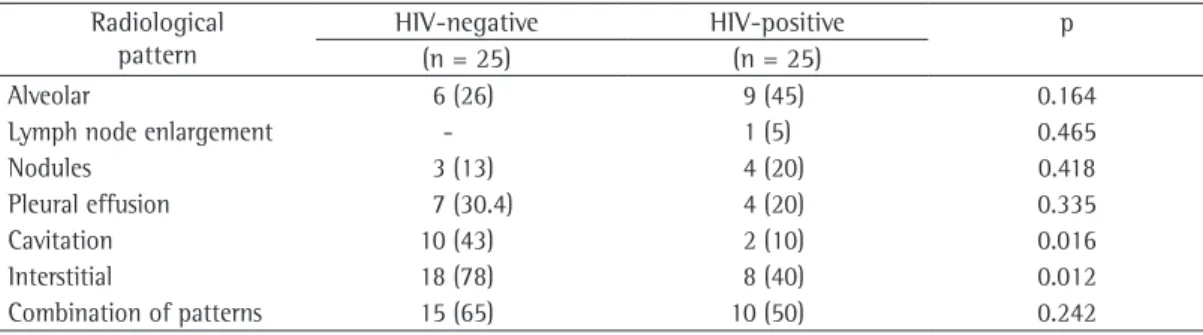
X-ray reports were available for 23 individuals, whereas they were available for only 20 of the individuals in the HIV-positive group. Radiological findings of cavitation and an interstitial pattern were more common in the individuals in the HIV-negative group (p < 0.05). Table 3 shows the distribution of radiological patterns.
Among the HIV-negative patients, culture for M. tuberculosis was positive, negative, contaminated, or not performed in 56%, 16%, 4%, and 24%, respectively, compared with 56%, 36%, 4%, and 1%, respectively, among the HIV-positive patients (p = 0.115). Among the HIV-negative patients, sputum smear microscopy results were negative, 1+, 2+, and 3+ in 12%, 28%, 20%, and 40%, respectively, compared with 28%, 36%, 16%, and 20%, respectively, among the HIV-positive patients (p = 0.336).
Mean hemoglobin levels were 10.2 ± 2.7 g/ dL for the sample as a whole, 11.1 ± 2.9 g/dL in the HIV-negative group, and 9.3 ± 2.2 g/dL in the HIV-positive group (p = 0.015 between the two groups).
Fever was reported by 40 patients (80%), and sweating was reported by 43 patients (86%). In addition, 9 (18%) of the participants reported day sweats, and 41 (82%) reported night sweats.
Weight loss was reported by 49 individuals (98%), the mean weight loss being 10.5 ± 7.1 kg, with no significant difference between the two groups. Loss of appetite was reported by 40 patients (80%), and asthenia was reported by 47 patients (94%).
Regarding the signs investigated, lymph node enlargement was found in 22 patients (44%), arthritis was found in 7 (14%), ascites was found in 3 (6%), digital clubbing was found in 10 (20%), hepatomegaly was found in 10 (20%), and splenomegaly was found in 12 (24%). There were no significant differences between the two groups in terms of the abovementioned signs. As can be seen in Table 2, there were no statistical differences between the two groups in terms of the mean duration of signs and symptoms.
We evaluated the chest X-ray reports that were available in the medical records of the participants. In the HIV-negative group, chest
Table 2 ‑ Distribution of the duration (in months) of the reported signs and symptoms by HIV status.a
Signs and symptoms HIV-negative HIV-positive p (n =25) (n = 25)
Cough 4.1 (5.9) 1.7 (1.2) 0.082
Wheezing 4.4 (8.6) 1.9 (1.8) 0.296
Fever 8 (19.3) 1.4 (0.9) 0.127
Weight loss 4.2 (7.2) 2.2 (1.8) 0.194
Loss of appetite 4.7 (8.3) 2 (1.5) 0.154
Asthenia 3.3 (5.1) 1.3 (0.8) 0.074
Chest pain 5.8 (9.7) 1.6 (1.0) 0.145
Dyspnea 10.4 (20.3) 1.7 (1.6) 0.097
Irritability 6.2 (9.7) 1.7 (1.3) 0.187
aValues expressed as mean ± SD.
Table 3 ‑ Distribution of the radiological patterns by HIV status.a
Radiological pattern
HIV-negative HIV-positive p (n = 25) (n = 25)
Alveolar 6 (26) 9 (45) 0.164
Lymph node enlargement - 1 (5) 0.465
Nodules 3 (13) 4 (20) 0.418
Pleural effusion 7 (30.4) 4 (20) 0.335
Cavitation 10 (43) 2 (10) 0.016
Interstitial 18 (78) 8 (40) 0.012
Combination of patterns 15 (65) 10 (50) 0.242
study might be due to possible associated factors, such as smoking (which was not explored), as well as to the possibility of concomitant airway obstructive diseases (asthma or COPD) that were not reported by the participants or that had not been diagnosed.
Two studies(12,13) reported a higher prevalence of fever and fatigue among HIV-positive patients, a result that is in disagreement with those of the present study. However, those studies were based on data collected from medical records rather than on those obtained by focused history taking, which might explain the difference.
In the present study, sweating was reported by 86.0% of the patients. Of those, 82.0% reported night sweats. The reported prevalence of sweating was found to be 50.5-84.0%.(8,14,15) The data of the present study confirm that sweating, principally night sweats, is a classic symptom of tuberculosis.
In the present study, weight loss was the most common symptom (observed in 98.0%). In another study conducted in Brazil,(16) weight loss was found in 74.5% of the patients. One group of authors(17) reported that malnutrition is related to tuberculosis, meaning that malnutrition is a risk factor for tuberculosis and a consequence of the disease. The consumptive nature of tuberculosis is related to anorexia and inflammatory cytokines, which induce a catabolic state.
In the present study, asthenia was the second most prevalent symptom, having been reported by 94.0% of the patients. In another study involving HIV-positive individuals,(18) asthenia was found in 51.6%. There are few studies showing objective data regarding asthenia in patients with tuberculosis. However, because of the high prevalence of the symptom, more studies should address the theme, despite its subjective nature.
In the present study, chest pain was reported by 58.0% of the participants. The reported prevalence of chest pain among tuberculosis patients ranges from 13.1% to 76.0%.(16,18) Such chest pain is related to pleural involvement. (4) Because we found a significant prevalence of pleural effusion in the present study, a high prevalence of chest pain was expected.
Dyspnea was reported by 68% of the patients, a result that is in agreement with those of one study(9) but in disagreement with those of others,
Discussion
Among the regions of Brazil, the southern region has the lowest tuberculosis incidence rates. The tuberculosis incidence rate reported for 2009 in the state of Santa Catarina was 27.63/100,000 population, lower than the 37.99/100,000 population reported for Brazil as a whole. However, the proportion of tuberculosis patients co-infected with HIV is 20.3%, which is higher than the 15.0% reported for Brazil as a whole.(5,6) The proportion of tuberculosis patients co-infected with HIV found in the present study (i.e., 50.0%) is in agreement with the findings of another group of researchers,(7) who reported that proportion to be 48.9% among inpatients at hospitals in southern Brazil. The fact that the proportion of HIV-positive individuals among patients hospitalized with tuberculosis is higher than is that of those among all of the reported cases of tuberculosis might denote greater disease severity in that group or reflect the trend toward hospitalization at referral hospitals for the treatment of infectious and parasitic diseases.
The clinical manifestations of tuberculosis are varied and nonspecific, and there is no clinical sign or symptom that is exclusive to the disease. Cough (dry or productive) for more than three weeks is a sentinel of tuberculosis. (8) One group of authors(9) found that symptom in 96% of a group of patients with pulmonary tuberculosis, a proportion that is similar to that found in the present study.
We found expectoration in 68% of our patients, a finding that is in agreement with data in the literature, according to which expectoration is found in 65-72% of patients. (10,11) In the present study, expectoration was more common among the individuals in the HIV-negative group than among those in the HIV-positive group. This might reflect the impaired immune status of the latter, with a reduced number of defense cells and, consequently, less expectoration. The presence of unproductive cough in HIV-positive patients contributes to the difficulty in diagnosing tuberculosis through sputum smear microscopy and culture, invasive tests being sometimes required.
with pleural effusion has been reported to be 5.3-18.0%.(7,14,15,23) We identified nodules in 16.3% of the cases. In one study,(15) X-rays revealed nodules in only 1.3% of tuberculosis patients co-infected with HIV. In another study,(24) nodules constituted the most common CT finding in patients with active tuberculosis, having been found in 82.0% of the cases. This disparity can be attributed to the difference in sensitivity between the two methods.
The chest X-ray findings in patients with tuberculosis/HIV co-infection have often been described as being uncharacteristic of tuberculosis. In our study sample, chest X-ray findings of cavitation were less common in the patients in the HIV-positive group (p = 0.016). In studies analyzing the degree of immunosuppression in individuals with tuberculosis/HIV co-infection, cavitation was reported to be most common in those with a CD4+ T-cell count > 200 cells/mm3. (12,25) Another different finding of the present study was a higher prevalence of interstitial lesions among the patients in the HIV-negative group (p = 0.012). One group of authors(26) found that, among patients with tuberculosis/ HIV co-infection, interstitial lesions were most common in those with a CD4+ T-cell count < 200 cells/mm3.
Various authors have reported that mediastinal lymph node enlargement is a common radiological finding in immunocompromised patients with tuberculosis.(12,25) In the present study, mediastinal lymph node enlargement was found in 5% of the X-rays of HIV-positive individuals and in none of those of HIV-negative individuals (p = 0.465), similar to the findings of another study.(15) In the evaluation of the degree of immunosuppression, intrathoracic lymph node enlargement is the radiological finding that is related to the lowest CD4+ T-cell counts.(26) The small number of patients in our study sample, as well as their relatively high CD4+ T-cell counts, can explain the differences between our findings and those reported in the literature.
In the literature, pleural effusion is described as a finding that is more common among HIV-positive patients than among HIV-negative patients. One group of authors(23) found that the prevalence of pleural effusion among HIV-positive and HIV-negative patients was 16.0% and 6.8%, respectively (p < 0.001). in which the proportion of dyspnea was reported
to be approximately 26%.(16-18) The possibility of association with other opportunistic infections, such as pneumocystosis, cannot be completely ruled out. Although the presence of opportunistic infections, as registered in the medical records, constituted an exclusion criterion, no tests were performed in order to identify such infections.
In patients with tuberculosis, involvement of the joints results from hematogenous dissemination or contiguous bone lesion.(19) In one study,(10) the proportion of bone or joint involvement was reported to be 0.2%, markedly lower than the 14.0% found in the present study.
The peritoneum ranks sixth among the extrapulmonary sites that are most affected by tuberculosis. The principal manifestations are ascites, found in 73.0% of patients, and abdominal pain, found in 64.5%.(20) In one study,(20) the prevalence of ascites among the cases of tuberculosis was reported to be 3.5%, a proportion that is close to the proportion of 6.0% found in the present study.
Digital clubbing is a finding that indicates severe disease. In one study,(21) the prevalence of digital clubbing was reported to be 34%. Improvements in the treatment of tuberculosis and early treatment initiation might have led to a reduction in the prevalence of digital clubbing. We found no significant differences between the groups under study in terms of constitutional symptoms, such as weight loss, loss of appetite, and irritability. A review of tuberculosis and malnutrition reported greater weight loss among the tuberculosis patients who were co-infected with HIV.(17) One group of authors(22) found that, among HIV-positive patients, those with a CD4+ T-cell count above 200 cells/mm3 lost the least amount of weight.
The predominance of the interstitial and alveolar patterns over the cavitation pattern can be explained by the large proportion of immunocompromised individuals in our study sample, the mean CD4+ T-cell count being lower than 200 cells/mm3. The predominance of consolidation over cavitation might also be due to the fact that the disease was diagnosed in a timely manner.(8,14)
presentation of pulmonary tuberculosis. A study of the relationship between these factors in patients with human immunodeficiency virus infection. Chest. 1995;107(1):74-80.
5. Severo NP, Leite CQ, Capela MV, Simões MJ. Clinical and demographic characteristics of patients hospitalized with tuberculosis in Brazil between 1994 and 2004. J Bras Pneumol. 2007;33(5):565-71.
6. Menezes AM, Costa JD, Gonçalves H, Morris S, Menezes M, Lemos S, et al. Incidência e fatores de risco para tuberculose em Pelotas, uma cidade do Sul do Brasil. Rev Bras Epidemiol. 1998;1(1):50-60.
7. Picon PD, Caramori ML, Bassanesi SL, Jungblut S, Folgierini M, Porto Nda S, et al. Differences in the clinical and radiological presentation of intrathoracic tuberculosis in the presence or absence of HIV infection. J Bras Pneumol. 2007;33(4):429-36.
8. Domínguez Del Valle F, Fernández B, Pérez de Las Casas M, Marín B, Bermejo C. Clinical manifestations and radiology of thoracic tuberculosis [Article in Spanish]. An Sist Sanit Navar. 2007;30 Suppl 2:33-48.
9. Job JR, Prado PE, Vranjac S, Duarte PC. Comparison of epidemiological data on pulmonary tuberculosis in Sorocaba, Säo Paulo, Brazil, (1986-1996) [Article in Portuguese]. Rev Saude Publica. 1998;32(6):596-7. 10. Aktoğu S, Yorgancioglu A, Cirak K, Köse T, Dereli SM.
Clinical spectrum of pulmonary and pleural tuberculosis: a report of 5,480 cases. Eur Respir J. 1996;9(10):2031-5. 11. Mello FC. Modelos Preditivos para o Diagnóstico da
Tuberculose Pulmonar Paucibacilar [dissertation]. Rio de Janeiro: Universidade Federal do Rio de Janeiro; 2001. 12. Henn L, Nagel F, Dal Pizzol F. Comparison between
human immunodeficiency virus positive and negative patients with tuberculosis in Southern Brazil. Mem Inst Oswaldo Cruz. 1999;94(3):377-81.
13. Richter C, Perenboom R, Mtoni I, Kitinya J, Chande H, Swai AB, et al. Clinical features of HIV-seropositive and HIV-seronegative patients with tuberculous pleural effusion in Dar es Salaam, Tanzania. Chest. 1994 ;106(5):1471-5.
14. Burrill J, Williams CJ, Bain G, Conder G, Hine AL, Misra RR. Tuberculosis: a radiologic review. Radiographics. 2007;27(5):1255-73.
15. de Albuquerque Mde F, Albuquerque SC, Campelo AR, Cruz M, de Souza WV, Ximenes RA, et al. Radiographic features of pulmonary tuberculosis in patients infected by HIV: is there an objective indicator of co-infection? Rev Soc Bras Med Trop. 2001;34(4):369-72.
16. Oliveira HM, Brito RC, Kritski AL, Ruffino-Netto A. Epidemiological profile of hospitalized patients with TB at a referral hospital in the city of Rio de Janeiro, Brazil. J Bras Pneumol. 2009;35(8):780-7.
17. Schwenk A, Macallan DC. Tuberculosis, malnutrition and wasting. Curr Opin Clin Nutr Metab Care. 2000;3(4):285-91.
18. Boffo MM, Mattos IG, Ribeiro MO, Oliveira Neto IC. Tuberculosis associated to AIDS: demographic, clinical and laboratory characteristics of patients cared for at a reference center in the south of Brazil [Article in Portuguese]. J Pneumol. 2004;30(2):140-6.
19. Harisinghani MG, McLoud TC, Shepard JA, Ko JP, Shroff MM, Mueller PR. Tuberculosis from head to toe. Radiographics. 2000;20(2):449-70; quiz 528-9, 532. 20. Sanai FM, Bzeizi KI. Systematic review: tuberculous
peritonitis--presenting features, diagnostic
In another study,(25) the prevalence of pleural effusion was found to be higher in patients with a CD4+ T-cell count > 200 cells/mm3. However, another group of authors(22) found that pleural effusion was more common in individuals with a CD4+ T-cell count < 200 cells/mm3. The HIV-positive patients analyzed in the present study had mean CD4+ T-cell counts that were quite close to the cut-off point of 200 cells/ mm3, and this might have influenced the results obtained. An analysis of a larger number of patients with CD4+ T-cell counts significantly lower and significantly higher than the cut-off point might yield data that are more robust.
Of the total number of cultures performed, 65.1% were positive, a proportion that is lower than is that found in another study (i.e., 78.6%). (10) The difference might be due to the large proportion of HIV-positive patients in the present study. It is known that HIV-positive patients are unable to produce a quantity of spontaneous sputum that is sufficient for testing.(27)
Anemia is a common finding in patients with tuberculosis. In one study,(28) the prevalence of anemia among tuberculosis patients was reported to be 31.9%. In the present study, mean hemoglobin levels were significantly lower in the HIV-positive patients, which probably reflects the fact that HIV infection worsens anemia.
Our comparative study of clinical, radiological, and laboratory characteristics of individuals with pulmonary tuberculosis co-infected or not with HIV revealed that expectoration was significantly less prevalent, hemoglobin levels were significantly lower, and cavitation was significantly less common, as was an interstitial pattern, among those co-infected with HIV than among those without HIV co-infection.
References
1. Ministério da Sáde. Fundação Nacional de Sáde. Centro de Referência Professor Hélio Fraga; Sociedade Brasileira de Pneumologia e Tisiologia. Controle da Tuberculose: uma proposta de integração ensino-serviço. 5th ed. Rio de Janeiro: FUNASA; 2002.
2. Pitchenik AE, Rubinson HA. The radiographic appearance of tuberculosis in patients with the acquired immune deficiency syndrome (AIDS) and pre-AIDS. Am Rev Respir Dis. 1985;131(3):393-6.
3. Haramati LB, Jenny-Avital ER. Approach to the diagnosis of pulmonary disease in patients infected with the human immunodeficiency virus. J Thorac Imaging. 1998;13(4):247-60.
25. da Silva RM, da Rosa L, Lemos RN. Radiographic alterations in patients presenting human immunodeficiency virus/ tuberculosis coinfection: correlation with CD4+ T cell counts. J Bras Pneumol. 2006;32(3):228-33.
26. Post FA, Wood R, Pillay GP. Pulmonary tuberculosis in HIV infection: radiographic appearance is related to CD4+ T-lymphocyte count. Tuber Lung Dis. 1995;76(6):518-21.
27. Silva RM, Teixeira PJ, Moreira JS. O escarro induzido no diagnóstico das doenças pulmonares em pacientes positivos ao vírus da imunodeficiência humana. J Pneumol. 2004;30(5):452-8.
28. Lee SW, Kang YA, Yoon YS, Um SW, Lee SM, Yoo CG, et al. The prevalence and evolution of anemia associated with tuberculosis. J Korean Med Sci. 2006;21(6):1028-32.
strategies and treatment. Aliment Pharmacol Ther. 2005;22(8):685-700.
21. Reeve PA, Harries AD, Nkhoma WA, Nyangulu DS, Wirima JJ. Clubbing in African patients with pulmonary tuberculosis. Thorax. 1987;42(12):986-7.
22. Garcia GF, Moura AS, Ferreira CS, Rocha MO. Clinical and radiographic features of HIV-related pulmonary tuberculosis according to the level of immunosuppression. Rev Soc Bras Med Trop. 2007;40(6):622-6.
23. Tshibwabwa-Tumba E, Mwinga A, Pobee JO, Zumla A. Radiological features of pulmonary tuberculosis in 963 HIV-infected adults at three Central African Hospitals. Clin Radiol. 1997;52(11):837-41.
24. Bombarda S, Figueiredo CM, Funari MB, Soares Jr J, Seiscento M, Filho MT. Imagem em tuberculose pulmonar. J Pneumol. 2001;27(6):329-40.
About the authors
Aline Besen
Medical Student. Federal University of Santa Catarina, Florianópolis, Brazil.
Guilherme Jönck Staub
Medical Student. Federal University of Santa Catarina, Florianópolis, Brazil.
Rosemeri Maurici da Silva