A modified method by differential adhesion for enrichment
of bladder cancer stem cells
_______________________________________________
Yong-tong Zhu
1,2, Shi-yu Pang
2, Yang Luo
2, Wei Chen
2, Ji-ming Bao
2, Wan-long Tan
21 Department of Obstetrics and Gynecology, Center for Reproductive Medicine, Nanfang Hospital, Southern
Medical University, Guangzhou, China; 2 Department of Urology, Nanfang Hospital, Southern Medical
University, Guangzhou, China
ABSTRACT
ARTICLE
INFO
______________________________________________________________ ______________________
Purpose: In a previous study the vaccine was effective against bladder cancer in a mouse model. However, a small portion of tumors regrew because the vaccine could not eliminate bladder cancer stem cells (CSCs). In this study, we showed a modified method for the isolation of bladder CSCs using a combination of differential adhesion method and serum-free culture medium (SFM) method.
Materials and Methods: Trypsin-resistant cells and trypsin-sensitive cells were isolated from MB49, EJ and 5637 cells by a combination of differential adhesion method and SFM method. The CSCs characterizations of trypsin-resistant cells were verified by the flow cytometry, the western blotting, the quantitative polymerase chain reaction, the resistance to chemotherapy assay, the transwell assay, and the tumor xenograft forma-tion assay.
Results: Trypsin-resistant cells were isolated and identified in CSCs characters, with high expression of CSCs markers, higher resistance to chemotherapy, greater migration in vitro, and stronger tumorigenicity in vivo.
Conclusion: Trypsin-resistant cells displayed specific CSCs properties. Our study showed trypsin-resistant cells were isolated successfully with a modified method using a combination of differential adhesion method and SFM method.
Keywords:
Urinary Bladder Neoplasms; Neoplastic Stem Cells; Trypsin
Int Braz J Urol. 2016; 42: 817-24
_____________________
Submitted for publication: July 24, 2015
_____________________
Accepted after revision: October 15, 2015
INTRODUCTION
The human interleukin-2 surface modified MB49 bladder cancer cells vaccine induced speci-fic antitumor immunity and was effective against metastatic bladder cancer in our previous study (1). However, a small portion of the mouse bladder tumors underwent regression and regrew after a period of time because the cancer stem cells (CSCs) were not eliminated. Recurrence of solid tumors may be due to the inability of traditional chemo-therapy and radiochemo-therapy to eliminate CSCs (2, 3). The vaccine used in our previous study was not
the CSCs vaccine and thus could not induce speci-fic immunity directed against CSCs.
purity of cell sorting. The enrichment of bladder cancer stem cells would promote development of our vaccine research. Thus, we provide a modified method here by combining the differential adhe-sion and SFM methods to enrich bladder CSCs.
MATERIALS AND METHODS
Cell lines
The murine bladder cancer cell line, MB49, was a gift from Dr. I. C. Summerhayes (1). The hu-man bladder cancer cell lines, EJ and 5637, were provided and preserved in Pathology Lab, Sou-thern Medical University. These cells were cultu-red in RPMI1640 that contained 10% fetal bovine serum (FBS, Thermo Scientific HyClone, Logan, Utah) at 37ºC in a 5%CO2 humidified incubator.
The differential adhesion method
Cells were cultured to confluency in a 6-well plate, washed with phosphate buffered saline (PBS), and digested with trypsin solution (eBioscience, San Diego, California) at 37ºC. After several minutes, cells were divided and collected by washing with PBS. Trypsin was added in the attached cells and the digested and collected pro-cess repeated several times with the same times. Cells collected after different times were cultured in a 6-well plate for 3 days. Then the trypsin--sensitive cells and trypsin-resistant cells were digested with trypsin again, divided and collected as former, separately. Such step was repeated for 3 cycles. Finally, the trypsin-resistant cells were cultured with SFM in a 6-well plate. By the 15th day, these cells had grown to spheres and were considered to be CSCs.
The constitutes of SFM were RPMI1640, fi-broblast growth factor basic (20ng/mL), epidermal growth factor (20ng/mL), B-27 serum-free supple-ment (20µL/mL), leukemia inhibitory factor (20ng/ mL) and bovine serum albumin (4µg/mL).
Flow cytometry (FCM)
The MB49, EJ and 5637 cells and their rele-vant CSCs were harvested respectively. They were dissociated in autoMACS running buffer (Miltenyi Biotec, Bergisch Gladbach, Germany), labeled with FITC antiCD44 (Miltenyi Biotec) and PE
anti-pro-minin-1 (Miltenyi Biotec), incubated at 4°C for 20 minutes, and washed twice with PBS. The FITC rat IgG2bκ isotype control (eBioscience) and the PE rat IgG1κ isotype control (eBioscience) were used as a control. The portion of CD133+CD44+ cells was calculated using a BD FACSAria cell sorter (Becton-Dickinson, San Jose, California).
Quantitative polymerase chain reaction (qPCR) Total RNA was isolated using Arcturus Pi-coPure RNA isolation kit (Arcturus, Life Techno-logies, CA, USA). The RNA quality was verified using Bioanalyzer RNA Pico Chip (Agilent Tech-nologies, CA, USA). cDNAs were synthesized by reverse transcription using the Superscript III re-verse transcriptase (Invitrogen, CA, USA). cDNAs were amplified using SYBR green PCR master mix (Bio-Rad, California) on a 7500 real time PCR sys-tem (AB Applied Biosyssys-tems, Singapore). The se-quences of the primers used are listed in Table-1. GAPDH was used as a control. Normalization and fold changes were calculated using the ∆∆Ct me-thod (6).
Western blotting (WB)
The protein extracts were separated by electrophoresis and transferred to polyvinylide-ne difluoride membrapolyvinylide-nes (Millipore, MA, USA). Membranes were blocked and incubated using the primary antibody anti-CD133 (Abcam, MA, USA), anti-CD44 (Abcam) and anti-β-actin anti-body (Abcam). Then membranes were incubated with anti-mouse secondary antibodies (Abcam) (7). Finally, protein bands were detected using Fluor Chem FC2 (Alpha Innotech, CA, USA) and their intensity was analyzed using the Image Lab software.
Chemotherapy-resistance ability
Migration abilities in vitro
Cells were seeded, in pure RPMI1640 (1×10⁴ cells/0.25mL/well), onto the upper well, and a 6.5-mm pore-size polycarbonate membrane chamber was inserted into the transwell appara-tus (Costar, MA, USA). RPMI1640 containing 10% FBS was added into the lower well. Cells were in-cubated and migrated to the bottom surface after 24 hours, fixed, stained, rinsed and examined by inverted microscopy.
Tumorigenic abilities in vivo
All animal experiments performed were approved by the Ethics Committee of Southern Medical University under Contract 1116904. The MB49, EJ and 5637 cells and their relevant CSCs were injected subcutaneously into 4-week-old nude mice (Guangzhou, China). The volume of the tumor xenograft was observed every week, remo-ved at week 8 and measured. The volume of tu-mors was measured by using the formula d2×D/2, where d and D were the shortest and the longest diameters respectively (8).
Statistical analysis
All analyses were performed by the SPSS19.0 software, setting significance at P<0.05. All of the data was expressed as the mean±standard deviation and analyzed using one-way ANOVA.
RESULTS
Trypsin-resistant cells separable by differential adhesion method
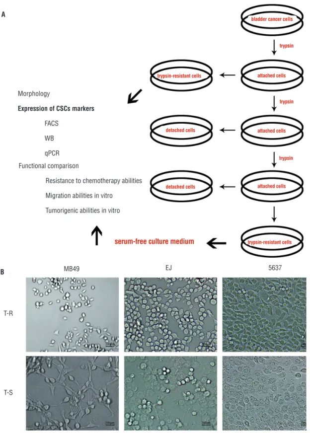
The differential adhesion method showed that the digested time of MB49, EJ and 5637 cells
was different, average time was 2, 3 and 5 mins, respectively (Figure-1A). It is found that cells de-tached by fewer time of trypsinization retained original shaped morphologies. On the other hand, cells detached by more time exhibited round sha-ped morphologies, and cells detached by middle time had mixed morphologies (Figure-1B).
Characterizations of CSCs
Compared with the trypsin-sensitive cells, trypsin-resistant cells could enrich for CSCs. And these cells cultured with SFM method could gain the purity of CSCs sorting. To confirm this conclu-sion, it is necessary to identify trypsin-resistant cells in CSCs characters with high expression of CSCs markers, higher resistance to chemotherapy, greater migration in vitro, and stronger tumorige-nicity in vivo.
Expression of CSCs markers
The FCM analysis revealed that the frac-tion of CD44+CD133+ cells in trypsin-resistant cells was more than in trypsin-sensitive cells of MB49, EJ and 5637 cells (Figure-2A). The WB analysis indicated that the CD133 and CD44 proteins were abundantly expressed in trypsin-resistant cells, but much less in trypsin-sensitive cells (Figure--2B). The qPCR analysis showed that the relative levels of CD133 and CD44 mRNAs in trypsin-re-sistant cells were higher than those observed in trypsin-sensitive cells (Figure-2C).
Functional comparison
Compared to trypsin-sensitive cells, tryp-sin-resistant cells displayed higher cell viabilities after being exposed to different concentrations of doxorubicin, which suggested that
trypsin-re-Table 1 - Primers of selected genes.
Gene name Primers (forward/reverse) Base pairs of product
CD133 F: 5’-CGGGATCCGAAAAACTGATCTGT-3’
615bp R: 5’-CCGCTCGAGTTACCTAGTTACTCTCTCC-3’
CD44 F: 5’-CCCTGCTACCAGAGACCAAGAC-3’
401bp R; 5’-GCAGGTTCCTTGTCTCATCAGC-3’
GAPDH F: 5’-CCATGGAGAAGGCTGGGG-3’
Figure 1 - Isolation of cells by differential adhesion method and serum-free culture medium (SFM) method. (A) Diagram illustration to the proposed model for isolation of CSCs by differential adhesion method and SFM method. (B) Morphology of trypsin-resistant cells (T-R) and trypsin-sensitive cells (T-S) in MB49, EJ and 5637 cells.
A
B MB49
T-R
T-S
EJ 5637
bladder cancer cells
attached cells
attached cells
attached cells
serum-free culture medium detached cells detached cells
trypsin
trypsin
trypsin
Morphology
Expression of CSCs markers
FACS
WB
qPCR
trypsin-resistant cells trypsin-resistant cells
Functional comparison
Resistance to chemotherapy abilities
Migration abilities in vitro
T-R T-S MB49 CD44-FITC CD44 T-S T-R CD133
CD133 CD44 CD133 CD44 CD133 CD44
ß-actin CD44-FITC CD44-FITC CD44-FITC CD44-FITC CD44-FITC EJ 5637 5637 5637 12.4% 5.1% 30.5% 103 103 103 103 6.0 4.0 2.0 .0 103 103 102 102 102 102 102 102 101 101 101 101 101 101 100 100 100 100 100
100 100 100
100
100
100
101 101 101
101
101
101
102 102 102
102
102
102
103 103 103
103 103 103 100 15.2% 28.4% 9.3% CD133-PE CD133-PE T-R MB49 MB49 EJ EJ T-R T-R
T-S T-S T-S
CD133-PE
CD133-PE
CD133-PE
CD133-PE
sistant cells had lower susceptibility to traditio-nal anticancer agents (Figure-3A). The results of the transwell migration assay indicated that more trypsin-resistant cells invaded the bottom cham-ber when compared to trypsin-sensitive cells un-der the same incubation conditions, which sugges-ted that trypsin-resistant cells had higher invasion ability than trypsin-sensitive cells (Figure-3B). Regarding xenograft formation, trypsin-resistant
cells produced tumors with larger volumes than trypsin-sensitive cells did with the same number of injections (Figure-3C).
DISCUSSION
Enrichment of CSCs was an absolute neces-sity when using CSCs in vaccine applications. Three methods were mostly used to isolate CSCs from
can-Figure 2 - Comparison of specific markers in trypsin -resistant cells (T-R) and trypsin-sensitive cells (T-S) of MB49, EJ and 5637 cells. (A) The FCM analysis revealed that the fraction of CD44+CD133+ cells in trypsin-resistant cells was more than in trypsin-sensitive cells. (B) The WB analysis indicated that the CD133 and CD44 proteins were abundantly expressed in trypsin-resistant cells, but much less in trypsin-sensitive cells. (C) The qPCR analysis showed that the relative levels of CD133 and CD44 mRNAs in trypsin-resistant cells were higher than those observed in trypsin-sensitive cells.
A
B C
Figure 3 - Functional characteristics comparison in trypsin-resistant cells (T-R) and trypsin-sensitive cells (T-S). (A) Comparison of resistance to chemotherapy using CCK-8. Compared to trypsin-sensitive cells, trypsin-resistant cells show higher cell viabilities after treatment with various concentrations of anti-cancer drugs doxorubicin. (B) In the transwell migration assay, the number of invaded trypsin-resistant was higher than that of trypsin-sensitive cells. (C) In xenograft formation experiments, trypsin-resistant produced larger tumor volumes than trypsin-sensitive cells did.
A
B
C
Cell Viability (%)
MB49 EJ 5637
100
80
60
40
20
T-S T-R
5637
5637 MB49
MB49
EJ
EJ
10nM 10nM 1µM 10µM 10nM 10nM 1µM 10µM 10nM 10nM 1µM 10µM
T-S
T-S T-R
cers: specific CSCs surface markers, side population cells, and SFM. The shortcoming of these methods was the lack of purity and the purity was not enough for CSCs (5). Repeated cycles of differential trypsini-zation could gather CSCs by 20-fold in breast cancer cells, keratinocytes and human mammary epithelial cells. So the differential adhesion method was able to isolate CSCs from bladder cancer cells. Considering that the serum caused irreversible differentiation of stem cells, SFM selection might be useful for CSCs expansion and would allow for maintenance of an undifferentiated stem cell status (9). To our know-ledge, this is the first report about the isolation and expansion of CSCs via a combination of differential adhesion method and SFM method, which is modi-fied to improve the purity of CSCs.
The CD133 and CD44 markers were used to ascertain CSCs in most tumor tissues (10, 11). Additionally, our study found elevated expression levels of CD133+ and CD44+ in trypsin-resistant cells. Targeting CD44+ and CD133+ cancer cells in-volving a CD133+CD44+ cell subpopulation might be a way for colorectal cancer therapy (12). Thus CD133+CD44+ cells may be the concentrated CSC subpopulation in bladder cancer cell populations. Only cells expressing CD133+ and CD44+ were con-sidered as CSCs by FCM analysis (13). In addition, the expression of both markers was found elevated in trypsin-resistant cells not only at the mRNA pression (qPCR) level, but also at the protein ex-pression (WB) level.
We functionally characterized the trypsin-re-sistant cells populations by different techniques (14, 15). Specifically, trypsin-resistant cells had a greater ability to penetrate wells. Moreover, although che-motherapy killed most tumor cancer cells, it could not kill CSCs. Additionally, trypsin-resistant cells exhibited a lower sensitivity to doxorubicin, which meet the theory of resistance to chemotherapy (16, 17). Tumorigenicity in nude mice was the standard experiment used to evaluate the tumorigenic abili-ty of CSCs (18). Trypsin-resistant cells had a greater ability to form subcutaneous tumors in nude mice. Taken these above results together, trypsin-resistant cells showed specific CSC properties.
Taken together, this study showed that cultured trypsin-resistant bladder cancer cells dis-played specific CSC properties.
CONCLUSIONS
In conclusion, bladder CSCs were isolated successfully with a modified method using a com-bination of differential adhesion method and SFM method. Trypsin-resistant cells contained characte-ristics resembling CSCs such as chemotherapy re-sistance and in vivo tumorigenic capacity. Trypsin--resistant cells may provide an ideal model for the development of bladder cancer vaccine research.
ACKNOWLEDGEMENT
Yong-tong Zhu, Shi-yu Pang and Yang Luo contri-buted equally to this work.
This study was supported by the Science and Tech-nology Innovation Project of Education Depart-ment in Guangdong Province (No. 2013KJCX0039) and the Scientific Initiative Research Foundation of Southern Medical University (No.PY2014N031).
CONFLICT OF INTEREST
None declared.
REFERENCES
1. Zhang X, Shi X, Li J, Hu Z, Guo F, Huang X, et al. Novel
immunotherapy for metastatic bladder cancer using vaccine of human interleukin-2 surface-modified MB 49 cells. Urology. 2011;78:722.
2. McDermott SP, Wicha MS. Targeting breast cancer stem
cells. Mol Oncol. 2010;4:404-19.
3. Ho PL, Kurtova A, Chan KS. Normal and neoplastic urothelial
stem cells: getting to the root of the problem. Nat Rev Urol. 2012;9:583-94.
4. Walia V, Elble RC. Enrichment for breast cancer cells with
stem/progenitor properties by differential adhesion. Stem Cells Dev. 2010;19:1175-82.
5. Li L, Li B, Shao J, Wang X. Chemotherapy sorting can be
used to identify cancer stem cell populations. Mol Biol Rep. 2012;39:9955-63.
6. Wang L, Mezencev R, Bowen NJ, Matyunina LV, McDonald
JF. Isolation and characterization of stem-like cells from a human ovarian cancer cell line. Mol Cell Biochem. 2012;363:257-68.
7. Liao WT, Jiang D, Yuan J, Cui YM, Shi XW, Chen CM, et
8. Zhu YT, Lei CY, Luo Y, Liu N, He CW, Chen W, et al. A modified method for isolation of bladder cancer stem cells from a MB49 murine cell line. BMC Urol. 2013;13:57.
9. Yanamoto S, Kawasaki G, Yamada S, Yoshitomi I, Kawano T,
Yonezawa H, et al. Isolation and characterization of cancer stem-like side population cells in human oral cancer cells. Oral Oncol. 2011;47:855-60.
10. Brescia P, Richichi C, Pelicci G. Current strategies for identification of glioma stem cells: adequate or unsatisfactory? J Oncol. 2012;2012:376894.
11. Han ME, Jeon TY, Hwang SH, Lee YS, Kim HJ, Shim HE, et al. Cancer spheres from gastric cancer patients provide an ideal model system for cancer stem cell research. Cell Mol Life Sci. 2011;68:3589-605.
12. Ou J, Deng J, Wei X, Xie G, Zhou R, Yu L, et al. Fibronectin extra domain A (EDA) sustains CD133(+)/CD44(+) subpopulation of colorectal cancer cells. Stem Cell Res. 2013;11:820-33.
13. Zhu YT, Zhao Z, Fu XY, Luo Y, Lei CY, Chen W, et al. The granulocyte macrophage-colony stimulating factor surface modified MB49 bladder cancer stem cells vaccine against metastatic bladder cancer. Stem Cell Res. 2014;13:111-22. 14. Dalerba P, Cho RW, Clarke MF. Cancer stem cells: models
and concepts. Annu Ver Med. 2007;58:267-84.
15. Visvader JE, Lindeman GJ. Cancer stem cells in solid tumours: accumulating evidence and unresolved questions. Nat Rev Cancer. 2008;8:755-68.
16. Sung JM, Cho HJ, Yi H, Lee CH, Kim HS, Kim DK, et al. Characterization of a stem cell population in lung cancer A549 cells. Biochem Biophys Res Commun. 2008;371:163-7.
17. Okamoto A, Chikamatsu K, Sakakura K, Hatsushika K, Takahashi G, Masuyama K. Expansion and characterization of cancer stem-like cells in squamous cell carcinoma of the head and neck. Oral Oncol. 2009;45:633-9.
18. Lobo NA, Shimono Y, Qian D, Clarke MF. The biology of cancer stem cells. Annu Rev Cell Dev Biol. 2007;23:675-99.
_______________________
Correspondence address: