BOVINE HERPESVIRUS 5 DETECTION BY VIRUS ISOLATION IN CELL CULTURE AND MULTIPLEX-PCR IN CENTRAL NERVOUS SYSTEM FROM CATTLE WITH NEUROLOGICAL
DISEASE IN BRAZILIAN HERDS
Marlise Pompeo Claus; Alice Fernandes Alfieri; Kerlei Cristina Médici; Michele Lunardi; Amauri Alcindo Alfieri*
Laboratório de Virologia Animal, Departamento de Medicina Veterinária Preventiva, Centro de Ciências Agrárias, Universidade Estadual de Londrina, Londrina, PR, Brasil
Submitted: January 04, 2007; Returned to authors for corrections: April 02, 2007; Approved: July 27, 2007.
ABSTRACT
Bovine herpesvirus 5 (BoHV-5) is an important cause of meningoencephalitis in young and adult cattle. The multiple etiology of neurological disturbances in cattle makes the quick and conclusive diagnosis of BoHV-5 infection important for animal and public health, mainly because of herbivore rabies that is endemic in Brazilian cattle herds. The objective of this retrospective study was to use a multiplex-polymerase chain reaction (multiplex-PCR) for BoHV-5 and BoHV-1 glycoprotein C gene detection from stored central nervous system (CNS) tissue fragments of cattle with neurological clinical signs. Forty-seven frozen CNS samples of young and adult cattle from 31 herds in three Brazilian geographical regions (South, Southeast, and Center-west) were evaluated. Eighteen (38.3%) of these CNS samples were BoHV-positive by virus isolation in cell culture. By multiplex-PCR 30 (63.8%) CNS samples were BoHV-5 positive. All 18 positive samples by virus isolation were confirmed as BoHV-5 by the multiplex-PCR, that provided a increase of 25.5% (12/47) in the BoHV-5 diagnosis rate. BoHV-1 was not detected in any CNS sample. This retrospective study demonstrated the wide regional distribution of BoHV-5 infection in Brazilian cattle herds since positive results were obtained in CNS samples of cattle with neurological disease from Paraná, São Paulo, Minas Gerais, Mato Grosso, and Mato Grosso do Sul States.
Key words: cattle, meningoencephalitis, bovine herpesvirus 5, multiplex-PCR
*Corresponding Author. Mailing address: Rodovia Celso Garcia Cid - Caixa Postal 6001 - cep 86051-990 Londrina, PR - Brasil. Tel.: (43) 3371-4714. E-mail: alfieri@uel.br
INTRODUCTION
The diseases that cause neurological disturbances in dairy and beef cattle can be responsible for significant economic losses due to high death rates. Infectious agents such as virus, bacteria, and protozoa can be involved in central nervous system (CNS) diseases in cattle (17). A range of very similar clinical signs, imply that definitive diagnosis can only be made with the utilization of laboratory techniques that identify the etiological agent (3). In Brazil, herbivore rabies occurs throughout the country (16). Then, in addition to their importance for animal health, diseases that affect the CNS of cattle also have importance on public health making it very important in the elaboration of conclusive laboratorial diagnosis.
Bovine herpesvirus 5 (BoHV-5), classified in the Herpesviridae family, Alphaherpesvirinae sub-family, is a neurovirulent strain of bovine herpesvirus (BoHV) that is the etiological agent of bovine non-suppurative meningoencephalitis (12). BoHV-5 has been detected in various regions of the world, and also in Brazil (10,20,22) causing a neurological disease in young and adult cattle. The most characteristic signs of the infection include anorexia, respiratory and ocular secretions, muscle trembling, circling, opisthotonus, nystagmus, teeth grinding, ataxia, and seizures (4).
troublesome technique that requires the maintenance of cell cultures and a long time to obtain conclusive results. Furthermore, the need of the infectiousness of the virus that can be reduced by collection, transport and storage conditions of biological samples suggest that false-negative results can be generated in many situations. On the other hand, molecular techniques such as PCR detect the viral DNA from different types of specimens, are highly sensitive, specific, fast and do not require the infectiousness of the viral particle (18).
This retrospective study describes the use of the multiplex-PCR assay for BoHV-5 and BoHV-1 detection in a CNS fragments collection, previously assessed in cell culture, from cattle with clinical neurological signs in seven Brazilian states.
MATERIALS AND METHODS Virus and Cells
MDBK (Madin Darby bovine kidney) cells were amplified in Dulbecco’s modified essential medium (D-MEM, Gibco BRL, USA) supplemented with 10% fetal bovine serum (FBS, Gibco BRL, USA), 55 µg/ml gentamicine (Sigma Co., USA) and 2.5 µg/ml amphotericin B (Sigma Co., USA). The Los Angeles strain (LA) of BoHV-1 and the field strain AA 01, characterized as BoHV-5 by immunoperoxidase with monoclonal antibodies (19) and by Restriction Fragment Length Polymorphism (RFLP) analysis of the viral DNA (9), were used as positive controls in the multiplex-PCR technique. The MDBK cells were used for both, amplification of the LA and AA 01 virus strains, and for attempts of BoHV-5 isolation from clinical specimens.
CNS fragments
From 1999 to 2004, 47 CNS samples, including fragments of brain, cerebellum, and brainstem were submitted for viral diagnosis. The samples were collected in 31 outbreaks of neurological disease in young and adult animals in dairy (n=5) and beef (n=26) cattle herds from seven Brazilian states (Paraná, São Paulo, Minas Gerais, Goiás, Mato Grosso, Mato Grosso do Sul, and Rondônia). Infection by the rabies virus was excluded in all cases by the negative results in the diagnosis performed by direct immunofluorescence and by inoculation in mice according to the standard procedure in a reference laboratory.
Virus isolation in cell culture
Suspension (10-20% w/v) of the CNS fragments in phosphate-buffered saline pH 7.2 (137 mM NaCl; 3 mM KCl; 8 mM Na2HPO4; 15 mM KH2PO4) was centrifuged at 3000g for
15 min at 4ºC. A fraction of the supernatant was immediately used for virus isolation in cell culture and the other was stored at -20ºC for subsequent analysis. Aliquots of 500 µl of the supernatant were treated with gentamicine (55 µg/ml) and
amphotericin B (2.5 µg/ml) and inoculated in MDBK cells maintained in FBS-free D-MEM in a roller system for attempted virus isolation. All specimens were inoculated in cell culture for at least three consecutive blind passages. In the samples that induced cytopathic effect in cell culture the presence of BoHV was evaluated by a semi-nested PCR to detect the BoHV glycoprotein B gene (23).
Nucleic Acid Extraction
For the nucleic acid extraction aliquots of all 47 CNS fragments that had been kept in storage at -20ºC for more than five years were used. Frozen suspensions aliquots (500 µl) of each sample were treated with sodium dodecyl sulphate (SDS) and Proteinase K (Invitrogen, Life Technologies, USA) at final concentration of 1% and 0.2 mg/ml, respectively. After homogenization, the samples were kept at 56ºC for 30 min. For DNA extraction a combination of phenol/chloroform/isoamyl alcohol and silica/guanidine isothiocyanate methods (1) was performed with slight modification. Briefly, the samples were treated with an equal volume of phenol/chloroform/isoamyl alcohol (25:24:1), homogenized and heated at 56ºC for 15 min (21). After centrifugation at 10,000g for 10 min the aqueous phase was processed with silica/guanidine isothiocyanate (5). The DNA was eluted in 50 µl ultrapure (MilliQ) sterile water and kept at -20ºC until use. Aliquots of ultrapure sterile water were included as negative controls in all the DNA extraction procedures.
Multiplex-PCR
Multiplex-PCR was carried out according to Claus (6). Briefly, the reaction was performed in a solution containing 5 µl of the extracted DNA and 45 µl of PCR-mix consisting of 20 pmol of each primers B1 (5‘CAA CCG AGA CGG AAA GCT CC 3‘ nt 185-204); B5 (5‘CGG ACG AGA CGC CCT TGG 3‘- nt 322-339) and Bcon [5‘AGT GCA CGT ACA GCG GCT CG 3‘ nt 519-538 (BoHV-1) and nt 461-480 (BoHV-5)]; 1.6 mM of each dNTP (InvitrogenTM Life Technologies, USA); 2.5 units Platinum
Taq DNA polymerase (InvitrogenTM Life Technologies, USA);
1x PCR buffer (20 mM Tris-HCl pH 8.4 and 50 mM KCl); 1.5 mM MgCl2; 8% dimetilsulfoxide (DMSO, Sigma Co., USA) and
ultrapure sterile water to a final volume of 50 ml. Amplification was performed in a thermocycler (PTC 200, MJ Research Co., USA) according to the following conditions of time and temperature: an initial step of 3 min at 94ºC followed by 40 cycles of 1 min/94ºC; 1 min/58ºC, 1 min/72ºC and a final extension step of 7 min/72ºC.
Statistical analysis
The kappa statistic (κ) was calculated to evaluate the agreement between virus isolation in cell culture technique and multiplex-PCR assay. When κ=0 there is no agreement, κ<0.3 the agreement is poor, κ between 0.3 and <0.5 is acceptable, κ between 0.5 and 0.7 is good, and κ>0.7 is excellent (15). A value of p < 0.05 was considered statistically significant. The statistical analysis was made by using the EpiInfo 6.04 program (8).
RESULTS AND DISCUSSION
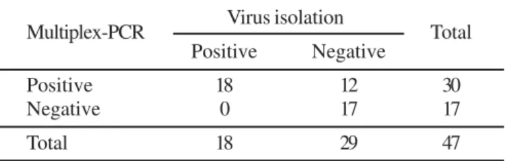
The BoHV isolation in cell culture was obtained in 38.3% (18/47) of the assessed CNS samples. A 159 bp DNA fragment was amplified by multiplex-PCR in 30 (63.8%) CNS samples collected from dairy (n=13) and beef (n=17) cattle herds (Fig. 1). All 18 positive samples in cell culture were also positive by multiplex-PCR for the BoHV-5. A 354 bp DNA fragment of the BoHV-1 glycoprotein C gene was not amplified in any CNS sample. The agreement between the two assays was good (κ=0.52; p < 0.001). Table 1 shows the distribution of the concordant and discordant results obtained between the virus isolation in cell culture technique and multiplex-PCR assay.
During this retrospective study the use of multiplex-PCR demonstrated a increase in the BoHV-5 diagnosis rate in CNS fragments from cattle with neurological disease, compared with the results obtained by virus isolation in cell culture technique.
In 25.5% (12/47) of the samples, where BoHV isolation in cell culture was not possible in previous studies, the use of multiplex-PCR detected the glycoprotein C gene of the BoHV-5 in CNS fragments stored at freezing temperatures, some for up to five years.
The greater number of the BoHV-5 positive samples obtained with the use of multiplex-PCR was mainly due to the requirement of the infectiousness of the virus for isolation in cell culture. Due to the specimens collection under field conditions the virus can be inactivated (24). This characteristic is very important in veterinary medicine, especially for cattle farms in Brazil, because specimens collected in the field for diagnosis are often improperly conserved and require a long transport time from the farms to the laboratory.
The results of this study showed that the use of only one diagnostic method for BoHV-5, and in particular virus isolation in cell culture may result in the generation of false-negative results. Besides reducing the efficiency of the diagnosis, the false-negative results also induce incorrect information that has direct consequences on epidemiological studies and can also be responsible for the non-adoption of control and prophylaxis measures. Nevertheless, virus isolation in cell culture, especially in a research laboratory, is an essential technique because it supplies the availability of the wild-type virus strains for antigenic and molecular analysis.
Ely (11) also described that the PCR assay was more sensitive than virus isolation for diagnosis of BoHV infection. This virus was isolated in only one of the five bovine brain samples submitted to cell culture, while by PCR all samples were positive. A greater number of BoHV-5 positive samples were also detected by the PCR technique in nasal swabs from experimentally infected cattle (2). Takiuchi (24) used virus isolation and semi-nested PCR to detect BoHV-1 in organs fragments of aborted fetuses and found positive results in 5 and 14 samples, respectively.
In the present study the BoHV-5 was identified in CNS specimens from cattle herds in five Brazilian States (Mato Grosso Table 1. Distribution of the positive and negative results for bovine herpesvirus 5 obtained by virus isolation in cell culture and multiplex-PCR techniques in central nervous system samples from cattle with neurological disease.
Multiplex-PCR Virus isolation Total
Positive Negative
Positive 18 12 30
Negative 0 17 17
Total 18 29 47
κ = 0.52; p < 0.001.
do Sul, Mato Grosso, Minas Gerais, São Paulo, and Paraná). Negative results were obtained only from samples obtained from Goiás and Rondônia States from which only one and two samples, respectively, were analyzed.
In Brazil, meningoencephalitis by BoHV-5 was also been described in some of these States, and others such as Rio de Janeiro and Rio Grande do Sul (7,14,20,22) indicating that this virus is widely distributed in cattle herds in geographically distant Brazilian regions.
The remainder 17 (36.2%) multiplex-PCR negative-CNS specimens from cattle with neurological signs may result from other common diseases that cause neurological disturbances in Brazilian cattle herds, such as polyencephalomalacia or plant intoxications, that were not assessed in this study. Another aspect to be reported is the set of CNS fragments with low virus titre or even without virus. BoHV-5 is not uniformly distributed in all the CNS, and the processing of fragments from different CNS regions are very important when etiological diagnostic techniques are used (4). In this study only BoHV-5 was identified by multiplex-PCR. Souza (22) using monoclonal antibodies to characterize the type of BoHV isolated from clinical specimens related that all samples from cattle with encephalitis presented BoHV-5 antigenic profile. However, Ely (11) characterized a virus strain from cattle with encephalitis as BoHV-1.
With the samples used in the present study the category of cattle (suckling/weaned) where the infection was most frequent could not be determined. The distribution of the positive results for BoHV-5 obtained by multiplex-PCR showed that 50% (15/30) of the positive samples were from suckling calves (21 days to 5 months). The others 15 (50%) positive samples were obtained from weaned animals, and was most frequent in 15 to 24 months old cattle. Two cows with 5-years-old and other one with 6-years-old were the older animals with BoHV-5 infection (Table 2).
This retrospective study showed the importance of the use of alternative diagnostics methods that enable the BoHV-5 identification even in situations where there are failures of collection, storage, and transport of specimens. It was also shown that BoHV-5 is an important pathogen of encephalitis in cattle and due to its wide geographical distribution in Brazil, this virus should be included in the differential diagnosis of
neurological disturbances in young and adult animals from beef and dairy cattle herds.
ACKNOWLEDGEMENTS
The authors thank the Brazilian Institutes CNPq, CAPES, FINEP, and Fundação Araucária (FAP/PR) for the financial support. Alfieri, A.A. and Alfieri, A.F. are recipient of CNPq fellowship. Table 2. Cell culture isolation and multiplex-PCR results for bovine herpesvirus 5 detection in central nervous system samples from cattle with neurological disease.
outbreak bred age nº animals positivessamples
n° year state dead evaluated VICC (#) M-PCR
01 1999 MS Nelore 24 m 21 4 4 4
02 1999 MG Holstein 60 m 1 1 1 1
03 1999 SP Holstein 21 d 9 2 2 2
04 1999 SP Holstein 60 d 11 3 3 3
05 2000 MT Nelore 45 d NI 1 1 1
06 2000 MT Nelore 70 d NI 1 1 1
07 2000 MS Canchim 4 m NI 1 -
-08 2000 SP Nelore 20 d NI 2 -
-09 2001 MS Nelore 24 m NI 1 -
-10 2001 PR ½ A.Angus 40 m 1 1 -
-11 2001 MT Nelore 15 m 11 1 1 1
12 2001 MS ½ Marchigiana 22 m 5 2 2 2
13 2001 MS ½ Marchigiana 24 m 5 2 2 2
14 2001 MG ½ A.Angus 18 m 20 1 -
-15 2001 PR Nelore 18 m 4 1 - 1
16 2001 RO Nelore 60 m 2 1 -
-17 2001 PR ½ Limousin 15 m 3 1 - 1
18 2002 PR Simenthal 30 m 5 1 -
-19 2002 GO ½ Simenthal 18 m 1 1 -
-20 -2002 PR Nelore 42 m 3 1 -
-21 2002 RO Nelore 72 m 3 1 -
-22 2002 PR Limousin 5 d NI 1 -
-23 2002 MG Simenthal 60 d 2 1 - 1
24 2003 MT Nelore 22 m 4 1 1 1
25 2003 MS Nelore 18 m 1 1 -
-26 2003 MT Nelore 36 m 6 1 -
-27 2003 PR Nelore 24 m 4 1 - 1
28 2003 PR Nelore 40 m 1 1 -
-29 2004 MG Holstein 6 m NI 2 -
-30 2004 PR Swiss Brown 4 m 7 7 - 7
31 2004 PR Nelore 24 m 8 1 - 1
Total 47 18 30
RESUMO
Detecção do herpesvírus bovino 5 por isolamento viral e multiplex-PCR em SNC de bovinos com doença
neurológica em rebanhos brasileiros
O herpesvírus bovino 5 (BoHV-5) é um importante agente etiológico de meningoencefalite em bovinos jovens e adultos. A etiologia múltipla dos distúrbios neurológicos em bovinos torna o diagnóstico conclusivo do BoHV-5 importante tanto em termos de sanidade animal quanto de saúde pública, principalmente pela característica endêmica da raiva dos herbívoros nos rebanhos bovinos brasileiros. O objetivo desse estudo retrospectivo foi utilizar a reação em cadeia da polimerase (multiplex-PCR) para a detecção do gene da glicoproteína C do BoHV-5 e do BoHV-1 em fragmentos estocados de sistema nervoso central (SNC) de bovinos com sinais clínicos neurológicos. Foram avaliadas 47 amostras congeladas de fragmentos de SNC de bovinos jovens e adultos pertencentes a 31 rebanhos de três regiões geográficas brasileiras (Sul, Sudeste e Centro-oeste). Por meio do isolamento viral em cultivo celular foi possível o isolamento do BoHV em 18 (38,3%) amostras. Pela técnica de multiplex-PCR 30 (63,8%) amostras de SNC foram positivas para o BoHV-5. Todas as 18 amostras positivas no isolamento viral foram confirmadas como BoHV-5 pela multiplex-PCR, proporcionando um incremento na taxa de diagnóstico do BoHV-5 de 25,5% (12/47). Em nenhuma das amostras avaliadas foi possível a identificação do BoHV-1 pela multiplex-PCR. Esse estudo retrospectivo demonstrou a ampla distribuição da infecção pelo BoHV-5 nos rebanhos bovinos brasileiros uma vez que resultados positivos foram obtidos em amostras de SNC colhidas de bovinos com doença neurológica, provenientes dos estados do Paraná, São Paulo, Minas Gerais, Mato Grosso e Mato Grosso do Sul.
Palavras-chave: bovino, meningoencefalite, herpesvírus bovi-no 5, multiplex-PCR
REFERENCES
1. Alfieri, A.F.; Alfieri, A.A.; Barreiros, M.A.B.; Leite, J.P.G.; Richtzenhain, L.J. (2004). G and P genotypes of group A rotavirus strains circulating in calves in Brazil, 1996-1998. Vet. Microbiol., 99: 167-173.
2. Ashbaugh, S.E.; Thompson, K.E.; Belknap, E.B.; Schultheiss, P.C.; Chowdhury, S.; Collins, J.K. (1997). Specific detection of shedding and latency of bovine herpesvirus 1 and 5 using a nested polymerase chain reaction. J. Vet. Diag. Invest., 9: 387-394.
3. Barros, C.S.L.; Marques, G.H.F. (2003). Procedimentos para o Diagnóstico das Doenças do Sistema Nervoso Central de Bovinos. Departamento de Defesa Animal, Ministério da Agricultura, Pecuária e Abastecimento (MAPA), Brasília, p.1-50.
4. Belknap, E.B.; Collins, J.K.; Ayers, V.K.; Schultheiss, P.C. (1994). Experimental infection of neonatal calves with neurovirulent bovine herpesvirus type 5 (BHV-5). Vet. Pathol., 31: 358-365.
5. Boom, R.; Sol, C.J.A.; Salimans, M.M.M.; Jansen, C.L.; Wertheim-van Dillen, P.M.E.; Noordaa, J. Wertheim-van der. (1990). Rapid and simple
method for purification of nucleic acids. J. Clin. Microbiol., 28: 495-503.
6. Claus, M.P.; Alfieri, A.F.; Folgueras-Flatschart, A.V.; Wosiacki, S.R.; Médici, K.C.; Alfieri, A.A. (2005). Rapid detection and differentiation of bovine herpesvirus 1 and 5 glycoprotein C in clinical specimens by multiplex-PCR. J. Virol. Meth., 128: 183-188.
7. Colodel, E.M.; Nakazato, L.; Weiblen, R.; Mello, R.M.M.; Silva, R.R.P.; Souza, M.A.; Filho, J.A.O.; Caron, L. (2002). Meningoencefalite necrosante causada por herpesvírus bovino no Estado de Mato Grosso, Brasil. Ciência Rural, 32: 293-298.
8. Dean, A.G.; Dean, J.A.; Coulombier, D.; Burton, A.H.; Brendel, K.A.; Smith, D.C.; Dicker, R.C.; Sullivan, K.M.; Fagan, R.F.; Arne, T.G. (1997). Epi Info versão 6.04b disponível em http://www.cdc.gov/ epiinfo/.
9. D’Arce, R.C.F.; Almeida, R.S.; Silva, T.C.; Franco, A.C.; Spilki, F.; Roehe, P.M.; Arns, C.W. (2002). Restriction endonuclease and monoclonal antibody analysis of Brazilian isolates of bovine herpesvirus types 1 and 5. Vet. Microbiol., 88: 315-324.
10. D’Offay, J.M.; Mock, R.E.; Fulton, R.W. (1993). Isolation and characterization of encephalitic bovine herpesvirus type 1 isolates from cattle in North America. Am. J. Vet. Res., 54: 534-539. 11. Ely, R.W.; D’Offay, J.M.; Ruefer, A.H.; Cash, C.Y. (1996). Bovine
herpesviral encephalitis: a retrospective study on archived formalin-fixed, paraffin-embedded brain tissue. J. Vet. Diag. Invest.,8: 487-492.
12. Fauquet, C.M.; Mayo, M.A.; Maniloff, J.; Desselberger, U.; Ball, L.A. (2004). Virus Taxonomy. The eighth report. Academic Press, San Diego, p.1162.
13. Giavedoni, L.D.; Ruiz, M.; Fijtman, N.; Schudel, A.A.; Rodriguez, M. (1988). Rapid diagnosis of bovine herpesvirus encephalitis: comparasion of nucleic acid hybridization and immunoperoxidase methods using clinical samples. J. Vet. Med.,35: 280-285. 14. Gomes, L.I.; Rocha, M.A.; Costa, E.A.; Lobato Z.I.P.; Mendes,
L.C.N.; Borges, A.S.; Leite, R.C.; Barbosa-Stancioli, E.F. (2002). Detecção de herpesvírus bovino 5 (BoHV-5) em bovinos do Sudeste Brasileiro. Arq. Bras. Med. Vet. Zootec.,54: 217-220.
15. Martin, S.W.; Bonnet, B. (1987). Clinical epidemiology. Can. Vet. J.,28: 318-325.
16. MAPA (2005). Boletim da Defesa Sanitária Animal, DSA, SDA, Ministério da Agricultura, Pecuária e Abastecimento, Brasil. http:// www.agricultura.gov.br/portal/page?_pageid=33,3271386&_dad= portal&_schema=PORTAL
17. Radostitis, O.M.; Gay, C.C.; Blood D.C.; Hinchcliff, W. (2000). Veterinary Medicine. A textbook of diseases of cattle, sheep, goats and horses. 9th edition. WB Saunders, London, UK.
18. Ros, C.; Riquelme, M.E.; Forslund, K.Ö.; Belák, S. (1999). Improved detection of five related ruminant alphaherpesviruses by specific amplification of viral genomic sequences. J. Virol. Meth.,83: 55-65. 19. Roehe, P.M.; Silva, T.C.; Nardi, N.B.; Oliveira, L.G.; Rosa, J.C.A. (1997). Diferenciação entre os vírus da rinotraqueíte infecciosa bovina (BHV-1) e vírus da encefalite bovina (BHV-5) com anticorpos monoclonais. Pesq. Vet. Bras.,17: 41-44.
20. Salvador, C.S.; Lemos, R.A.A.; Riet-Corrêa, F.; Roehe, P.M.; Osório, A.L.A.R. (1998). Meningoencefalite em bovinos causada por herpesvírus bovino-5 no Mato Grosso do Sul e São Paulo. Pesq. Vet. Bras., 18: 76-83.
21. Sambrook, J.; Russel, D.W.(2000). Molecular Cloning. A Laboratory Manual. 3th edition. Cold Spring Harbor Laboratory Press, New York,
USA, 999 p.
23. Takiuchi, E.; Médici, K.C.; Alfieri, A.F.; Alfieri, A.A. (2003). Otimização da reação em cadeia pela polimerase (semi-nested PCR) para a detecção do herpesvírus bovino tipo 1 em fragmentos de órgãos fetais e em sêmen de bovinos naturalmente infectados. Semina: Ci. Agr., 24: 43-56.