INTRODUCTION
Address to: Dra Cíntia de Moraes Borba. Lab. de Taxonomia, Bioquímica e Bioprospecção de Fungos/IOC/FIOCRUZ. Av. Brasil 4365, 21045-900 Rio de Janeiro, RJ, Brasil.
Phone: 55 21 3865-8114; Fax: 55 21 3865-8113 e-mail:cborba@ioc.fi ocruz.br; [email protected] Received 7 May 2014
Accepted 11 September 2014
Interaction of an opportunistic fungus
Purpureocillium
lilacinum
with human macrophages and dendritic cells
Mariana Lima Perazzini Peixoto
[1],
Dilvani Oliveira Santos
[2],
Ivy de Castro Campos de Souza
[2],
Eloah Christina Lyrio Neri
[2],
Danielly Correa Moreira de Sequeira
[1],
Paula Mello De Luca
[3]and
Cíntia de Moraes Borba
[1][1]. Laboratório de Taxonomia, Bioquímica e Bioprospecção de Fungos, Instituto Oswaldo Cruz, Fundação Oswaldo Cruz, Rio de Janeiro, RJ. [2]. Laboratório de Biopatógenos e Ativação Celular, Universidade Federal Fluminense, Niterói, RJ. [3]. Laboratório de Imunoparasitologia, Instituto Oswaldo Cruz, Fundação Oswaldo Cruz, Rio de Janeiro, RJ.
ABSTRACT
Introduction:Purpureocillium lilacinum is emerging as a causal agent of hyalohyphomycosis that is refractory to antifungal drugs; however, the pathogenic mechanisms underlying P. lilacinum infection are not understood. In this study, we investigated the interaction of P. lilacinum conidia with human macrophages and dendritic cells in vitro. Methods: Spores of a P. lilacinum
clinical isolate were obtained by chill-heat shock. Mononuclear cells were isolated from eight healthy individuals. Monocytes were separated by cold aggregation and differentiated into macrophages by incubation for 7 to 10 days at 37°C or into dendritic cells by the addition of the cytokines human granulocyte-macrophage colony stimulating factor and interleukin-4. Conidial suspension was added to the human cells at 1:1, 2:1, and 5:1 (conidia:cells) ratios for 1h, 6h, and 24h, and the infection was evaluated by Giemsa staining and light microscopy. Results: After 1h interaction, P. lilacinum conidia were internalized by
human cells and after 6h contact, some conidia became infl ated. After 24h interaction, the conidia produced germ tubes and hyphae, leading to the disruption of macrophage and dendritic cell membranes. The infection rate analyzed after 6h incubation
of P. lilacinum conidia with cells at 2:1 and 1:1 ratios was 76.5% and 25.5%, respectively, for macrophages and 54.3% and 19.5%, respectively, for cultured dendritic cells. Conclusions: P. lilacinum conidia are capable of infecting and destroying both macrophages and dendritic cells, clearly demonstrating the ability of this pathogenic fungusto invade human phagocytic cells.
Keywords: Purpureocillium lilacinum. Macrophages. Dendritic cells. Interaction in vitro.
Purpureocillium lilacinum (Thom) Luangsa-ard, Houbraken, Hywel-Jones & Samson, comb. nov 2011, previously called
Paecilomyces lilacinus (Thom) Samson 1974 is a fi lamentous,
asexual hyaline fungus. The new genera Purpureocillium was established after a recent molecular and morphological study suggested that P. lilacinus was not related to the Paecilomyces genus1.
Purpureocillium lilacinus is widely considered as a cosmopolitan, saprophytic fungus frequently detected in the environmental soil samples; it can cause deterioration of grains, food, and paper. The fungus can also be recovered from contaminated skin creams and lotions used clinically, and from clinical materials such as catheters and plastic implants.
Currently, it is considered an important opportunistic pathogen in both immunocompromised and immunocompetent hosts2,3. It has been found parasitizing insects and nematodes, and hence,
some researchers have described a potential use of this fungus as a biocontrol agent4. It can also cause infection in other animals
such as cats5.
Purpureocillium lilacinum is one of the causal agents of hyalohyphomycosis, a mycotic infection caused by a group of fungi including Acremonium spp, Beauveria spp, Fusarium spp,
Scopulariopsis spp,and Paecilomyces spp. In this condition, the fungi are observed in the affected tissues as septate hyphae with pigmentless cell walls6,7. Most clinical manifestations of
P. lilacinum hyalohyphomycosis are associated with ocular, cutaneous, or subcutaneous infections and the major risk factors are organ transplantations, corticosteroid therapy, primary immunodeficiency, diabetes mellitus, acquired
immunodefi ciency syndrome, intraocular lens implantation,
and ophthalmic surgery2,3,8,9. No effective treatment has
been established for this infection, and antifungal agents,
including amphotericin B, fl ucytosine, and fl uconazole, have
often provided unsatisfactory results3. However, some of the so-called new azoles, such as posaconazole, ravuconazole, and voriconazole, have recently demonstrated good activity
METHODS
antifungal agents or application of these agents together with surgical treatment was necessary to induce remission2,10.
An in vivo study using an animal model has demonstrated that
P. lilacinum virulence is generally low, as evidenced by the high inoculum (106-107 conidia per animal) and immunosuppression
required to establish a successful infection3. On the other hand,
we have previously shown that both immunocompetent and immunosuppressed mice that intravenously received about 104
P. lilacinum conidia developed the infection: fungal structures were observed and fungal cells were recovered from different organs2,11. These data indicate the need of additional studies to
better comprehend the real invasive capability of P. lilacinum. The antifungal immune response, although exhibiting certain
species-specifi c variations, is generally initiated by phagocytic
cells. Neutrophils, macrophages, and monocytes are important antifungal effector cells. Additional effector cells, including neutrophils and monocytes, are recruited to the sites of infection
by infl ammatory signals such as cytokines, chemokines, and
complement components12.
Professional antigen-presenting cells (APCs) belong to the host innate immune system and are represented mainly by macrophages and dendritic cells (DCs). These cells capture and process antigens, express lymphocyte costimulatory molecules, migrate to lymphoid organs, and secrete cytokines to initiate immune response13. DCs are important components of the immune system; they provide the fi rst line of defense and are
therefore essential for the onset of a strong immune response to several incoming pathogens14,15. DCs play an instrumental role
in linking innate and adaptive responses against a variety of pathogenic fungi including Aspergillus fumigatus, Cryptococcus neoformans, and Candida albicans12.
This study aimed to analyze the in vitro interaction between
P. lilacinum conidia and two types of human APCs, macrophages and DCs, to help elucidate the pathogenesis of infection caused by this fungus.
A clinical P. lilacinum isolate from the nasal sinus, which was kindly provided by Dr. Annette W. Fothergill (Fungus Testing Laboratory, University of Texas Health Science Center, San Antonio, USA), was grown on potato-dextrose agar (Difco, Detroit, MI, USA) at room temperature for 14 days. Spores were collected by scraping the colonies, suspended in 50mM phosphate-buffered saline (PBS), pH 7.2, chilled to 4°C, and heated to 37°C. The suspension was then centrifuged at 200 × g for 30min, and the number of conidia in the resultant supernatant (rich in conidia but free of hyphae) was estimated by microscopy using a Neubauer hemocytometer16. All the
conidial suspensions were freshly prepared for each experiment. Peripheral blood mononuclear cells (PBMCs) were obtained from buffy coats of eight peripheral blood samples from healthy
donors screened for human immunodefi ciency virus (HIV) and
hepatitis B virus (kindly provided by Serviço de Hemoterapia,
Hospital Universitário Clementino Fraga Filho, RJ, Brazil).
The cells were isolated using a Ficoll-Hypaque 1077 gradient (Sigma, St. Louis, MO, USA)17. Briefl y, cells were washed,
resuspended in Roswell Park Memorial Institute (RPMI) medium containing L-glutamine and penicillin-streptomycin
(Sigma), and quantifi ed using the Neubauer hemocytometer.
Monocytes were separated from lymphocytes by cold aggregation during 30min17. The cells were resuspended in fresh
RPMI medium containing 10% fetal bovine serum (Hyclone®;
Thermo Scientifi c, South Logan, UT, USA) and seeded at
2 × 10⁵ cells/well into eight-well chamber slides (Lab-TekTM
Nunc International, Rochester, NY, USA) and at 1 × 10⁶/tube
into Falcon® polystyrene tubes (Becton Dickinson Company, Franklin Lakes, NJ, USA) for cell phenotype evaluation by flow cytometry. For differentiation into macrophages,
monocytes were incubated at 37°C in a humidifi ed incubator
with a 5% CO2/95% air mixture (model MC0-19AIC-UV; Etten Leur, The Netherlands) for 7 to 10 days. For differentiation into DCs, monocytes were incubated in the presence of 100U/mL recombinant human granulocyte-macrophage colony stimulating factor (rhGM-CSF; Peprotech, Rocky Hill, NJ, USA) and 1,000U/mL recombinant human interleukin-4 (rhIL-4; Peprotech) as described above17,18. For phenotypic
evaluation, cells were washed with 200µl PBS containing
0.1% bovine serum albumin and 0.01% sodium azide and stained with fl uorescein isothiocyanate (FITC)-conjugated
monoclonal antibodies against cluster of differentiation 14 (CD14) and peridinin-chlorophyll protein-cyanine dye
(PerCP-Cy5.5)-conjugated CD209, also known as dendritic cell-specifi c
intercellular adhesion molecule-3-grabbing nonintegrin (DC-SIGN)19-21,on ice for 60min. Cells were washed and analyzed using an Accuri C6 fl ow cytometer (Accuri
Cytometers Inc., Ann Arbor, MI, USA) and the FlowJoTM
software (Tree Star, Ashland, OR,USA).
The interaction experiments were conducted with three different ratios of conidia to human cells (5:1, 2:1, and 1:1).
Briefl y, the conidial suspension was added to each well of the
chamber slides at the desired concentration, and incubated with APCs (macrophages or DCs) differentiated from monocytes of each donor for 1h, 6h, and 24h at 37°C in a 5% CO2 atmosphere. Subsequently, the cells were washed gently with sterile PBS at
room temperature to remove extracellular conidia, fi xed with
methanol for 3min, stained with Giemsa solution (Sigma) for 15min, and then examined under a light microscope (model Axiophot, Carl Zeiss Microscopy GmbH, Göttingen, Germany). Control APCs without conidia were treated and evaluated in the
same manner. Quantifi cation was performed by counting 100 fi elds on duplicate coverslips and the results were expressed
as follows: % of infected cells = [(APC with fungus - control) ÷ control] × 100%22. The data were analyzed by the Student’s
t-test and the difference at p < 0.05 was considered statistically
signifi cant.
Ethical considerations
This study was reviewed and approved by the Research
RESULTS
Immunophenotyping of the cells was performed to monitor the differentiation of monocytes to macrophages and DCs. The
differentiated DCs were characterized by the high expre ssion of
CD209 (DC-SIGN) and low expression of CD14 (Figure 1). In contrast, the differentiated macrophages displayed high expression of CD14 and low levels of CD209 (Figure 1).
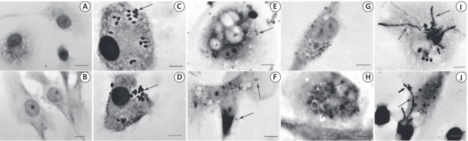
For all three tested ratios of conidia to human cells (5:1, 2:1, and 1:1), phagosome-like structures containing conidia could be observed inside the cells, indicating that conidia were
phagocytized by APCs (Figure 2). The control APCs presented typical morphology (Figures 2A and 2B). The results obtained
with the 5:1 ratio showed that conidia were phagocytized by APCs, similar to the fi ndings for the 2:1 sample (Figures 2C and 2D). However, because of the excessive number of conidia inside and
outside APCs, it was not possible to quantify the internalized
conidia.
For the 1:1 ratio of conidia to human cells, the infection could be followed for 1h, 6h, and 24h (Figures 2E to 2J). Within 1h of interaction, P. lilacinum conidia were internalized
by macrophages (Figure 2E) and DCs (Figure 2F); at 6h,
the internalization gradually increased and some conidia became infl ated (Figures 2G and 2H). After 24h interaction,
macrophages and DCs presented infl ated conidia that formed
germ tubes and hyphae (Figures 2I and 2J); in many cells,
they developed into septate hyphae and fi nally destroyed both
macrophages and DCs (data not shown). This pattern of infection was observed for APCs from all the donors.
We also analyzed the percentage of infected APCs
(macrophages and DCs) after 6-h interaction, because at this
FIGURE 2 -Macrophages (A, C, E, G, I) and dendritic cells (B, D, F, H, J) derived from monocytes isolated from human mononuclear cells were infected in vitro with conidia of a Purpureocillium lilacinum human isolate, stained using Giemsa staining, and analyzed by light microscopy at ×1,000 magnifi cation. Macrophages (A) and dendritic cells (DCs; B) incubated without the fungi were used as negative controls and did not show any changes throughout the experiment. The conidia were phagocytized by macrophages and DCs, as evidenced by the presence of phagosome-like structures containing conidia (→) after 1h of interaction at the 2:1 (C and D) and 1:1 (E and F) ratios of conidia to human cells. At 6h of contact using the 1:1 ratio, the internalization gradually increased and some conidia became infl ated (→) inside macrophages (G) and DCs (H) and at 24h of interaction, the conidia with germ tubes () and hyphae (→) were observed in macrophages (I) and DCs (J). Scale bar, 10µm.
60 CD14
CD209
40
20
0
Macrophages Dendritic cells
Positive cells (%)
FIGURE 1 - Percentages of CD14- and CD209-expressing cells among macrophages and dendritic cells differentiated from monocytes isolated from buffy coat samples of eight healthy donors. CD: cluster of differentiation.
time point, the infection was well established and conidia were clearly observed inside the cells (Figures 2G and 2H).
No signifi cant differences between the infected macrophages
and DCs containing P. lilacinum conidia were detected at both
ratios analyzed (1:1 and 2:1, conidia:human cells) (Figure 3).
A C E G I
B D F H J
DISCUSSION
ACKNOWLEDGMENTS
The authors are grateful to Dr. A.W. Fothergill (Fungus Testing Laboratory, University of Texas Health Science Center, USA) for kindly providing P. lilacinum isolates and Dr. Carmem Nogueira (Hemotherapy Service of Hospital Universitário Clementino
Fraga Filho, RJ, Brazil) for kindly providing buffy coats.
The authors declare that there is no confl ict of interest. CONFLICT OF INTEREST
FINANCIAL SUPPORT
This study was partly fi nanced by the Programa de Fomento à Pesquisa (FOPESQ) Universidade Federal Fluminense (UFF) and
Conselho Nacional de Desenvolvimento Científi co e Tecnológico
(CNPq). The authors MLPP and DCMS are MSc students at
Instituto de Pesquisa Clínica Evandro Chagas Evandro Chagas
Clinical Research Institute, Fundação Oswaldo Cruz (FIOCRUZ).
The authors ICCS and ECLN are MSc students at UFF.
REFERENCES
1. Luangsa-Ard JJ, Houbraken J, van Doorn T, Hong SB, Borman AM, Hywel-Jones NL, et al. Purpureocillium, a new genus for the medically important Paecilomyces lilacinus. FEMS Microbiol Lett 2011; 321:141-149.
Infected cells (%)
100
Macrophages Dendritic cells 80
60
40
20
0
Ratio (conidia: human cells)
2:1 1:1
FIGURE 3 - Phagocytosis of Purpureocillium lilacinum conidia by human macrophages and dendritic cells (DCs). The results are presented as the mean ± standard deviation of the percentage of infected cells after 6-h interaction between conidia and human cells at the ratios of 2:1 and 1:1. No signifi cant differences were detected between the percentages of infected macrophages and DCs.
After a 1h interaction period, we found conidia inside human APCs as described previously with Penicillium marneffei, Fusarium solani, F. oxysporum, and Verticillium nigrescens23,24.
A recent study showed that phagocytosis and cell death of
Aspergillus fumigatus, A. terreus, and A. fl avus occurred within macrophages and DCs after 30min of interaction25.
Here, we observed that, once internalized, the conidia swelled
over time and started producing germ tubes in an attempt to generate mycelia. The ability to produce mycelia and sporulate in the infected tissue is a peculiar feature of P. lilacinum26.
According to Latgé, conidial swelling is a prerequisite step for the development of hyphae27. This researcher showed that
inhaled conidia of A. fumigatus could reach the alveoli and that, at this stage of infection, Aspergillus germinated and showed the growth of small germ tubes, followed by the generation of hyphal fragments28. Furthermore, conidia can germinate in monocytes,
suggesting an essential role of phagocytic cells such as neutrophils, in containing conidia that resist intracellular killing29-31.
Previous studies have also demonstrated the ability of DCs to ingest latent A. fumigatus that develop swollen conidia and hyphae32,33. Macrophages and DCs have been described
as efficient phagocytic cells with regard to Histoplasma capsulatum, Cryptococcus neoformans, Candida albicans, and
A. fumigatus34 and, until this study, also to P. lilacinum.
After 24h of interaction with human cells, the conidia of
P. lilacinum destroyed macrophages and DCs, demonstrating that the fungus was able to remain viable and active inside both types of APCs, evading the innate cell defense responses by an as-yet unknown mechanism. A similar phenomenon has been observed for Fusarium solani, one of the species phenotypically related to P. lilacinum1, after phagocytosis by
human macrophages (unpublished observations).
Purpureocillium lilacinum is generally described as a fungus of low virulence, but our group has demonstrated its capacity to infect murine macrophages and to produce mycelium within 24h of in vitro interaction16. The present observations with human
cells indicate the rapid germination of P. lilacinum conidia and
complete destruction of all macrophages and DCs, in sharp
contrast to the data obtained for Aspergillus spp., including
A. fumigatus, which were effi ciently killed by human
monocyte-derived macrophages within 120min29. This difference is
interesting considering that A. fumigatus is likely more invasive
than P. lilacinum, causing infection that often leads to fatal
invasive aspergillosis in humans27.
Previous studies on hyalohyphomycosis caused by
P. lilacinum have focused on clinical manifestations, treatment,
prognosis, and drug-susceptibility testing2, while little is
known about the fungus interaction with the host. To the best of our knowledge, this is the fi rst study to address the fate of
P. lilacinum after phagocytosis by human APCs. We are
currently investigating the immune evasion mechanisms of
P. lilacinum, in particular the factors that support its survival within the host cells and are responsible for the pathogenicity of infection. The elucidation of these mechanisms will certainly help in the development of new antifungal treatment strategies.
In conclusion, P. lilacinum conidia were capable of
infecting and destroying both macrophages and DCs, clearly
demonstrating the ability of this fungus to invade human
2. Antas PRZ, Brito MMS, Peixoto E, Ponte CGG, Borba CM. Neglected and emerging fungal infections: review od hyalohyphomycosis by Paecilomyces lilacinus focusing in disease burden, in vitro antifungal susceptibility and management. Microbes and Infect 2012; 14:1-8.
3. Pastor FJ, Guarro J. Clinical manifestations, treatment and outcome of Paecilomyces lilacinus infection. Clin Microbiol Infect 2006; 12:948-960.
4. Prabhu S, Kumar S, Subramanian S, Senthil K. Mass production and commercial formulation of Paecilomyces lilacinus. Indian J Nematol 2008; 32:350-358.
5. Pawloski DR, Brunker JD, Singh K, Sutton DA. Pulmonary Paecilomyces lilacinus infection in a cat. J Am Anim Hosp Assoc 2010; 46:197-202.
6. Vartivarian SE, Anassie EJ, Bodey GP. Emerging fungal pathogens in immunocompromised patients: classifi cation, diagnosis and management. Clin Infect Dis1993; 17 (supl II):487-S491.
7. Matsumoto T, Ajello L, Matsuda T, Szaniszlo PJ, Walsh TJ. Developments in hyalohyphomycosis and phaeohyphomycosis. J Med Vet Mycol 1994; 32 (supl I):329-349.
8. Hall VC, Goyal S, Davis MDP, Walsh JS. Cutaneous hyaloyphomycosis caused by Paecilomyces lilacinus: report of three cases and review of literature. Int J Dermatol 2004; 43:648-653.
9. Walsh TJ, Groll A, Hiemenz J, Fleming R, Roilides E, Anaissie E. Infections due to emerging and uncommon medically important fungal pathogens. Clin Microbiol Infect 2004; 10 (supl I):48-66.
10. Yuan X, Wilhelmus KR, Matoba AY, Alexandrakis G, Miller D, Huang AJ. Pathogenesis and outcome of Paecilomyces keratitis. Am J Ophthalmol 2009; 147:691-696.
11. Brito MMS, Lima MS, Morgado FN, Raibolt P, Menezes RC, Conceição-Silva F, et al. Characteristics of Paecilomyces lilacinus infection comparing immunocompetent and immunosupressed murine model. Mycoses 2011; 54:e513-e521.
12. Shoham S, Levitz SM. The immune response to fungal infections. British J Haematol 2005; 129:569-582.
13. Banchereau J, Steinman RM. Dendritic cells and the control of immunity. Nature 1998; 392:245-252.
14. Santos DO, Van Heuverswyn H, Nery JÁ, Bourguignon SC, Castro HL, Ragel CR, et al. Expression of B7-1 co-stimulatory molecule in lepromatous leprosy and reactional episodes. Clin Exp Dermatol 2007; 32:75-80. 15. Santos DO, Miranda A, Suffys P, Bourguignon SC, Rodrigues CR,
Castro HC. Current understanding of dendritic cells and their co-stimulatory molecules as a key in generating effi cient T cell responses in lepromatous leprosy. Curr Immunol Rev 2007; 3:77-85.
16. Peixoto E, Oliveira JC, Antas PRZ, Borba CM. In vitro study of the host–parasite interactions between mouse macrophages and the opportunistic fungus Paecilomyces lilacinus. Ann Trop Med Parasitol 2010; 104:529-534.
17. Santos DO, Santos SL, Esquenazi D, Nery JÁ, Defruyt M, Lorré K, et al. Evaluation of B7-1 (CD80) e B7-2 (CD86) costimulatory molecules and dendritic cells on the immune response in leprosy. Nihon Hansenbyo Gakkai 2001; 70:15-24.
18. Sallusto F, Cella M, Danieli C, Lanzavecchia A. Dendritic cells use macropinocytosis and the mannose receptor to concentrate
macromolecules in the major histocompatibility complex class II compartment: downregulation by cytokines and bacterial products.
J Exp Med 1995; 182:283-238.
19. Slakek Z, Rysanek D. Expression of macrophage CD14 receptor in the course of experimental infl ammatory responses induced by lipopolysaccharide and muramy dipeptide. Vet Med-Czech 2008; 53:347-357.
20. Steinman RM, Banchereau J. Taking DC into medicine. Nature 2007; 449:1-8.
21. Sallusto F, Lanzavecchia A. Monocytes in the dendritic cell family. Cell 2010; 143:339-340.
22. Pinheiro L, Castro HC, Bernardino A, Rangel CR, Bourguignon SC, Santos DO. Searching for new antileishmanial lead drug candidates: Synthesis, biological and theoretical evaluations of promising thieno [2,3-b] pyridine derivatives. J Microbiol Antimicrob 2012; 4:32-39. 23. Rongrungruang Y, Levitz S. Interactions of Penicillium manerffei with
human leukocytes in vitro. Infec Immun 1999; 67:4732-4736.
24. Winn, RM, Gil-Lamaignerer C, Maloikou AVGI, Roilides E. The Eurofung Network. Interactions of human phagocytes with mould
Fusarium spp. and Verticillium nigrescens possessing different pathogenicity. Med Mycol 2003; 41:503-509.
25. Perkhofer S, Speth C, Dierich MP, Lass-Florl C. In vitro determination of fagocytosis and intracellular killing of Aspergillus species by mononuclear phagocytes. Mycopathologia 2007; 63: 303-307.
26. Liu K, Howell DN, Perfect JR, Schell WA. Morphologic criteria for the preliminary identifi cation of Fusarium, Paecilomyces, and Acremonium species by histopathology. Microbiol Infect Dis 1998; 109:45-54.
27. Latgé J. Aspergillus fumigatus and aspergillosis. Clin Microbiol Rev 1999; 12:310-350.
28. Latgé JP. The pathobiology of Aspergillus fumigatus. Trends in Microbiol 2001; 9:382-389.
29. Perkhofer S, Speth C, Dierich MP, Lass-Florl C. In vitro determination of fagocytosis and intracellular killing of Aspergillus species by mononuclear phagocytes. Mycopathologia 2007; 163:303-307.
30. Schaffner, A, Douglas H, Braude A I, Davis CE. Killing of Aspergillus spores depends on the anatomical source of the macrophages. Infect Immun 1983; 42:1109-1115.
31. Washburn RG, Gallin JI, Bennett JE. Oxidative killing of Aspergillus fumigatus proceeds by parallel myelo peroxidase-dependent and – independent pathways. Infect Immun 1987; 55:2088-2092.
32. Gafa V, Remoli ME, Giacomini E, Gagliardi MC, Lande R, Severa M, et al. In vitro infection of human dendritic cells by Aspergillus fumigatus conidia triggers the secretion of chemokines for neutrophil and Th1 lymphocyte recruitment. Microbes Infect 2007; 9:971-980.
33. Bozza S, Gaziano R, Spereca A, Bacci A, Montagnoli C, Di Francesco P, et al. Dendritic cells transport conidia and hyphae of Aspergillus fumigatus from the airways to the draining lymph nodes and initiate disparate Th resposnses to the fungus. J Immunol 2002; 168:1362-1371.