DOI: 10.5935/2359-4802.20170043
ORIGINAL ARTICLE
Mailing Address: Tania Pavão Oliveira Rocha
Estrada da pimenta, nº 100, Condomínio Costa do Sauípe, Casa 04. Alto do Calhau. Postal Code: 65071-760. São Luís, MA – Brazil E-mail: [email protected]
Evaluation of Endothelial Function in Pre-Menopausal Women with Coronary Arterial Disease
Wilma Karlla dos Santos Farias, Tania Pavão Oliveira Rocha, Jorgileia Braga de Melo, Erika Joseth Nogueira da Cruz Fonseca, Darci Ramos Fernandes, Leticia Prince Pontes, Maria Valneide Gomes Andrade, José Albuquerque de Figueiredo NetoHospital Universitário da Universidade Federal do Maranhão (HUUFMA), MA – Brazil
Manuscript received December 11, 2016; revised February 20, 2017; accepted March 06, 2017.
Introduction
Cardiovascular diseases (CVD) are the leading cause of overall mortality in women, accounting for 34% of total deaths in 2011. In Brazil, it is the primary cause of death in women over 60, accounting for 39% of deaths in this age group in 2011.1
The incidence of CVD significantly increases in women after menopause. Hormonal changes that occur
Abstract
Background: The endothelium plays an important vascular regulatory function. Its dysfunction is an early marker
of cardiovascular risk. However, there are few studies in our community that assess endothelial function in pre‑menopausal women.
Objective: To assess endothelial function in pre‑menopausal women in the presence or absence of coronary artery
disease, using a biophysical method (carotid intima media thickness) and a biochemical method (serum levels of hsCRP).
Methods: Cross‑sectional study that evaluated carotid intima‑media thickness and serum levels of hsCRP of 31 pre‑ menopausal women undergoing coronary angiography at the Hemodynamics Service of Hospital Universitário da Universidade Federal do Maranhão from March 2012 to July 2013. The data were sent to statistical analysis and a statistical significance level of 5% was considered.
Results: The sample was divided into two groups according to the presence of coronary artery disease (CAD): CAD
group (n = 13) and group without CAD (n = 18). The average ages for the groups were 57.92 ± 5.17 and 51.72 ± 4.63 years, respectively (p = 0.001). CIMT was abnormal in 29.03% in the general population. Carotid intima‑media thickness was 1.55 ± 0.78 mm in the general group, 1.92 ± 0.94 mm in the CAD group and 1.18 ± 0.71 mm in the group without CAD (p = 0.001). CAD patients had predominance of abnormal CMIT compared those without CAD: 36.46% vs. 22.22%, respectively. There was a sensitivity of 38%, specificity of 77% with a positive predictive value of 0.55 and a negative predictive value of 0.63 with likelihood ratio of 1.73. Patients with abnormal CIMT presented higher levels of hsCRP, but without statistical significance. CAD patients had higher levels of hsCRP, but without statistical significance.
Conclusion: In the population studied, assessment of endothelial function using the CIMT method showed higher
sensitivity and specificity for the diagnosis of CAD compared to the measurement of hsCRP levels in menopausal women. (Int J Cardiovasc Sci. 2017;30(3):227‑234)
Keywords: Coronary Artery Disease; Endothelium/dysfunction; Women; Premenopause; Atherosclerosis;
Cineangiography.
during this period and the vascular and blood effects associated are recognized as participants in the onset and progression of CVD and therefore relate to this increased risk of illness.2
to current stimuli, coagulation, leukocyte adhesion and
vascular proliferation.3,4 It is one of the main regulators
of vascular biology. Its dysfunction plays a central role in the pathophysiology of atherosclerosis development.5,6
Endothelial dysfunction is the endothelial inability to appropriately react to stimuli, especially with respect to its function on the vascular tone. Such dysfunction is one of the earlier markers for the risk of developing CVD,7,8 possibly contributing to increased cardiovascular
morbidity in the menopausal period.
The study of endothelial function is done mainly through non‑invasive biophysical and biochemical methods. Among the biophysical methods, ultrasound measurement of the carotid intima‑media thickness (CIMT) is a strong predictor of future cardiovascular events,9,10 serving to monitor the progress or regress
of atherosclerotic lesions,11 and is useful in identifying
subclinical vascular diseases,12 being an excellent factor
in the assessment of cardiovascular risk.
Among the biochemical markers, platelet activation markers and inflammation, especially ultrasensitive C‑reactive protein (hsCRP), also prove useful in the assessment of endothelial function.13 HsCRP is an acute
phase protein synthesized by the liver in a systemic response to an inflammatory condition. Some studies have shown that this is a useful marker to evaluate the presence of vascular inflammatory processes, being related to the development of vascular adverse events and CVD.14,15
This study aimed to evaluate endothelial function in pre‑menopausal women in the presence or absence of coronary artery disease, using CIMT measurements and hsCRP levels.
Methods
Cross‑sectional analytical study that evaluated 31 pre‑menopausal women aged between 40 and 65, who underwent coronary angiography at Hospital Universitário da Universidade Federal do Maranhão (HUUFMA) between March 2012 and July 2013, and agreed to participate in the study after signing the Informed Consent Form.
During the study period, 2046 coronary angiography procedures were performed, of which 815 were in women. Of these, 447 were in the menopausal period, and 368 did not meet the inclusion criteria. Of the 79 women able to participate in the study, 48 have abandoned the research protocol for several reasons, and were not
considered in the sample, which was set at 31 participants who completed all the assessments.
The study did not include pregnant patients on statins, those undergoing coronary angioplasty or with coronary stents, or those with history of acute myocardial infarction. Based on the results of coronary angiography, patients with coronary artery disease (CAD) were identified. The sample was then divided into two groups: group I, with CAD (n = 13); group II, without CAD (n = 18).
For sociodemographic characteristics, we collected their age, self‑reported skin color, education and household income. All data collected from patients were recorded on standardized protocol forms for this study. To measure the carotid intima‑media thickness (CIMT), we used the method first described by Pignoli et al.16 With
the patient in the supine position, the neck was exposed, inclined and turned to the opposite side to improve visualization of the vessels. Placing the transducer, the carotid wall is then viewed, and the thickness measured by the distance between two well‑defined echogenic lines separated by a discrete anechoic strip.16 Any values greater
than 0.9 mm17 were considered abnormal.
CIMT measurements were taken by an experienced sonographer, blind to the results of coronary angiographies, who used a two‑dimensional ultrasound device with pulsed Doppler, color flow mapping and a linear transducer operating at 7.5 MHz (Philips Ultrasound®, HD7, software revision 2.0.1, Bothell ‑ USA).
HsCRP measurements were taken at the HU‑UFMA laboratory, and the samples were collected after the patients went through a 12‑hour fasting.
The cutoff values for hsCRP normality were low cardiovascular risk (< 1 mg/L), moderate risk (1 to 3 mg/L) and high risk (> 3 mg/L).18
Statistical analysis
This manuscript is part of a set of projects from a larger cross‑sectional study called “Disfunção Endotelial e Avaliação do Risco Cardiovascular em Mulheres Climatéricas” [Endothelial Dysfunction and Cardiovascular Risk Assessment in Pre‑Menopausal Women,” approved by the institution’s ethics committee under opinion no. 182/11, following Resolution 196/96 and other complementary resolutions issued by the Brazilian National Council of Health (CNS/MS).
Results
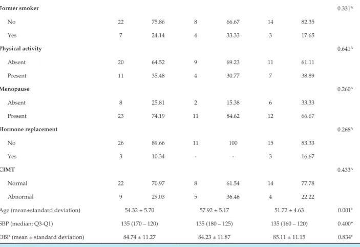
In this study, 31 pre‑menopausal women met the inclusion criteria and participated in all of its stages. The average age of the entire group was 54.32 ± 5.70. The mean age of the groups with and without CHD was 57.92 ± 5.17 and 51.72 ± 4.63, respectively, statistical significance (p = 0.001).
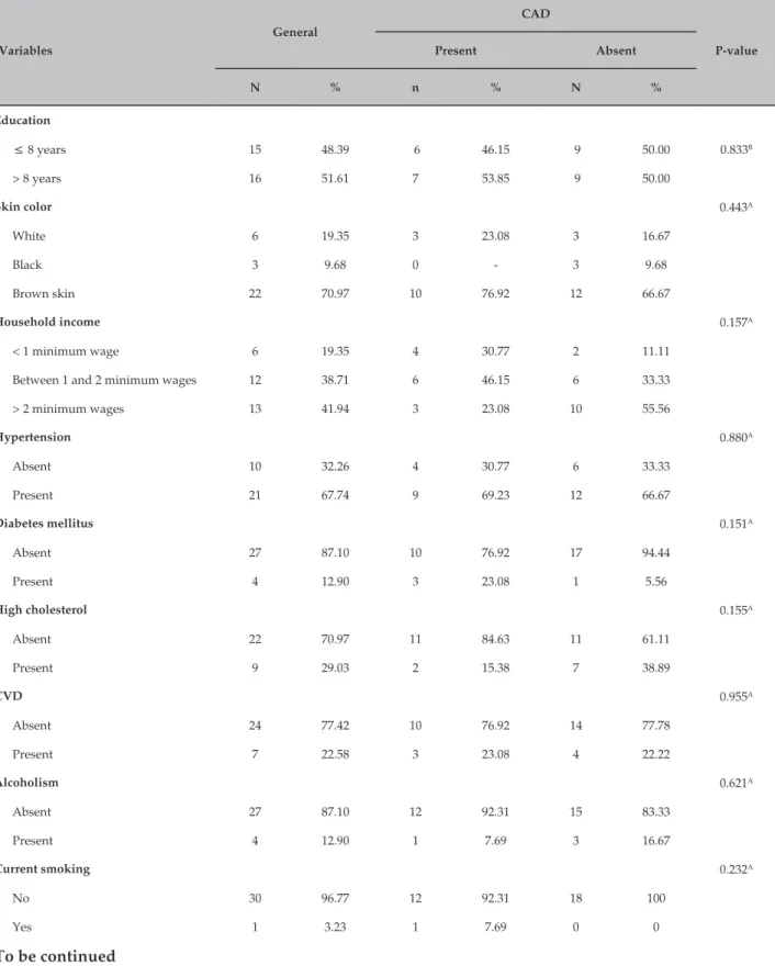
There was a predominance of brown women (70.97%), with monthly household income lower than two minimum wages (58.06%) and education of more than 8 years (51.61%) (Table 1).
CIMT was abnormal in 29.03% in the general population. The intima‑media thickness was 1.55 ± 0.78 mm in the general group, 1.92 ± 0.94 mm in the CAD group and 1.18 ± 0.71 mm in the group without CAD (p = 0.001) (Table 2). CAD patients had predominance of abnormal CMIT compared to those without CAD: 36.46% vs. 22.22%, respectively (Table 1).
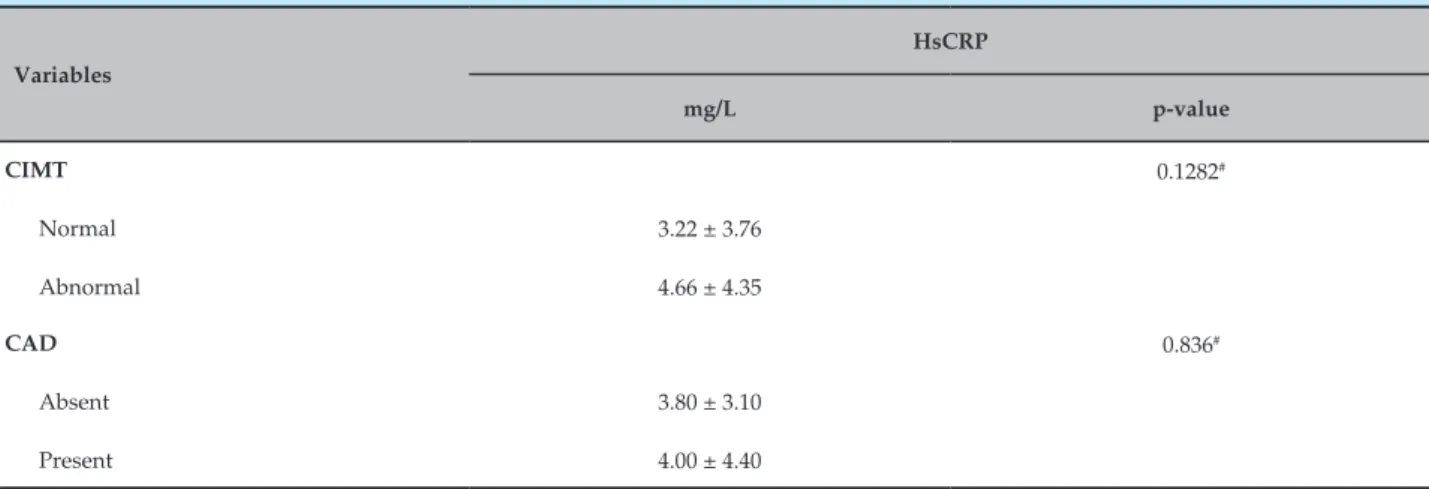
Patients with abnormal EMI had higher levels of hsCRP, but with no statistical significance (Table 3).
CAD patients had higher levels of hsCRP, but with no statistical significance (Table 3).
Discussion
Atherosclerotic disease is the leading cause of morbidity and mortality in Western communities.19
Its incidence in women is increasing along with changes in lifestyle.20
Another peculiarity related to CAD affecting women is that the onset of the disease is typically late, especially after menopause. This characteristic requires reliable diagnostic tools that may indicate early dysfunction and risk factors for cardiovascular disease.
A study conducted in Europe in 2008 by Allender et al.21 showed that coronary disease was responsible for 23%
of deaths in women. Several studies have demonstrated
the increased incidence of CAD with increasing age and menopause. Tremollieres et al. (1999) found a 36% prevalence of CAD in most women after menopause.22
Endothelium dysfunction is extremely important in the pathophysiology of atherosclerosis.23 The main
methods currently available for assessing endothelial function consist in measuring endothelial response to physical or pharmacological stimuli.
Platelet activation markers and inflammation, especially ultrasensitive C‑reactive protein (hsCRP), also proved useful in the evaluation of endothelial health.24
CIMT has been considered a useful, low‑cost technique for early detection of atherosclerosis and as a predictor of risk of cardiovascular events.25
Kablak‑Ziembicka et al.,26 in a study involving
558 patients of both genders, including 120 women, with a mean age of 58.8 ± 9.2, showed that there is an association of greater CIMT abnormality in patients with angiographically confirmed CAD than in patients who had normal coronary arteries.
Several studies have suggested a correlation between the levels of hsCRP and CMIT.28‑31 Trinidad et al.,27 in a study
conducted at Universidade Estadual do Rio de Janeiro with 116 hypertensive women aged 40 to 65, showed hsCRP correlated to CIMT. Similarly, the findings of a work developed by Blackburn et al. with 1,051 individuals with dyslipidemia showed correlation between hsCRP and CIMT.28 The studies of Wang et al.29 and Sitzer et al.30 also
showed correlations between these variables.
Kawamoto et al.,31 in a study of 440 patients of both
genders, including 201 women aged 75±10, found that hsCRP levels were associated with increased CIMT.
Parildar et al.,32 found a positive and significant
correlation between CIMT and hsCRP in a study involving 110 pre‑diabetic patients and 76 healthy patients with mean age of 51.1 ± 9.9, with a percentage of female patients of 68.1%.
Besides this, Amer et al.,33 in a case‑control study
involving elderly hypertensive patients, found that CIMT had a positive and significant correlation with hsCRP levels.
On the other hand, the association between CIMT and hsCRP levels were not significant in some studies. Folsom et al.,34 examined the association of hsCRP with
Table 1 – Sociodemographic and health characteristics of pre-menopausal women – HUUFMA. São Luís – MA, 2013
Variables
General
CAD
P-value
Present Absent
N % n % N %
Education
≤ 8 years 15 48.39 6 46.15 9 50.00 0.833B
> 8 years 16 51.61 7 53.85 9 50.00
Skin color 0.443A
White 6 19.35 3 23.08 3 16.67
Black 3 9.68 0 ‑ 3 9.68
Brown skin 22 70.97 10 76.92 12 66.67
Household income 0.157A
< 1 minimum wage 6 19.35 4 30.77 2 11.11
Between 1 and 2 minimum wages 12 38.71 6 46.15 6 33.33
> 2 minimum wages 13 41.94 3 23.08 10 55.56
Hypertension 0.880A
Absent 10 32.26 4 30.77 6 33.33
Present 21 67.74 9 69.23 12 66.67
Diabetes mellitus 0.151A
Absent 27 87.10 10 76.92 17 94.44
Present 4 12.90 3 23.08 1 5.56
High cholesterol 0.155A
Absent 22 70.97 11 84.63 11 61.11
Present 9 29.03 2 15.38 7 38.89
CVD 0.955A
Absent 24 77.42 10 76.92 14 77.78
Present 7 22.58 3 23.08 4 22.22
Alcoholism 0.621A
Absent 27 87.10 12 92.31 15 83.33
Present 4 12.90 1 7.69 3 16.67
Current smoking 0.232A
No 30 96.77 12 92.31 18 100
Yes 1 3.23 1 7.69 0 0
Continuation
Former smoker 0.331A
No 22 75.86 8 66.67 14 82.35
Yes 7 24.14 4 33.33 3 17.65
Physical activity 0.641A
Absent 20 64.52 9 69.23 11 61.11
Present 11 35.48 4 30.77 7 38.89
Menopause 0.260A
Absent 8 25.81 2 15.38 6 33.33
Present 23 74.19 11 84.62 12 66.67
Hormone replacement 0.268A
No 26 89.66 11 100 15 83.33
Yes 3 10.34 ‑ ‑ 3 16.67
CIMT 0.433A
Normal 22 70.97 8 61.54 14 77.78
Abnormal 9 29.03 5 36.46 4 22.22
Age (mean±standard deviation) 54.32 ± 5.70 57.92 ± 5.17 51.72 ± 4.63 0.001#
SBP (median; Q3‑Q1) 135 (170 – 120) 135 (180 – 125) 135 (160 – 120) 0.400*
DBP (mean ± standard deviation) 84.74 ± 11.27 84.23 ± 11.87 85.11 ± 11.15 0.834#
# t-test; * Mann-Whitney test; A Fisher exact test; B Qui-square test; Q3-Q1 - quartile 3 and quartile 1.
Table 2 – Association of CIMT with CAD in pre-menopausal women – HUUFMA. São Luís – MA, 2013
Characteristic
General
CAD
P-value
Present Absent
Mean ± SD Mean ± SD Mean ± SD
CIMT 1.55 ± 0.78 1.92 ± 0.94 1.8 ± 0.71 0.001#
HUUFMA: Hospital Universitário da Universidade Federal do Maranhão; CIMT: carotid intima-media thickness; CAD: coronary artery disease; # t-test.
Similarly, the results of the study by Hak et al.35
conducted in 186 healthy middle‑aged women selected from the general population, indicated that hsCRP is not significantly associated with CIMT.
Jie Cao J. et al,36 conducted a cohort of 5417 participants
to evaluate the correlation between hsCRP and CIMT in elderly patients with high risk of stroke. They concluded
that high levels of hsCRP represent an independent risk factor for stroke, not correlating with the severity of atherosclerotic plaque as measured by CIMT.
Table 3 – Association of HsCRP with CIMT and CAD in pre-menopausal women – HUUFMA. São Luís – MA, 2013
Variables
HsCRP
mg/L p-value
CIMT 0.1282#
Normal 3.22 ± 3.76
Abnormal 4.66 ± 4.35
CAD 0.836#
Absent 3.80 ± 3.10
Present 4.00 ± 4.40
HUUFMA: Hospital Universitário da Universidade Federal do Maranhão; HsCRP: ultrasensitive C-reactive protein; CIMT: carotid intima-media thickness; CAD: coronary artery disease; # t-test.
by the Emerging Risk Factors Collaboration group, hsCRP levels were linearly associated with the presence of several cardiovascular risk factors and inflammatory markers and were strongly associated with the risk of ischemic vascular diseases.14
In this study, although serum levels of hsCRP are higher among women with CAD, no relationship was found between these variables. It should be considered that, in both groups, the patients already had cardiovascular risk factors, which may have led to the convergence of hsCRP values.
Several studies have shown baseline levels of hsCRP as independent predictors for coronary artery disease.15,38 A prospective case‑control study conducted
by Boekholdt et al.37 with 25,663 men and women aged
between 45 and 79, who were part of the EPIC‑Norfolk study, found that hsCRP levels were among the main predictors of coronary artery disease and mortality.
A prospective case‑control study conducted by Ridker et al.39 evaluated, for three years, the levels of
inflammatory markers of 28,263 apparently healthy women in post‑menopause. The study also found cardiovascular events during the study period. HsCRP levels proved to be the most important independent predictors of risk of cardiovascular events in this group.15
The disagreements between the medical literature available and some of our results can be explained by the sample of limited size, resulting from losses during the study, and the fact that our sample is composed
solely of patients with clinical indication for coronary angiography, having different characteristics compared to the general population of pre‑menopausal women.
Clinical implications
This study fills a gap of few national studies assessing endothelial function in pre‑menopausal patients.
Study limitations
The main limination of this study was a non‑probabilistic sample limited to a relatively small number of individuals, making studies with larger sample sizes necessary.
Conclusion
In the population studied, assessment of endothelial function using the CIMT method showed higher sensitivity and specificity for the diagnosis of CAD compared to the measurement of hsCRP levels in pre‑menopausal women.
Author contributions
1. Ministério da Saúde. Indicadores e dados básicos. Brasilia; 2012. (online) .[Citado em 2016 out 10].Disponível em:tabnet.datasus.gov.br/cg1/ idb2012/matriz.htm – 2012.
2. De Lorenzi DRS, Basso E, Fagundes PdO, Saciloto B. Prevalence of overweight and obesity among climacteric women. Rev Bras Ginecol Obstet. 2005;27(8):479‑84.
3. Caramori PRA, Zago AJ. Disfunção endotelial e doença arterial coronariana. Arq Bras Cardiol. 2000;75(2):163‑82.
4. Vita JA, Keaney JF. Endothelial function a barometer for cardiovascular risk? Circulation. 2002;106(6):640‑2.
5. Davignon J, Ganz P. Role of endothelial dysfunction in atherosclerosis. Circulation. 2004;109(23 Suppl 1):III‑27‑III‑32.
6. Gimbrone Jr MA, García‑Cardeña G. Vascular endothelium, hemodynamics, and the pathobiology of atherosclerosis. Cardiovasc Pathol. 2013;22(1):9‑15.
7. Verma S, Buchanan MR, Anderson TJ. Endothelial function testing as a biomarker of vascular disease. Circulation. 2003;108(17):2054‑9.
8. Garcia MM, Lima PR, Correia LC. Prognostic value of endothelial function in patients with atherosclerosis: systematic review. Arq Bras Cardiol. 2012;99(3):857‑65.
9. Bots ML, Hoes AW, Koudstaal PJ, Hofman A, Grabee DE. Common carotid intima‑media thickness and risk of stroke and myocardial infarction the Rotterdam Study. Circulation. 1997;96(5):1432‑7.
10. Lorenz MW, Markus HS, Bots ML, Rosvall M, Sitzer M. Prediction of clinical cardiovascular events with carotid intima‑media thickness a systematic review and meta‑analysis. Circulation. 2007;115(4):459‑67.
11. De Groot E, van Leuven SI, Duivenvoorden R, Mewese MC, Akdim F, Boots ML, et al. Measurement of carotid intima–media thickness to assess progression and regression of atherosclerosis. Nature Clin Pract Cardiovasc Med. 2008;5(5):280‑8.
12. Stein JH, Korcarz CF, Hurst RT, Lonn E, Kendall CB, Mohler ER,et al. Use of carotid ultrasound to identify subclinical vascular disease and evaluate cardiovascular disease risk: a consensus Statement from the American Society of Echocardiography Carotid Intima‑Media Thickness Task Force. Endorsed by the Society for Vascular Medicine. J Am Soc Echocardiogr.2008;21(2):93‑111.
13. Kusche‑Vihrog K, Urbanova K, Blanque A, Wilhelni M, Schillers H, Kliche K, et al.. C‑reactive protein makes human endothelium stiff and tight. Hypertension. 2011;57(2):231‑7.
14. Emerging Risk Factors Collaboration (ERFC). C‑reactive protein concentration and risk of coronary heart disease, stroke, and mortality: an individual participant meta‑analysis. Lancet. 2010;375(9709):132.
15. Ridker PM, Hennekens CH, Buring JE, Rifai N. C‑reactive protein and other markers of inflammation in the prediction of cardiovascular disease in women. N Engl J Med. 2000;342(12):836‑43.
16. Pignoli P, Tremoli E, Poli A, Oreste P, Paoletti R. Intimal plus medial thickness of the arterial wall: a direct measurement with ultrasound imaging. Circulation. 1986;74(6):1399‑406.
17. Sociedade Brasileira de Cardiologia/Sociedade Brasileira de Hipertensão/ Sociedade Brasileira de Nefrologia. VI Diretrizes Brasileiras de Hipertensão. Arq Bras Cardiol. 2010;45(1 supl 1):1‑51.
18. Fernandes DdC, Laurindo FRM. Endothelial function and oxidative stress biomarker. Rev Soc Cardiol Estado de São Paulo. 2010;20(2):182‑94.
19. Chan DC, Watts GF. Apolipoproteins as markers and managers of coronary risk. QJM. 2006; 99(5)277‑87.
20. Da Luz PL, Solimene MC. Peculiaridades da doença arterial coronária na mulher. Rev Ass Med Bras. 1999;45(1):45‑54.
21. Allender S, Scarborough P, Viv P, Rayner M. European Cardiovascular Disease Statistics. 8th ed. Boston: British Heart Foundation Health
Promotion Research Group; 2008.
22. Trémollières FA, Pouilles JM, Cauneille C, Ribot C. Coronary heart disease risk factors and menopause: a study in 1684 French women. Atherosclerosis. 1999;142(2):415‑23.
23. Deanfield JE, Halcox JP, Rabelink TJ. Endothelial function and dysfunction: testing and clinical relevance. Circulation. 2007;115(10):1285‑95.
24. Kusche‑Vihrog K, Urbanova K, Blanqué A, Wilhelmi M, Schillers H, Kliche K, Pavenstadt H, et al. C‑reactive protein makes human endothelium stiff and tight. Hypertension. 2011;57(2):231‑7.
25. Chua SK, Kilung A, Ong TK, Fong AY, Yew KL, Khiew NZ, et al. Carotid intima media thickness and high sensitivity C‑reactive protein as markers of cardiovascular risk in a malaysian population. Med J Malaysia. 2014;69(4):166‑74.
26. Kablak‑Ziembicka A, Tracz W, Przewlocki T, Pieniazek P, Sokolowski A, Konieczynska M. Association of increased carotid intima‑ media thickness with the extent of coronary artery disease. Heart. 2004;90(11):1286–90.
27. Trindade M, Martucci RB, Burlá AK, Oigman W, Neves MF, Araújo DV. Avaliação de fatores de risco para o espessamento médio‑intimal da carótida em mulheres hipertensas. Revista Hospital Universitário Pedro Ernesto ( HUPE). 2012;11(1):55‑63.
28. Blackburn R, Giral P, Bruckert E, André JM, Gonbert S, Bernard M, et al. Elevated C‑reactive protein constitutes an independent predictor of advanced carotid plaques in dyslipidemic subjects. Arterioscler Thromb Vasc Biol, 2001.21(12):1962‑8.
References
Statistical analysis: Fernandes DR. Obtaining financing: Figueiredo Neto JA. Writing of the manuscript: Farias WKS. Critical revision of the manuscript for intellectual content: Farias WKS, Rocha TPO, Melo JB, Fonseca EJNC, Figueiredo Neto JA.
Potential Conflict of Interest
No potential conflict of interest relevant to this article was reported.
Sources of Funding
This study was funded by Fundação de Amparo à Pesquisa e ao Desenvolvimento Científico e Tecnologia do Maranhão.
Study Association
29. Wang TJ, Nam B‑H, Wilson PW, Wolf PA, Levy D, Polak JF, et al. Association of C‑reactive protein with carotid atherosclerosis in men and women: the Framingham Heart Study. Arterioscler Thromb Vasc Biol. 2002;22(10):1662‑7.
30. Sitzer M, Markus HS, Mendall MA, Liehr R, Knorr U, Steinmetz H. C‑reactive protein and carotid intimal medial thickness in a community population. Eur J Cardiovasc Risk. 2002;9(2):97‑103.
31. Kawamoto R, Tomita H, Inoue A, Ohtsuka N, Kamitani A. Impact of C‑reactive protein on the likelihood of carotid atherosclerosis in Japanese adults. J Atheroscler Thromb. 2006.13(4):175‑82.
32. Parildar H, Gulmez O, Cigerli O, Dogruk Unal A, Erdal R, Guvener Demirag N. Carotid artery intima media thickness and HsCRP; predictors for atherosclerosis in prediabetic patients? Pak J Med Sci. 2013;29(2):495‑9.
33. Amer MS, Elawam AE, Khater MS, Omar OH, Mabrouk RA, Taha HM. Association of high‑sensitivity C‑reactive protein with carotid artery intima‑media thickness in hypertensive older adults. J Am Soc Hypertens. 2011;5(5):395–400.
34. Folsom A, Pankow JS, Tracy RP, Arnett DK, Peacock JM, Hong JM, et al. Association of C‑reactive protein with markers of prevalent atherosclerotic disease. Am J Cardiol, 2001.88(2):112‑7.
35. Hak AE, Stehouwer CD, Bots ML, Polderman KH, Schalkwijk CG, Westendorp IC. Associations of C‑reactive protein with measures of obesity, insulin resistance, and subclinical atherosclerosis in healthy, middle‑aged women. Arterioscler Thromb Vasc Biol. 1999;19(8):986‑91.
36. Cao JJ, Thach C, Manolio TA, Psaty BM, Kuller LH, Chaves PH, et al. C‑reactive protein, carotid intima‑media thickness, and incidence of ischemic stroke in the elderly the cardiovascular health study. Circulation. 2003;108(2):166‑70.
37. Boekholdt SM, Hack CE, Sandhu MS, Luben R, Bingham SA, Wareham NJ, et al. C‑reactive protein levels and coronary artery disease incidence and mortality in apparently healthy men and women: the EPIC‑Norfolk prospective population study 1993–2003. Atherosclerosis. 2006;187(2):415‑22.
38. Ridker PM, Buring JE, Shih J, Matias M, Hennekens CH. Prospective study of C‑reactive protein and the risk of future cardiovascular events among apparently healthy women. Circulation. 1998;98(8):731‑3.