Carotid Intimal-Medial Thickening and Endothelial
Function in Coronary Artery Disease
Graziela Chequer, Bruno Ramos Nascimento, Túlio Pinho Navarro, Eduardo Belisário Falqueto, Maria Clara N. Alencar, Márcio Cristiano R. de Miranda, Ari Mandil, Jamil Abdalla Saad, Cirilo Fonseca, Antônio Luiz Pinho Ribeiro
Hospital das Clínicas da Faculdade de Medicina da UFMG, Hospital Socor e Hospital Felício Rocho - Belo Horizonte, MG - Brazil
M a i l i n g A d d r e s s : G r a z i e l a C h e q u e r • R u a M o n t e s C l a r o s , 4 7 5 / 7 0 2 – 3 0 3 1 0 - 3 7 0 – B e l o H o r i z o n t e , M G - B r a z i l
E-mail: [email protected] Received on 04/10/05 • Accepted on 05/04/05
O
BJECTIVETo investigate the correlation between the endothelial function and the carotid intimal-medial thickening (IMT) in a population of patients with coronary artery disease, as well as that between the endothelial function and carotid IMT with the severity of the coronary lesions.
M
ETHODSForty-three patients aged 60.5±9.2 years, (67.4% males) with coronary artery disease at the coronariography were studied. Endothelial function was assessed using the brachial artery reactivity test (BART), which measured the percentage of fl ow-mediated dilatation (%FMD). The carotid IMT was evaluated through vascular ultrasound.
R
ESULTSThe mean %FDM was 4.7 ± 3.6 and the mean carotid IMT was 1.08 ± 0.23 mm. The carotid MIT and %FMD measurements showed a statistically signifi cant correlation, with Spearman’s coefficient of 0.315, p= 0.042, demonstrating that lower %FMD values corresponded to an increased carotid IMT (r = -0.315, p = 0.042). There was no correlation between %FMD or IMT and the severity of coronary lesions.
C
ONCLUSIONThe presence of a correlation between carotid IMT and %FMD demonstrates a concomitance of anatomical and functional vascular alterations in coronary artery disease, regardless of the severity of the atherosclerotic lesions.
K
EY WORDSEndothelial dysfunction has an important role in plaque formation and the clinical course of atherosclerosis. The intimal-medial thickening (IMT) of the carotid is an early vascular alteration that precedes plaque formation. However, the correlation between these parameters in coronary artery disease is yet to be defi ned.
Coronary artery disease persists as the main cause of death worldwide. It is currently known that the endothelium has a vital participation in the pathogenesis of atherosclerosis, triggering an infl ammatory response that is responsible for the atherosclerotic plaque formation and instability, with a direct infl uence on the clinical course of atherosclerosis and other cardiovascular diseases, such as arterial hypertension and cardiac failure.
Until the beginning of the 90s, endothelial function was evaluated through invasive methods, with the intracoronary injection of acetylcholine1. In 1992, Celermajer et al2
standardized the brachial artery reactivity test (BART) , in which the endothelial function is assessed through a simple vascular ultrasonographic examination.
The vascular intimal-medial thickening (IMT) corres-ponds to the initial stage of atherosclerosis, preceding plaque formation. The ultrasonography of the carotid artery allows IMT to be easily measured, and the presence and severity of the carotid intimal-medial thickening have been correlated to coronary atherosclerosis3,4.
Although the presence of endothelial dysfunction and carotid intimal-medial thickening have a prognostic value that has been well demonstrated as independent predictors of future cardiovascular events (acute myocardial infarction, stroke and death), the value of these methods as indicators of possible coronary artery disease is yet to be defi ned and remains unclear.
The main objective of this study was to investigate a correlation between the brachial artery reactivity test (BART) and carotid intimal-medial thickening (IMT) in a population of patients with coronary artery disease. As a secondary objective, we investigated a correlation between these two tests and the severity of the atherosclerotic lesions detected by coronary angiography.
M
ETHODSThe sample consisted of patients referred to the Laboratory of Hemodynamics of the Felício Rocho and Socor Hospitals, in Belo Horizonte, Minas Gerais, Brazil, between March 2002 and March 2003. The coronary angiography was requested by the assistant physician, in order to investigate thoracic pain or ischemia detected through a functional test (ergometric stress test, myocardial scintillography or stress echocardiogram (STRESS ECHO)).
The patients were selected consecutively immediately after undergoing the elective coronary angiography. All
patients who presented evidence of any degree of luminal obstruction were included.
The exclusion criteria were: normal coronary angio-graphy (smooth coronary arteries); diagnosis of acute coronary syndrome (unstable angina or acute myocardial infarction) within seven days prior to the examination; clinical or laboratory evidence of any chronic disease such as hepatopathies, valvopathies, cardiac failure, thyroid disease, chronic renal failure with serum creatinine > 2.0 mg%, conjunctive tissue diseases and neoplasias.
After signing the written informed consent, the patients were referred to the Bias Fortes Outpatient Clinic, from Hospital das Clinicas of the Federal University of Minas Gerais, in order to fi ll out the research protocol, along with the assessment of the clinical picture and risk factors for coronary disease.
Smoking was defi ned as the habit of smoking during the previous three months. Dyslipidemia was defi ned as serum LDL cholesterol levels > 160 mg/dL and hereditary factor for coronary artery disease, such as the presence of at least one fi rst-degree relative with a diagnosis of the disease or sudden death, at an age younger than 55 yrs in the male sex and 65 yrs in the female sex. Systemic arterial hypertension was considered according to the information provided by the patient or use of anti-hypertensive medication. The presence of diabetes mellitus was defi ned according to the information provided by the patient, use of oral hypoglycemic drugs and/or insulin or through fasting glycemia levels (minimum of two measurements above 126 mg/dL).
All patients were refereed to the vascular laboratory of Ecograf, a Diagnostic Medicine Group in Belo Horizonte, Minas Gerais, Brazil, in order to undergo the endothelial function test and carotid ultrasonography.
Specifi c procedures – coronary angiography and left ventricle angiography - The coronary angiography was analyzed subjectively by two independent interventionist cardiologists. In case of discordance, a third specialist was called in for another evaluation. Lesions with a compromised luminal diameter < 50% were called parietal irregularities. The results were analyzed in patients with lesions < 50%, lesions between 50% and 70% and lesions > 70%.
Endothelial function test through the fl ow-mediated dilatation of the brachial artery - The brachial artery reactivity test (BART) was performed according to the original description by Celemajer et al2; drugs with effect
The patient remained in a resting position, in dorsal decubitus, for 10 minutes prior to the test. The diameter of the brachial artery was measured through a vascular ultrasound, using a linear transducer with a 7.0 MHz frequency in an ASPEN equipment (Acuson, Mountain View, California – USA). The images were obtained during resting, during reactive hyperemia, again during resting and after the administration of sublingual nitroglycerin.
With the transducer positioned 2 to 15 cm above the cubital fossa, the measurement of the diameter of the brachial artery was performed (in mm) in basal conditions. Next, the insuffl ation of a pneumatic cuff positioned 5 cm below the cubital fossa at 250 mmHg for 5 minutes was carried out, in order to produce distal ischemia. Sixty seconds after cuff desuffl ation, the recording of images in a state of reactive hyperemia was performed. After 15 minutes of wait, 400 µcg of sublingual nitroglycerin was administrated and 4 minutes later, the last recording of arterial diameter was performed. The variation of arterial diameter during resting and after reactive hyperemia was expressed as the percentage of fl ow-mediated dilatation (%FMD). The means of four measurements in each situation were obtained: basal, after reactive hyperemia, and after nitroglycerin. All images were obtained by the same examiner, who was blinded for the clinical picture and the coronary angiography results.
Measurement of the carotid intimal-medial thickening (IMT) - The measurement of the carotid intimal-medial thickening was performed according to the technique described in previous studies5,6, during the initial 10
minutes of the resting period, before the brachial artery reactivity test was performed, by the same examiner. The same vascular transducer described above was used, by positioning it longitudinally on the patient’s carotid artery, bilaterally. The highest of the values obtained from both carotids, expressed in millimeters (mm) was considered for analysis.
Statistical analysis - The quantitative variables were described by means and standard deviations. Qualitative variables were described by frequency and percentage. The correlation between the %FMD, carotid intimal-medial thickening and the severity of atherosclerotic lesions by coronary angiography was obtained through the Spearman’s rank correlation coeffi cient (rs), as the distribution of the variables was not normal. The results were considered signifi cant when p< 0.05.
The present study represents a sub-study of a broade project, and the sample calculation for such project was carried out considering an alpha error of 0.05 and a beta error of 0.20. Considering that the sample size was determined a priori, the power of this study was estimated at being 74.8% for an alpha error of 0.05, according to the PS Power and Sample Size Calculation Software (Dupont and Plummer, 1997, available at http://www. mc.vanderbilt.edu/prevmed/ps.htm).
Ethical aspects - All patients signed a written informed consent prior to the study. The study was carried out according to the norms in the 196/96 resolutions on research involving human beings published by the Health Ministry and National Council of Health, and approved by the Ethics Committee on Research of Hospital das Clinicas of the University of São Paulo School of Medicine.
Objectives - The main objective of this study was to investigate a correlation between BART and carotid intimal-medial thickening in a population of patients with an ample spectrum of coronary artery disease, demonstrated objectively through a cineangiocoronarygraphy.
A secondary objective was to evaluate a correlation between the endothelial function and the carotid intimal-medial thickening with the severity of the coronary artery disease expressed by the degree of luminal obstruction (in %), determined through an atherosclerotic plaque.
R
ESULTSForty-two patients were assessed with 66.7% of them being males. Mean age was 60.7 ± 9.2 yrs, arterial hypertension was present in 35 (83.3%) of the patients and dyslipidemia in 14 (33.3%) of them (Table I).
Clinical picture - Atypical thoracicpain was present in 19 (45.2%) of the patients, being the most frequent clinical picture, followed by stable angina in 12 patients (28.6%). Six patients (14.3%) were asymptomatic (Table I).
Coronary angiography - Regarding the extension of coronary disease, 12 (28.6%) of the patients presented just parietal coronary alterations, 5 (11.9%) had lesions between 50% and 70% and 25 (59.5%) had lesions ≥
70%. Of the patients with coronary artery disease and at least one lesion ≥50%, 10 (23.8%) were uniarterial lesions, 8 (19.0%) were biarterial and 10 (23.8%) were triarterial lesions. Sixteen patients (38.1%) presented
Table 1 - Risk factors and clinical picture
Characteristics n = 42 Porcentual
Hypertension 35 83.3
Diabetes 10 23.8
Dyslipidemia 14 33.3
Family history 9 21.4
Smoking 4 9.5
Previous event 13 31.0
Asymptomatic 6 14.3
Atypical pain 19 45.2
Dyspnea 7 16.7
Previous AMI 3 7.1
Previous unstable angina 2 4.8
Stable angina 12 28.6
Altered ET/SC 3 7.1
Others 4 9.5
some segmental defi cit assessed by ventriculography. The concordance among the interventionist cardiologists who performed the coronary angiographies was evaluated through Kappa method, with an 81.1 ± 8.5% rate.
Endothelial function - The mean of the percentage of vasodilation after the administration of sublingual nitroglycerin was 9.6 ± 4.2. Gender and age did not present a correlation with %FMD (r = -0.280, p = 0.07 and r = -0.088, p = 0.57, respectively).
The degree of coronary obstruction did not correlate with %FMD either (r = -0.008, p = 0.96). The mean of % FMD in the studied population was 4.7 ± 3.6. Figures 1 and 2 show BART images of one of the patients from the study group. There was a %FMD of 12, which represents normal endothelial function.
Carotid intimal-medial thickening - The mean of
the carotid intimal-medial thickening was 1.08 ± 0.23 mm (Fig. 2). The measurement of the carotid intimal-medial thickening did not show a correlation with gender and age (r = -0.099, p = 0.53 and r = 0.278, p = 0.074, respectively). The severity of coronary artery disease (degree of obstruction) did not correlate with the carotid intimal-medial thickening either (r = -0.063, p = 0.69).
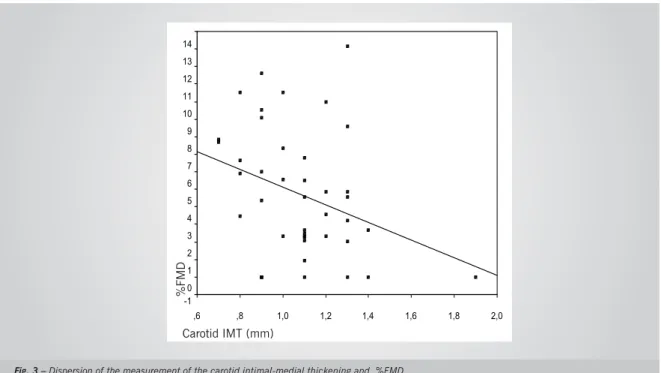
Correlation between %FMD and carotid intimal-medial thickening - The measurements of the carotid intimal-medial thickening and %FMD presented a statistically significant correlation, with Spearman’s coefficient of –0.315, p=0.042 (Fig.3). When we evaluate this correlation from another point of view, considering the carotid intimal-medial thickening as a binary variable and present if > 1.0 mm, we observe
Fig.1 – Transversal section of the brachial artery in a patient from the study sample, showing a basal diameter of 3.9 mm.
that age and gender still do not show a correlation with intimal-medial thickening, but in a concordant way with the previous analysis, %FMD was 6.0 ± 3.7 in the 18 patients without intimal-medial thickening, a statistically signifi cant difference with a p value = 0.04 (Table II).
These authors defi ned a cutoff through the ROC curve, fi nding the value of %FMD < 4.5 as being the highest sensitivity and specifi city value to predict coronary disease. The test sensitivity and specifi city were 71.3% and 81.0%, respectively. The highest sensitivity was that of the myocardial scintillography (100%) followed by the presence of angina pain (95.1%) and the ergometric test (82.4%). Regarding the test specifi city, the values for the ergometric test were 57.1% and for the presence of angina, 23.8%. The data above show a good diagnostic value for the brachial artery reactivity test in terms of sensitivity and specifi city and the authors suggested that the test can have, in the future, a use in the screening of coronary artery disease, but one must consider the possibility of a bias caused by patient selection in this study.
Although BART has been employed in several studies, there is no consensus regarding a reference value for the %FMD that would defi ne endothelial dysfunction. In most studies, the test is described in different groups of patients, with values that are compared to one another, without the objective to defi ne the presence or absence of endothelial dysfunction from a certain %FMD on. In 2001, a guideline on the use of BART was published containing important directions regarding the adequate methodology, but without establishing a value of %FMD that would defi ne endothelial function12.
In our sample of patients with a ample spectrum of coronary artery disease, the mean of %FMD of 4.7 ± 3.6 was decreased, which was expected considering the role of endothelium as an initial event of atherogenesis. This value is a little below those found by other authors while studying the endothelial function in patients with coronary disease demonstrated by coronary angiography. Chan
Table 2 – Age, gender and FMD percentage in patients without and with carotid IMT
Group Without carotid
IMT (n=18)
With carotid
IMT (n=24) p
Age (yrs) 58.7 ± 10.8 62.2 ± 7.7 0.22
Male sex (%) 12 (66.7) 16 (66.7) 1.00
% DMF 6.0 ± 3.7 3.7 ± 3.3 0.04
IMT - intimal-medial thickening > 1 mm; %FMD - percentage of fl ow-mediated dilation.
D
ISCUSSIONThe study of coronary artery disease in its more initial, sub-clinical phases has been the object of increasing interest. The importance of endothelial dysfunction as an independent predictor of cardiovascular events has been well demonstrated in previous studies7-9. Similarly,
the presence of carotid IMT is related to the occurrence of acute myocardial infarction and ischemic stroke, with a relative risk between 2 and 610.
The importance of these tests in the early diagnosis of atherosclerotic disease, however, remains indefi nite. Schroeder and cols.11 studied the value of the brachial
artery reactivity test in the screening of coronary artery disease in 122 patients who were referred to coronary angiography and established a comparison with other diagnostic methods, such as the presence of thoracic angina, the ergometric treadmill test and myocardial scintillography.
2,0 1,8 1,6 1,4 1,2 1,0 ,8 ,6
%DMF
14
13
12
11
10
9
8
7
6
5
4
3
2
1
0
-1
Carotid IMT (mm)
Fig. 3 – Dispersion of the measurement of the carotid intimal-medial thickening and %FMD.
and cols.13 found a mean of %FMD of 5.9 ± 3.8. It is
important to emphasize that the value found in our study, 4.7 ± 3.6, is much lower than the mean values found in healthy individuals without risk factors for atherosclerotic disease, between 8.2 ± 3.1 and 21.1 ± 2.02,14,15
.
In a study conducted by our group and published in 2003, we found a %FMD of 8.9 ± 5.7, studying 26 individuals with normal coronaries16.
There was no correlation between %FMD and the severity of coronary disease regarding the degree of obstruction of the atherosclerotic lesions, as also demonstrated by Enderle and cols.17, but in contrast
with the results by Neunteufl and cols.15. Demographic
differences, such as the percentage of symptomatic, diabetic, hypertensive, dyslipidemic, patients or smokers, might have determined these discrepant results. The criteria employed to defi ne the severity of coronary disease differ among the studies. Neunteufl and cols stratifi ed the patients in a larger number of subgroups according to the degree of obstruction of the atherosclerotic lesions: < 30, 30-50, 50-70, 70-90, 90-99 and 100%, whereas the stratifi cation employed by us consisted of only 3 subgroups: < 50%, between 50% and 70% and > 70%.
Especially important is the need to consider that, in all studies, the severity of the coronary artery disease was established considering only the degree of luminal obstruction, in a subjective way. Although the interobserver concordance was satisfactory in our study, with a Kappa value > 80%, the computerized quantitative coronary angiography (QCA) would have allowed a more appropriate evaluation and a comparison among the studies, eliminating the interventionist cardiologist’s subjectivity and the interobserver variation. Other angiographic characteristics, such as the number of signifi cant lesions by vessel, which would represent a more diffuse disease, could be employed, but there is no consensus or normative data regarding such use. The coronary angiography was used in our study and by the other authors as a reference pattern to defi ne coronary artery disease, a concept that has been discussed after the introduction of intracoronary ultrasound.
Carotid intimal-medial thickening mean in this studied sample was 1.08 ± 0.23 mm. This elevated value is expected and is in accordance with the coronary involvement previously established on the bedside. In contrast with Enderle and cols.17, there was no correlation
between the arterial intimal-medial thickening and the degree of coronary obstruction, but the same observations regarding the defi nition of disease severity discussed above must be considered.
The correlation between %FMD and carotid intimal-medial thickening has been evaluated in few studies, and always with a small sample of patients. The negative
correlation coefficient between %FMD and carotid intimal-medial thickening shows that, in this group of patients, lower values of %FMD were correlated with a higher degree of intimal-medial thickening (r=-0.315, p= 0.042). These data are in accordance with those demonstrated by Enderle and cols.17 in a group of patients
highly suspected of having coronary disease (r=-0.317, p= 0.0004). Campuzano and cols.18, studying a group
of 39 patients with risk factors for atherosclerosis and 13 without those factors, found a difference in %FMD between the groups (2.77 ± 2.57 versus 11.98 ± 4.61%, p value < 0.01), as well as a difference in IMT values (0.85 ± 0.24 versus 0.57 ± 0.14 mm, p value = 0.001), in addition to an inverse correlation between these variables (r = -0.357; p < 0.01). Juonala et al19,
studying adults between 24 and 39 yrs with no evidence of CAD, similarly found a negative and signifi cant correlation between %FMD and IMT in a multivariate analysis (r = -0.34, p = 0.001); additionally, the number of risk factors correlated with IMT in patients with lower %FMD. In the remaining studies, the evaluation of the correlation of the two methods was carried out in hypertensive patients and not in those with coronaropathies. Ghiadoni et al.5
evaluated the endothelial function and carotid IMT in 44 hypertensive patients and 30 normotensive individuals and found an inverse correlation between the two tests in the hypertensive group, only (r = -0.68, p = 0.0003). Different results were reported by Barenbrock and et al6, who did not fi nd a signifi cant correlation between
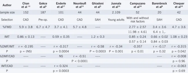
endothelial function and arterial IMT, when studying previously untreated hypertensive patients with no evidence of coronary disease (r = -0.17, p = 0.32). Table III shows a review of the outcomes of the main studies that evaluated these questions discussed here. Further studies are necessary to determine whether there is a correlation between these vascular alterations (endothelial dysfunction and IMT) and the severity of coronary artery disease.
C
ONCLUSIONR
EFERENCES1. Ludmer PL, Selwyn AP, Shook TL, et al. Paradoxical vasoconstriction induced by acetylcholine in atherosclerotic coronary arteries. N Engl J Med. 1986; 315: 1046-51.
2. Celermajer DS, Sorensen KE, Gooch VM, et al. Non-Invasive detection of endothelial dysfunction in children and adults at risk of atherosclerosis. Lancet. 1992;340(8828):1111-5.
3. Crouse JR III, Craven TE, Hagaman AP, et al. Association of coronary disease with segment-specific intimal-medial thickening of the extracranial carotid artery. Circulation. 1995;92:1141-7.
4. Kallikazaros I, Tsioufi s C, Sideris S, et al. Carotid artery disease as a marker for the presence of severe coronary artery disease in patients evaluated for chest pain. Stroke. 1999; 30:1002-7.
5. Ghiadoni L, Taddei S, Virdis A, et al. Endothelial function and common carotid artery wall thickening in patients with essential hypertension. Hypertension. 1998;32:25-32.
6. Barenbrock M, Hausberg M, Kosch M, et al. Flow-mediated vasodilation and distensibility in relation to intima-media thickness of large arteries in mild essential hypertension. Am J Hypertension. 1999;12:973-9.
7. Schächinger V, Britten MB, Zeiher AM. Prognostic impact of coronary vasodilator dysfunction on adverse long-term outcome of coronary heart disease. Circulation. 2000; 101:r1-r8.
8. Halcox J, Schenke W, Zalos G, et al. Prognostic value of coronary vascular endothelial dysfunction. Circ. 2002;106:653-58.
9. Gokce N, Keaney J, Hunter L, et al. Risk stratifi cation for postoperative cardiovascular events via noninvasive assessment of endothelial function – A prospective study. Circulation. 2002;105:1567-72.
10. Rothwell PM. Carotid artery disease and the risk of ischaemic stroke and coronary vascular events. Cerebrovasc Dis. 2000;10 Suppl 5:21-33.
11. Schroeder S, Enderle M, Ossen R, et al. Noninvasive determination of
endothelium-mediated vasodilation as a screening test for coronary artery disease: pilot study to assess the predictive value in comparison with angina pectoris, exercise electrocardiography, and myocardial perfusion imaging. Am Heart J. 1999;138(4):731-39.
12. Correti MC, Anderson TJ, Benjamín EJ, et al. Guidelines for the ultrassound assessment of endotelial-dependent fl ow-mediated vasodilation of the brachial artery. J Am Coll Cardiol. 2002;39:257-65.
13. Chan SY, Mancini J, Kuramoto L, et al. The prognostic importance of endothelial dysfunction and carotid atheroma burden in patients with coronary artery disease. J Am Coll Cardiol. 2003;42:1037-43.
14. Celermajer DS, Adams MR, Clarkson MB, et al. Passive smoking and impaired endothelium-dependent arterial dilatation in healthy young adults. N Engl J Med. 1996;334: 150-4.
15. Neunteufl T, Katzenschlager, Hassan A, et al. Systemic endothelial dysfunction is related to the extent and severity of coronary artery disease. Atherosclerosis. 1997;129:111-8.
16. Chequer G, Navarro T, Nascimento B. Função endotelial e teste de estresse em doença arterial coronariana. Arq Bras Cardiol. 2003;81(supl 3):60.
17. Enderle MD, Schroeder S, Ossen R, et al. Comparison of peripheral endothelial disfunction and intimal media thickness in patients with suspected coronary artery disease. Heart. 1998;80:349-54.
18. Campuzano R, Moya JL, García-Lledó A, et al. Endotelial disfunction and intimal-media thickness in relation to cardiovascular risk factors in patients without clinical manifestations of atherosclerosis. Rev Esp Cardiol. 2003;56(6):546-54.
19. Juonala M, Viikari JSA, Laitinen T, et al. Interrelations beetwen brachial endotelial function and carotid intima-media thickness in young adults – the cardiovascular risk in young Finns study. Circulation. 2004;110:2918-23.
Table 3 - Comparison among fi ndings from different studies regarding endothelial function and intimal-medial thickening
Author Chan
et al 13
Gokce et al9
Enderle et al17
Neunteufl et al15
Ghiadoni et al5
Juonala et al19
Campuzano et al18
Barenbrock et al6
Chequer et al19
Sample size 152 187 101 44 44 2.109 52 20 42
Selection CAD Pre-op. CAD CAD SAH Young adults With and without
risk factors SAH CAD
%FMD 5.9 ± 3.8 6.7 ± 4.7 3.7 ± 4.1 5.7 ± 4.8 --- 2.77 ± 2.57 3.4 ± 3.6 4.7 ± 3.6
11.98 ± 4.61 6.4 ± 1,.
IMT 0.86 ± 0.13 --- 0.59 ± 0.35 --- 1.2 ± 0.3 0.85 ± 0.24 0.66 ± 0.02 1.08 ± 0.23
0.57 ± 0.14 0.84 ± 0.03
%DMF/IMT r = -0.195 --- r = -0.317 --- r = -0.58 r = -0.34 -0.357 r = -0.17 r = -0.315
P p = (NS) p = 0.0004 P = 0.0003 P = 0.001 p < 0.01 p = 0.32 p = 0.042
%DMF/CAD --- --- NS r = -0.51 --- --- r = -0.008
P P < 0.0001 p = 0.96
IMT/CAD --- --- r = 0.324 --- --- --- r = -0.063
P p = 0.0003 p = 0.69
IMT - intimal-medial thickening > 1mm; %FMD - percentage of fl ow-mediated dilation; CAD - coronary artery disease; SAH - systemic arterial hypertension; pre-op - pre-operative.
Potencial Confl ict of Interest