A prospective multicenter European study on flexible
ureterorenoscopy for the management of renal stone
_______________________________________________
Francesco Berardinelli
1, Silvia Proietti
2, Luca Cindolo
1, Fabrizio Pellegrini
1, Roberto Peschechera
2,
Hennessey Derek
3, Orietta Dalpiaz
4, Luigi Schips
1, Guido Giusti
21 Dipartimento di Urologia, “S. Pio da Pietrelcina ‘’ Hospital, Vasto (CH), Italia; 2 Centro di pietra presso il
Dipartimento di Urologia, Humanitas centro clinico e di ricerca, Rozzano, Italia; 3 Department of Urology,
Craigavon Area Hospital, Portadown (UK); 4 Urologische Klinik, Medizinische Universität Graz, Austria
ABSTRACT
ARTICLE
INFO
______________________________________________________________ ______________________
Purpose: The aim of this study was to describe the outcomes and the complications of retrograde intrarenal surgery (RIRS) for renal stones in a multi-institutional working group.
Materials and Methods: From 2012 to 2014, we conducted a prospective study in-cluding all RIRS performed for kidney stones in 4 European centers. Demographic information, disease characteristics, and perioperative and postoperative data were gathered. Patients and stone data, procedure characteristics, results and safety outco-mes were analyzed and compared by descriptive statistics. Complications were reported using the standardized Clavien system.
Results: Three hundred and fifty-six patients underwent 377 RIRS with holmium laser lithotripsy for renal stones. The RIRS was completed in all patients with a mean opera-tive time of 63.5 min. The stone-free status was confirmed endoscopically and through fluoroscopic imaging after the first procedure in 73.6%. The second procedure was performed in twenty patients (5.6%) achieving an overall stone free rate of 78.9%. The overall complication rate was 15.1%. Intra-operative and post-operative complications were seen in 24 (6.7%) and 30 (8.4%) cases, respectively.
Conclusions: RIRS is a minimally invasive procedure with good results in terms of stone-free and complications rate.
Keywords:
Ureteroscopy; Kidney Calculi; Lithotripsy, Laser
Int Braz J Urol. 2016; 42: 479-86
_____________________
Submitted for publication: September 20, 2015
_____________________
Accepted after revision: January 23, 2016
INTRODUCTION
The management of kidney stones has evolved radically over the years. Previously, ex-tracorporeal shockwave lithotripsy (SWL) and per-cutaneous nephrolithotomy (PCNL) were the pre-ferred treatment modalities for renal calculi. The impressive technologic improvement in endosco-pic flexible equipment with the recent advent of digital technology made ureteroscopic approach to kidney calculi evolve from a mere diagnostic
tool to a real operative procedure capable to treat the vast majority of renal stone.
such as pregnancy, anatomic malformations or coagulopathy and solitary kidney (4, 5) although it still not is the standard of care.
In 2013, the European Association of Urology’s (EAU) Guidelines on Urolithiasis for the first time listed RIRS as a viable treatment option for all kidney stones, including stones larger than 2cm in diameter, in experienced hands in high--volume centers (6). It is noteworthy, that for sto-nes smaller than 2cm, SWL is no longer considered mandatory as first approach so that indications to RIRS have massively been broadened.
However, RIRS is associated with some disadvantages being the possible need for sta-ged procedures one of the major. Secondly, risk of ureteral injuries and the costs of acquisition and maintenance of the complex endourological armamentarium are other concerns that might have been limited the capillary diffusion of this endoscopic procedure (7). Herein we describe a multi-center prospectively study of RIRS for re-nal stones whose aim is to highlight the clinical outcomes with particular attention to the compli-cations rate.
MATERIALS AND METHODS
Clinical Data
A prospective study of all patients who underwent RIRS for kidney stone disease in four European referral centers was performed from 2012 to 2014. Data were recorded prospectively on each patient. Patient data obtained included: age, sex, body mass index (BMI), history and phy-sical examination findings, specific comorbidities, American Society of Anesthesiologists (ASA) class risk. The stone parameters evaluated were: the number of stones, stone location, previous treat-ments for stone, stone diameters and stone com-position. Double-J stent preoperatively, congenital renal anomalies (pelvi-ureteric junction obstruc-tion, horseshoe kidney), anticoagulant therapy were evaluated.
Stone location was classified as renal pel-vis/ureteropyelic junction, superior/middle/infe-rior major calyces and multiple caliceal location. The preoperative assessment included non-con-trast computed tomography (CT) or KUB
(kidney--ureter-bladder) plain radiograph and renal ultra-sound (US).
An antibiotic therapy, either as prophylaxis with cephalosporin or fluoroquinolone or adapted 5 days antibiotic therapy in patients with an intra-operative positive urine culture was administered. Informed consent was obtained from all patients, and the possible need for a staged procedure in order to obtain satisfactory stone clearance was mentioned. Exclusion criteria were as follows: pregnancy and cachexia.
Operative and Postoperative Data
The operative time was defined as the time that passed from insertion into the urethra of the cystoscope/semirigid ureteroscope for introducing the guidewire to the completion of ureteral stent placement. All the RIRS were performed using di-fferent types of flexible ureteroscopes: Flex-X2 or Flex-XC (Karl Storz Endoscope, Germany), the URF-P5 or URF-V (Olympus Europe, Germany). Lithotripsy was achieved by means of 200/273µm Holmium laser fibers. A guidewire was placed in the upper urinary tract through a rigid cystoscope or se-mirigid ureteroscope under fluoroscopic guidance. According to surgeon preference, visual assessment of the ureter and ureteropelvic junction was perfor-med with the semirigid ureteroscope. Alongside a second safety guidewire an Ureteral Access Sheath (UAS) (Flexor 9.5/11.5Fr or 12/14Fr, Cook Medical Bloomington, IL, USA, Navigator 11/13Fr, Boston Scientific, Natik, MA, USA or Retrace 10/12Fr, Co-loplast/Porges Humlebæk, Denmark) up to the pro-ximal ureter was placed. If the UAS placement was impossible, a sheathless procedure was attempted. If this last attempt failed, a pigtail, double J ureteral catheter (DJ) was left in situ for passive dilatation and the procedure was delayed.
for possible lesions. The ureteral injuries were classified in major and minor (8). The classification of ureteral wall injuries proposed by Traxer et al. (9) was not used as it was not available at the time of the beginning of the series. A DJ was applied at the end of procedure according to surgeon preference or after complicated procedures.
Follow up
The ‘‘stone-free’’ status was defined as no evidence of stones or stones less than 2mm on one-month postoperative CT and/or KUB and/or US, prescribed following the surgeon preferences. Patients with residual fragments, requiring a fur-ther RIRS, were routinely scheduled for the second treatment 30-45 days following the previous pro-cedure and were evaluated at 4 weeks from the last procedure. All patients were followed up to 6 months, with serial plain radiograph or renal ultrasound. Postoperative complications were as-sessed according to the modified Clavien classifi-cation (10).
RESULTS
Clinical Data and Patient Characteristics
Three hundred and fifty-six patients underwent 377 RIRS with holmium
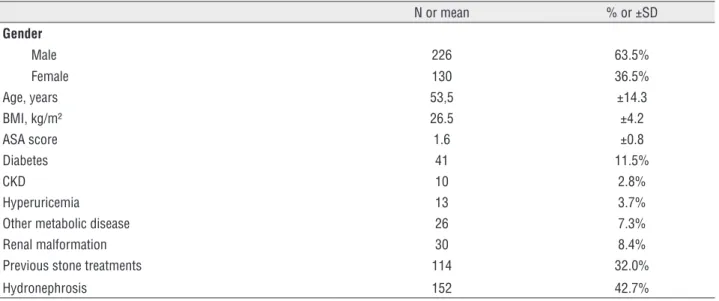
lasertrip-sy for renal stones. The cohort included 226 (63.5%) male and the mean age was 53.5 years (SD: 14.3). One third of patients (34.3%) had a history of urolithiasis, and the 32% already received a previous treatment. In 64% of pa-tients we found a single stone (mean diameters: 12.4x9.5mm) and in 165 cases (46%) the stone was located in the pelvis/ureteropyelic junc-tion. Demographic data and clinical characte-ristics of the cohort are listed in Tables 1 and 2.
Operative and Postoperative Data
RIRS was safely completed in all patients with a mean operative time of 63.5 min (range 13–250 min). The mean number of procedures per patient was 1.05. The UAS was placed in 283 pa-tients (79.5%), among these the 65% received a large UAS (11/13FR or 12/14F or larger) and the remaining 35% a smaller one (9.5/11.5F or 10/12F).
In 262 cases (73.6%) a complete stone-free status was confirmed endoscopically and through fluoroscopic imaging. Postoperatively a DJ was positioned in 332 patients (93.2%). Twenty pa-tients (5.6%) with a greater diameter than the overall average (mean diameters: 13x9.9mm) re-ceived a second retreatment and 1 of these (0.2%) required a third treatment. The second proce-dure achieved complete stone clearance for 281
Table 1 - Demographic data and clinical characteristics.
N or mean % or ±SD Gender
Male 226 63.5%
Female 130 36.5%
Age, years 53,5 ±14.3
BMI, kg/m² 26.5 ±4.2
ASA score 1.6 ±0.8
Diabetes 41 11.5%
CKD 10 2.8%
Hyperuricemia 13 3.7%
Other metabolic disease 26 7.3%
Renal malformation 30 8.4%
Previous stone treatments 114 32.0%
Hydronephrosis 152 42.7%
Table 2 - Operative and postoperative data.
N or mean % or ±SD Operative time (min)
UAS 63.5 ±32.4
Number of stone 283 79.5%
- Single 228 64.1%
- Multiple 128 35.9%
LOS 2 ±2
Stone size
- length (mm) 12.5 ±5.3
- width (mm) 9.4 ±4.5
Stone location
- upper calyx 9 2.5%
- mild calyx 29 8.1%
- lower calyx 58 16.2%
- pelvis 92 25.8%
- UPJ 73 20.5%
- pelvis + calyx 79 22.1%
- multiple calyces 34 9.5% Stone composition
- Ca oxalate 164 46%
- Uric acid 16 4.5%
- Mixed (Ca oxalate+Uric acid) 28 7.8%
- Brushite 43 12%
- Struvite 7 2%
- Cystine 18 5%
- Other 7 2%
- Unknown 92 25.8%
Pre-operative stent or nephrostomy 77 21.6% Stone X-ray characteristics
- radiopaque 274 76.9%
- radiolucent 90 23.1%
Post-operative stenting 332 93%
Re-intervention 20 5.5%
SFR
- First procedure 262 73.6% - Second procedure 281 78.9%
patients, for an overall SFR of 78.9%. Complete operative and postoperative data are provided in Table-2. If we analyze the subgroup of lower pole stone and sheatless procedures, the SFR is 68.9% and 77.4%, respectively.
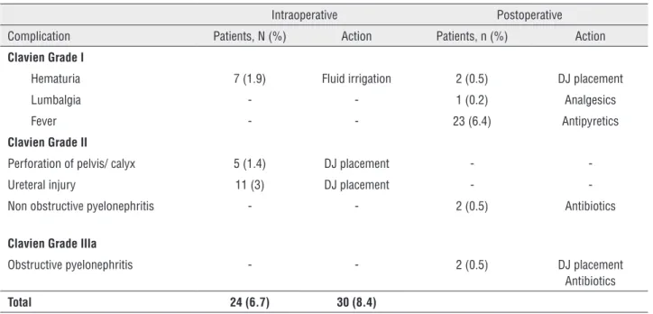
Complications
The overall complication rate was 15.1%. Intra-operative and post-operative complications were experienced in 24 (6.7%) and 30 (8.4%) ca-ses, respectively. A detailed description of the complications and the action taken are showed in Table-3. No major ureteral injuries occurred. Mi-nor ureteral wall injuries were noted in 11 patients (3%) and managed successfully with a stent pla-cement (for 3 to 6 weeks) (mucosal abrasion not reported). Two patients (0.5%) were re-admitted following discharge from hospital with non obs-tructive pyelonephritis: they were treated with intravenous antibiotics and bladder catheter. Two patients (0.5%), left unstented after the procedure, required DJ insertion after readmission for an obs-tructive pyelonephritis. No complications higher than Clavien grade IIIa were observed. No patients
complained late complications to follow-up visit after 6 months.
DISCUSSION
This prospective multi-institutional study on RIRS for renal calculi has shown this approa-ch to have excellent stone clearance rates with an acceptable complication profile. Major technical and surgical developments in endoscopic techno-logies and technique for the treatment of uroli-thiasis have led to changes in treatment approach, and subsequently to international guidelines (6). In this large study, we have demonstrated that the SFR achieved after the first treatment of RIRS to be as high as 73.6%. This finding is comparable to similar studies that demonstrated SFR of 65-79% (11-15).
We also noted that only 20 patients (5.6%), with a mean diameter greater than the overall average, required a second look RIRS to ensure the stone-free status. We do not regard this as a treatment failure of the first procedure, but rather as a necessary part of a planned staged procedure
Table 3 - Overall Complications according to Clavien classification.
Intraoperative Postoperative Complication Patients, N (%) Action Patients, n (%) Action Clavien Grade I
Hematuria 7 (1.9) Fluid irrigation 2 (0.5) DJ placement Lumbalgia - - 1 (0.2) Analgesics Fever - - 23 (6.4) Antipyretics Clavien Grade II
Perforation of pelvis/ calyx 5 (1.4) DJ placement - -Ureteral injury 11 (3) DJ placement - -Non obstructive pyelonephritis - - 2 (0.5) Antibiotics
Clavien Grade IIIa
Obstructive pyelonephritis - - 2 (0.5) DJ placement Antibiotics Total 24 (6.7) 30 (8.4)
N = number; DJ = double J stent; Clavien grade I = Any deviation from the normal postoperative course without the need for pharmacological treatment or surgical,
endoscopic and radiological interventions. Clavien grade II = Requiring pharmacological treatment with drugs other than such allowed for grade I complications. Clavien
to ensure stone clearance, in particular for large renal stones (eg. stones>2cm). This strategy allo-wed us to achieve an overall SFR equal of 79%.
Over the last 10 years, RIRS has become an increasingly important option for the treatment of the majority of kidney stones even in the most complicated clinical scenarios such as pregnancy, obesity, coagulopathy, large renal stones, calyce-al diverticula, and kidney mcalyce-alformations (4). RIRS is well accepted by patients, the convalescence is minimal, it generally does not require a prolonged hospital admission (2 days) or prolonged absence from work (2). In addition, using the endoscopic approach to the kidney, reduces the risk of blood loss, renal parenchyma damage and renal impair-ment. In contrast, in recent years we have seen a drift away from SWL and PCNL. This is in part due to SWL providing a low SFR (16) and a high re-tre-atment rate for large stones and for stones located in the lower pole calyx (17). Similarly, PCNL that is the gold standard for large kidney stones and is more effective than SWL for lower pole stones (18) is characterized by a risk of serious complications (19, 20). We noted that 77 patients (21.6%) had DJ stent placement prior to RIRS. DJ placement may facilitate passage of the UAS and extraction of the fragments (21), however this does not justify the routine DJ insertion before surgery except in case of a septic obstructed upper urinary tract or in case of ureter stricture that will not make possible the passage of access sheath (22). In contrast to this, a post-operative DJ stent was left in situ in 332 patients (93%). This is done to facilitate drainage, prevent post-operative ureteral obstruction. In 79% of cases the DJ stents were placed for 30 days or less. The uses of the UAS can significant facilitate RIRS and stone clearance by allowing multiple en-try and reenen-try to the kidney, decreasing intrarenal pressure and protect the scope from damage (23, 24). However, the routine use of a UAS is matter of debate (6). In this study, an UAS was used in 80% of the cases. The size of the UAS varied ac-cording to clinical conditions. In 52% of the ca-ses a larger UAS was employed. This probably re-flects the surgeon’s preference for lasertripsy. If the surgeon has a predilection for fragmentation and stone extraction, the preferred setting of laser has been low frequency/high energy using medium/
large diameter sheath. This allows the extraction of slightly larger fragments. If the surgeon prefers to dust by vaporization the preferred setting of la-ser is high frequency/low energy using a medium/ small UAS sufficient. However, the choice between vaporization and fragmentation is not only related to the surgeon preferences but also by stone size. For bigger stone could be better to start with the vaporization of the outer part of stone moving to the fragmentation of the residual part and the ex-traction of fragments.
The overall 30-day complication rate in this study was 15.1% being 6.7% intraoperative and 8.4% post-operative. These data are according with the data of EAU guidelines on Urolithiasis that report an overall complication rate of 9-25% (6). It’s noteworthy that the complications rate of pure RIRS is lacking. In fact, the complication rate repor-ted by EAU guidelines are based on surgical series of semirigid ureteroscopy for ureteral stone. A re-cent meta-analysis that included 2 randomized and 8 non-randomized studies showed an overall com-plication rate of RIRS of 10.4%. It was concluded by the authors, that the interpretation of complica-tions proved to be challenging because of a lot of key information (blood transfusion, antibiotic use, definition for sepsis, need for preoperative stenting, definition of ureteral injury, timing of post-opera-tive stenting, etc.) were not clearly stated (25). The complication rate in our study, that at first analysis could be considered slightly higher than expected, should be considered as a viable finding. In fact, in this prospective and standardized setting, we con-sidered any deviation from normal post-operative course as a complication and described it according to Dindo-modified Clavien classification (26).
minimizing the interobserver variability, it is use-less, from a clinical viewpoint, since variability is common in clinical practice (4). A second limitation would be the non-uniform treatment approach and the different experience of the surgeons. The multi--institutional nature of our cohort may be interpre-ted as limitation, however we believe that, in order to evaluate the generalizability of these findings, a certain grade of heterogeneity in baseline charac-teristics, rather than homogeneity, is advisable and desirable.
In our opinion, it is important to remind that, as stated by Giusti et al. (4), the “key to suc-cess is avoiding the start of RIRS on your own. Furthermore, detailed and frank counselling of the patients is strongly encouraged to inform them not only about the minimal invasiveness but also about outcomes of the surgeons/centers and the potential for staged multiple procedures in the most difficult cases and the possibility, although rare, of major complications”.
CONCLUSIONS
This European multicenter prospective stu-dy confirmed that RIRS performed with the newest generation of technical equipment allowed us to achieve a very high SFR without compromising on safety. Nevertheless, cost of acquisition and main-tenance and reimbursement policies by national health systems represent a critical issue that should be resolved to allow RIRS become a routine pro-cedure available in all urological departments and not just in a few tertiary centers.
CONFLICT OF INTEREST
Guido Giusti is a consultant for Boston Scien-tific, Cook Medical, Porges-Coloplast, Karl Storz.
All the other authors declare that they have no conflict of interest.
REFERENCES
1. Bozkurt OF, Resorlu B, Yildiz Y, Can CE, Unsal A. Retrograde intra-renal surgery versus percutaneous nephrolithotomy in the management of lower-pole renal stones with a diameter of 15 to 20 mm. J Endourol. 2011;25:1131-5.
2. Akman T, Binbay M, Ozgor F, Ugurlu M, Tekinarslan E, Kezer C, et al. Comparison of percutaneous nephrolithotomy and retrograde flexible nephrolithotripsy for the management of 2-4 cm stones: a matched-pair analysis. BJU Int. 2012;109:1384-9.
3. Akman T, Binbay M, Ugurlu M, Kaba M, Akcay M, Yazici O, et al. Outcomes of retrograde intrarenal surgery compared with percutaneous nephrolithotomy in elderly patients with moderate-size kidney stones: a matched-pair analysis. J Endourol. 2012;26:625-9.
4. Giusti G, Proietti S, Peschechera R, Taverna G, Sortino G, Cindolo L, et al. Sky is no limit for ureteroscopy: extending the indications and special circumstances. World J Urol. 2015;33:257-73.
5. Giusti G, Proietti S, Cindolo L, Peschechera R, Sortino G, Berardinelli F, et al. Is retrograde intrarenal surgery a viable treatment option for renal stones in patients with solitary kidney? World J Urol. 2015;33:309-14.
6. C. Türk, T. Knoll, A. Petrik, K. et al. Guidelines on Urolithiasis. European Association Guidelines. 2014. Available at http:// uroweb.org/wp-content/uploads/22-Urolithiasis_LR.pdf 7. Karaolides T, Bach C, Kachrilas S, Goyal A, Masood J,
Buchholz N. Improving the durability of digital flexible ureteroscopes. Urology. 2013;81:717-22.
8. de la Rosette JJ, Skrekas T, Segura JW. Handling and prevention of complications in stone basketing. Eur Urol. 2006;50:991-8; discussion 998-9.
9. Traxer O, Thomas A. Prospective evaluation and classification of ureteral wall injuries resulting from insertion of a ureteral access sheath during retrograde intrarenal surgery. J Urol. 2013;189:580-4.
10. Dindo D, Demartines N, Clavien PA. Classification of surgical complications: a new proposal with evaluation in a cohort of 6336 patients and results of a survey. Ann Surg. 2004;240:205-13.
11. Breda A, Ogunyemi O, Leppert JT, Schulam PG. Flexible ureteroscopy and laser lithotripsy for multiple unilateral intrarenal stones. Eur Urol. 2009;55:1190-6.
12. Aboumarzouk OM, Monga M, Kata SG, Traxer O, Somani BK. Flexible ureteroscopy and laser lithotripsy for stones >2 cm: a systematic review and meta-analysis. J Endourol. 2012;26:1257-63.
13. Aboumarzouk OM, Somani B, Monga M. Safety and efficacy of ureteroscopic lithotripsy for stone disease in obese patients: a systematic review of the literature. BJU Int. 2012;110:E374-80.
14. Sofer M, Watterson JD, Wollin TA, Nott L, Razvi H, Denstedt JD. Holmium:YAG laser lithotripsy for upper urinary tract calculi in 598 patients. J Urol. 2002;167:31-4.
16. Preminger GM, Assimos DG, Lingeman JE, Nakada SY, Pearle MS, Wolf JS Jr; AUA Nephrolithiasis Guideline Panel). Chapter 1: AUA guideline on management of staghorn calculi: diagnosis and treatment recommendations. J Urol. 2005;173:1991-2000.
17. Albala DM, Assimos DG, Clayman RV, Denstedt JD, Grasso M, Gutierrez-Aceves J, et al. Lower pole I: a prospective randomized trial of extracorporeal shock wave lithotripsy and percutaneous nephrostolithotomy for lower pole nephrolithiasis-initial results. J Urol. 2001;166:2072-80. Erratum in: J Urol 2002;167:1805.
18. Pardalidis NP, Andriopoulos NA, Sountoulidis P, Kosmaoglou EV. Should percutaneous nephrolithotripsy be considered the primary therapy for lower pole stones? J Endourol. 2010;24:219-22.
19. Matlaga BR, Jansen JP, Meckley LM, Byrne TW, Lingeman JE. Treatment of ureteral and renal stones: a systematic review and meta-analysis of randomized, controlled trials. J Urol. 2012;188:130-7.
20. de la Rosette J, Assimos D, Desai M, Gutierrez J, Lingeman J, Scarpa R, et al. The Clinical Research Office of the Endourological Society Percutaneous Nephrolithotomy Global Study: indications, complications, and outcomes in 5803 patients. J Endourol. 2011;25:11-7.
21. Rubenstein RA, Zhao LC, Loeb S, Shore DM, Nadler RB. Prestenting improves ureteroscopic stone-free rates. J Endourol. 2007;21:1277-80.
22. Shields JM, Bird VG, Graves R, Gómez-Marín O. Impact of preoperative ureteral stenting on outcome of ureteroscopic treatment for urinary lithiasis. J Urol. 2009;182:2768-74.
23. Stern JM, Yiee J, Park S. Safety and efficacy of ureteral access sheaths. J Endourol. 2007;21:119-23.
24. Bach C, Nesar S, Kumar P, Goyal A, Kachrilas S, Papatsoris A, et al. The new digital flexible ureteroscopes: ‘size does matter’—increased ureteric access sheath use! Urol Int. 2012;89:408-11.
25. De S, Autorino R, Kim FJ, Zargar H, Laydner H, Balsamo R, et al. Percutaneous nephrolithotomy versus retrograde intrarenal surgery: a systematic review and meta-analysis. Eur Urol. 2015;67:125-37.
26. Giusti G, Proietti S, Luciani LG, Peschechera R, Giannantoni A, Taverna G, et al. Is retrograde intrarenal surgery for the treatment of renal stones with diameters exceeding 2 cm still a hazard? Can J Urol. 2014;21:7207-12.
27. Sountoulides P, Metaxa L, Cindolo L. Is computed tomography mandatory for the detection of residual stone fragments after percutaneous nephrolithotomy? J Endourol. 2013;27:1341-8.
28. Macejko A, Okotie OT, Zhao LC, Liu J, Perry K, Nadler RB. Computed tomography-determined stone-free rates for ureteroscopy of upper-tract stones. J Endourol. 2009;23:379-82.
_______________________ Correspondence address:
Francesco Berardinelli, MD Dipartimento di Urologia, “S. Pio da Pietrelcina ‘’ Hospital, Vasto (CH), Italia