Acute lung injury induced by the intravenous
administration of cigarette smoke extract*
Lesão pulmonar aguda induzida pela administração endovenosa de extrato da fumaça do cigarro
Luciana Gomes Menezes, Juliana Alves Uzuelli, Cristiane Tefé-Silva, Simone Gusmão Ramos,
José Eduardo Tanus dos Santos, José Antônio Baddini Martinez
Abstract
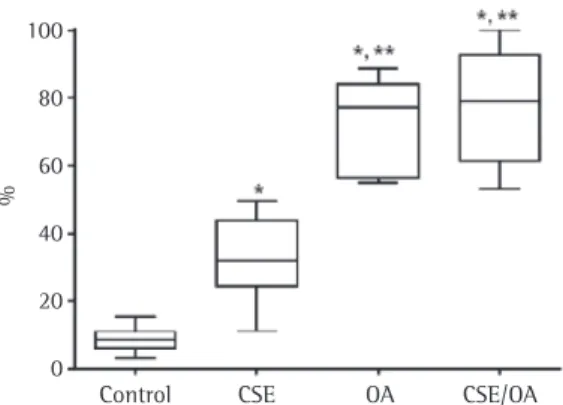
Objective: To investigate the acute effects of intravenous administration of cigarette smoke extract (CSE) on histological, inflammatory, and respiratory function parameters in rats, as well as to compare this potential acute lung injury (ALI) model with that with the use of oleic acid (OA). Methods: We studied 72 Wistar rats, divided into four groups: control (those injected intravenously with saline); CSE (those injected intravenously with CSE and saline); OA (those injected intravenously with saline and OA); and CSE/OA (those injected intravenously with CSE and OA). Results: Mean lung compliance was significantly lower in the OA and CSE/OA groups (2.12 ± 1.13 mL/cmH2O and 1.82 ± 0.77 mL/cmH2O, respectively)than in the control group (3.67 ± 1.38 mL/ cmH2O). In bronchoalveolar lavage fluid, the proportion of neutrophils was significantly higher in the OA and CSE/ OA groups than in the control group, as was the activity of metalloproteinases 2 and 9. Pulmonary involvement, as assessed by morphometry, was significantly more severe in the OA and CSE/OA groups (72.9 ± 13.8% and 77.6 ± 18.0%, respectively) than in the control and CSE groups (8.7 ± 4.1% and 32.7 ± 13.1%, respectively), and that involvement was significantly more severe in the CSE group than in the control group. Conclusions: The intravenous administration of CSE, at the doses and timing employed in this study, was associated with minimal ALI. The use of CSE did not potentiate OA-induced ALI. Additional studies are needed in order to clarify the potential role of this model as a method for studying the mechanisms of smoking-induced lung injury. Keywords: Smoking; Acute lung injury; Models, animal.
Resumo
Objetivo: Investigar os efeitos agudos da administração endovenosa de extrato da fumaça do cigarro (EFC) em parâmetros funcionais respiratórios, inflamatórios e histológicos em ratos e comparar esse potencial modelo de lesão pulmonar aguda (LPA) com aquele com o uso de ácido oleico (AO). Métodos: Foram estudados 72 ratos Wistar machos divididos em quatro grupos: tratados somente com soro fisiológico (SF; grupo controle); tratados com EFC e SF (grupo EFC); tratados com SF e AO (grupo AO); e tratados com EFC e AO (grupo EFC/AO). Resultados: As médias de complacência foram significantemente menores nos grupos AO e EFC/AO (2,12 ± 1,13 mL/cmH2O e 1,82 ± 0,77 mL/cmH2O, respectivamente) do que no controle (3,67 ± 1,38 mL/cmH2O). A proporção de neutrófilos e a atividade das metaloproteinases 2 e 9 em lavado broncoalveolar foram significantemente maiores nos grupos AO e EFC/AO que no controle. O acometimento pulmonar avaliado por morfometria foi significantemente maior nos grupos AO e EFC/AO (72,9 ± 13,8% e 77,6 ± 18,0%, respectivamente) do que nos grupos controle e EFC (8,7 ± 4,1% e 32,7 ± 13,1%, respectivamente), e esse acometimento foi significantemente maior no grupo EFC que no grupo controle. Conclusões: A administração endovenosa de EFC, nas doses e tempos deste estudo, associou-se à LPA mínima. O EFC não potencializou a LPA induzida por AO. Estudos adicionais são necessários para esclarecer o papel potencial desse modelo como método de estudo dos mecanismos de agressão pulmonar pelo tabaco.
Descritores: Tabagismo; Lesão pulmonar aguda; Modelos animais.
* Study carried out at the University of São Paulo at Ribeirão Preto School of Medicine, Ribeirão Preto, Brazil.
Correspondence to: José Antônio Baddini Martinez. Avenida Bandeirantes, 3900, Campus Universitário, CEP 14048-900, Ribeirão Preto, SP, Brasil.
Tel. 55 16 3602-2531. E-mail: [email protected]
Financial support: This study received financial support from the Foundation for the Support of Instruction, Research and Treatment of the University of São Paulo at Ribeirão Preto School of Medicine Hospital das Clínicas.
MMP-2 (gelatinase A) and MMP-9 (gelatinase B) have been associated with lung destruction and
even with the development of COPD.(11)
The objective of the present study was to investigate in rats the acute effects of i.v. administration of CSE on respiratory function parameters and on the cell profile and levels of MMP-2 and MMP-9 activity in bronchoalveolar lavage (BAL) fluid, as well as to investigate the presence of pulmonary histological changes. In addition, we compared the severity of the CSE-induced responses with those of a well-established model of ALI, i.e., systemic injection of oleic acid (OA).(12) Finally, we also investigated
whether previous administration of CSE has any potentiating effects on the model of OA-induced ALI.
The original working hypothesis was that i.v. administration of CSE would lead to significant histological changes and respiratory function changes, and that MMPs would participate in the genesis of these changes, the severity of which could be comparable to those induced by OA. In addition, we assumed that previous administration of CSE would lead to worsening of OA-induced ALI.
Methods
We used 72 male specific-pathogen-free Wistar
rats (≈ 240 g) obtained from the animal facility
of the University of São Paulo at Ribeirão Preto School of Pharmaceutical Sciences, located in the city of Ribeirão Preto, Brazil. The rats were divided into four groups of 18 animals each:
• Control group: i.v. administration of saline
at 0 min (T0) and at 30 min (T30)
• CSE group: i.v. administration of CSE at
T0 and i.v. administration of saline at T30
• OA group: i.v. administration of saline at
T0 and i.v. administration of OA at T30
• CSE/OA: i.v. administration of CSE at T0
and i.v. administration of OA at T30 The CSE was obtained with a “smoking machine”, which was developed by a company specializing in such services, under the guidance of the researchers involved in the present study. Its functioning required that a burning cigarette be attached to the end of an aspiration pump, whereas a tubular circuit, the tip of which was placed inside a disposable sterile plastic tube containing 2 mL of saline, was connected to the other end. Therefore, the smoke produced
Introduction
Recent years have seen the increasing use of a new model for the induction of lung injury in rats by intraperitoneal, endotracheal, or intranasal administration of a surrogate for tobacco smoke.
(1-4) This product, known as cigarette smoke extract
(CSE), is obtained by bubbling the smoke produced by a burning cigarette through a liquid medium such as saline. This element is described as being able to produce emphysematous injury after relatively short periods of repeated use, usually around 3 to 4 weeks. In addition, the pathological process is established without the need for animals to undergo lengthy daily exposure to smoke generated by smoking machines. Therefore, the mechanisms associated with the development of the inflammatory processes triggered by smoking could be studied more rapidly and in a simpler way.
A study investigating the effects of 3-hour administration of CSE on cultures of human fetal fibroblasts reported dose-dependent induction of oxidative stress. In addition, the product also induced apoptosis and cellular DNA fragmentation, suggesting that these mechanisms play a role
in the genesis of smoking-induced diseases.(5)
Therefore, systemic administration of high doses of CSE has the potential to produce substantial, acute, rapid onset lung injury. However, to date, there have been no in vivo studies evaluating the immediate effects of the use of i.v. CSE on pulmonary function parameters or pulmonary histological changes. The characterization of these effects can lead to the development of a new animal model that is useful for investigating the pathogenic mechanisms related to smoking-induced injury. Such a model would have the great advantage of characterizing the pathogenic mechanisms of smoking-induced injury more rapidly and in a more practical manner. In addition, depending on the degree of pulmonary inflammation induced, we could be faced with a new model for investigating mechanisms related to the development of acute lung injury (ALI).
In contrast, there is mounting evidence that matrix metalloproteinases (MMPs), a family of genetically related endopeptidases whose activity is zinc-dependent, mediate numerous physiological
and pathological processes.(6-9) Gelatinases
The BAL samples were processed in a
conventional manner, as described elsewhere.(13,14)
The supernatant obtained after ultracentrifugation
was frozen at −70°C for future studies of MMP-2
and MMP-9 activity, which was determined by zymography, i.e., analysis of the samples by sodium dodecyl sulfate polyacrylamide gel electrophoresis with the enzyme substrate (gelatin) incorporated into the separating gel. Gelatinase activity was semiquantified with the use of a common scanner, and the images obtained were measured via ImageJ software (NIH, Bethesda, NC, USA), as published elsewhere.(15,16)
Lung tissue samples were processed in a standard manner and stained with H&E. The extent of pulmonary parenchymal involvement was determined by morphometry, on the basis of analysis of conventional optical microscopy slides via Leica Qwin software (Leica Microsystems, Cambridge, UK), installed on a computer equipped
with a digital camera, as described elsewhere.(17)
Results are expressed as means, medians, and standard deviations. To compare the four groups on variables with normal distribution, we used ANOVA and, when necessary, the Student-Newman-Keuls post test. For variables with non-normal distribution, we used the Friedman test, followed by Dunn’s post test if indicated. The level of significance was set at 5% (p < 0.05) for all comparisons. The present study was approved by the Animal Experimentation Ethics Committee of the University of São Paulo at Ribeirão Preto School of Medicine.
Results
The mean weight of the animals did not differ among the groups (control: 233.0 ± 7.3 g; CSE: 242.7 ± 6.9 g; OA: 244.0 ± 6.8 g; and CSE/ OA: 242.6 ± 8.2 g). The mean RR and the mean
minute volume (VE) were significantly higher in
the CSE, OA, and CSE/OA groups than in the
control group (Figure 1). In addition, VE was
significantly higher in the OA group than in the CSE and CSE/OA groups. The CSE and CSE/OA groups had lower total lung compliance than did the control group.
Recovered BAL volumes were significantly lower in the CSE and CSE/OA groups than in the control group (Table 1). The proportion of macrophages in BAL fluid was significantly lower in the OA group than in the control and CSE groups. The proportion of neutrophils was higher within the circuit was bubbled into the saline
before being released into the atmosphere. An electronic device allowed controlling the duration of the suction and discharge cycles generated by the pump. Two commercial cigarettes were burned over a 6- to 8-min period for every 2 mL of saline. Throughout the study period, the same brand (Marlboro; Altria, Brazil) was used. The CSE obtained was brownish yellow and had a characteristic smell. The animals then received either CSE or saline via caudal vein puncture at a dose of 1 mL/kg of body weight.
The OA solution was produced by diluting 1 mL of OA (Sigma-Aldrich, St. Louis, MO, USA) with 4 mL of 70% ethanol and 5 mL of saline. This solution, at a volume of 2 mL/kg (corresponding to 178.2 mg of OA per kg of body weight), or saline at corresponding volumes, was slowly administered i.v. 30 min after the initial administration of CSE or saline.
Four hours after the administration of saline or OA, the animals were anesthetized i.p. with 2.5% tribromoethanol (Sigma Aldrich) and underwent tracheostomy for insertion of a plastic endotracheal tube. Subsequently, the tube was connected to a monitoring system (Quadra-t; SCIREQ, Montreal, QC, Canada) that made it possible to obtain respiratory function data in a spontaneous and minimally invasive manner. Once respiratory monitoring was completed, an abdominal incision was made to expose the large blood vessels, which were sectioned under direct vision, and the animals died from exsanguination. Subsequently, extensive thoracotomy was performed, and the heart-lung bloc, which remained in the chest, was infused with saline through the endotracheal tube at a volume of 25 mL/kg. Saline was injected and aspirated with a plastic syringe three times. The collected material was placed into a plastic tube and kept on ice until it was sent to the research laboratory for analysis.
The animals used in the anatomopathological study (6 animals in each group) were handled and treated in the same manner. However, they did not undergo pulmonary function evaluation or BAL sample collection. Once their chest had been open, they were infused with 10% buffered formalin through an endotracheal tube at a pressure of 20
cmH2O. Subsequently, the distal trachea, which
higher in the OA group than in the control and CSE groups.
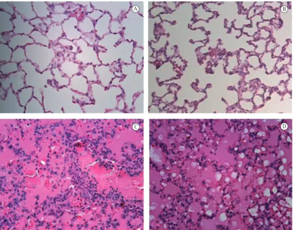
Histological examination of the treated lungs showed small inflammatory changes in the lungs of the animals in the CSE group, with some septal thickening and leukocyte infiltration (Figure 2). In contrast, the lungs of the animals in the OA in the OA and CSE/OA groups than in the control
group, and that proportion was significantly higher in the OA group than in the CSE group. Gelatinolytic activity of MMP-2 was found to be significantly higher in the OA and CSE/OA groups than in the control and CSE groups. As for gelatinolytic activity of MMP-9, values were
Figure 1 - Comparison of RR, tidal volume (VT), minute volume (VE), and total lung compliance (C) among the groups studied. CSE: group injected i.v. with cigarette smoke extract; OA: group injected i.v. with oleic acid; CSE/OA: group injected i.v. with cigarette smoke extract and, 30 min later, with oleic acid. *p < 0.05 for comparison with the control group by ANOVA and the Student-Newman-Keuls post test. **p < 0.05 for comparison with the OA group by ANOVA and the Student-Newman-Keuls post test.
Table 1 - Results of cellularity and metalloproteinase activity in bronchoalveolar lavage fluid.a
Variable Group
Control CSE OA CSE/OA
Volume, mL 3.1 ± 0.5 (3.0) 2.5 ± 0.4 (2.5)* 2.9 ± 0.3 (3.0) 2.6 ± 0.2 (2.5)* Total cell count × 103/mL 261 ± 215 (170) 353 ± 262 (352) 271 ± 176 (210) 294 ± 181 (336) Macrophages, % 95.4 ± 6.6 (99.0)** 91.5 ± 10.1 (96.5)** 56.7 ± 27.6 (51.5) 80.9 ± 26.6 (94.5) Lymphocytes, % 2.4 ± 3.0 (1.1) 3.4 ± 4.6 (1.1) 4.8 ± 3.9 (4.5) 1.6 ± 1.4 (1.3) Neutrophils, % 2.0 ± 3.7 (0.4) 5.0 ± 7.5 (2.1)** 39.6 ± 27.4 (43.9)* 17.4 ± 25.2 (4.0)* Eosinophils, % 0.0 ± 0.1 (0.0) 0.0 ± 0.0 (0.0)** 0.3 ± 0.4 (0.2) 0.1 ± 0.2 (0.0) MMP-2 activity 0.62 ± 0.29 (0.69) 0.50 ± 0.44 (0.77) 1.49 ± 0.43 (1.48)*.*** 1.19 ± 0.53 (0.97)* .*** MMP-9 activity 0.04 ± 0.13 (0)** 0.00 ± 0.00 (0.00)** 0.70 ± 0.23 (0.68) 0.36 ± 0.36 (0.34)
investigation of the smoking-related mechanisms of lung injury. To have an idea of the severity of the acute changes triggered by the use of i.v. CSE, we compared the new model with the traditional model of OA-induced ALI. In addition, we investigated the possible potentiating effects of previous administration of CSE on the model of OA-induced ALI.
Because intraperitoneal administration of CSE over weeks leads to the development of emphysema, it would be natural to assume that administration of a large volume of CSE directly into the venous system could lead to the development of a significant acute pulmonary inflammatory response.
Given that there are no previous studies with a similar design, the time interval between administration of the two offending agents was and CSE/OA groups were found to be severely
edematous and congested, with some peripherally located areas of necrosis. Tissue involvement was significantly more severe in the CSE, OA, and CSE/OA groups than in the control group, and that involvement was significantly more severe in the OA and CSE/OA groups than in the CSE group (Figure 3).
Discussion
In the present study, we investigated the acute effects that i.v. administration of CSE as an offending agent to the lungs has on physiological, morphological, and inflammatory parameters. Given that cigarette burning releases more than
4,000 chemicals(18) and CSE is rich in oxidants,
such an animal model could become a new, rapid, and practical instrument for original
In the present study, the most consistent finding related to BAL cellularity was that the proportion of neutrophils was significantly higher in the OA and CSE/OA groups than in the control group. In addition, the levels of neutrophils in the OA group were significantly different from those in the CSE group. These results indicate an important participation of neutrophils in the genesis of OA-induced ALI. In contrast, the increase in neutrophils in the CSE group was so slight that the neutrophil levels in this group did not differ significantly from those in the control group. Although the OA and CSE/OA groups did not differ significantly in terms of the proportion of neutrophils in BAL fluid, the previous i.v. administration of CSE was accompanied by a trend toward a reduction in neutrophils after the use of OA. Therefore, we can suggest that the previous use of CSE led to a reduction in OA-induced neutrophil extravasation.
Most data from animal models of inflammation using two consecutive injuries indicate that the responses and the risk of infection are greater
after the second injury.(20,21) However, a study
in mice had shown that previous induction of a low-grade systemic inflammatory response protected the animals from developing sepsis induced by Enterococcus faecalis or methicillin-resistant Staphylococcus aureus.(22) When initially
a severe systemic inflammatory response was induced, severe sepsis developed after infection with microorganisms. The mechanisms responsible for these phenomena seem to involve distinct macrophage activation phenotypes. On the basis of these findings, we can speculate that, in the present study, similar effects occurred in response to the initial use of CSE. Further investigation is needed to elucidate this issue.
The significant increases in MMP-2 activity in BAL fluid in the OA and CSE/OA groups in comparison with the two other groups suggest that this mediator plays an important role in the development of OA-induced ALI. In contrast, the use of CSE alone was not accompanied by an increase in this gelatinase activity. It is equally interesting that, in comparison with the use of OA alone, the previous use of CSE tended to cause a reduction in MMP-2 activity. However, once again, this comparison did not show a significant difference. The gelatinase MMP-2 is produced by a variety of cell types, including endothelial cells—the primary target of OA-induced injury, established empirically. In contrast, the time interval
between the last administration of the agents and data collection was based on values reported in
studies involving the model of OA-induced ALI.(19)
It is recognized that a 4-hour interval between OA infusion and collection of samples for analysis is sufficient for the full development of ALI and, therefore, in the present study, this time interval was used for data and sample collection after the second administration of the substances.
The pulmonary function results indicate that the animals in the CSE, OA, and CSE/OA groups tended to show a restrictive respiratory pattern after administration of the different treatments. This becomes more evident when we analyze the total lung compliance values, although significant differences in comparison with the control group were found only for the groups that received OA. Surely this is the type of pulmonary function pattern that is expected for conditions that are accompanied by edema and diffuse inflammatory infiltration of the lungs. However, the effects of i.v. CSE-induced ALI on respiratory function were quite modest. In addition, the previous use of CSE does not seem to potentiate the harmful effects of OA on respiratory function substantially, although
VE was found to be significantly lower in the
CSE/OA group than in the OA group.
group. Taken together, the results confirm that OA is an extremely toxic agent to the lungs,(19,25)
and that pre-treatment with a single i.v. dose of CSE does not seem to affect the development of this process significantly. These results point in the same direction as that of the interpretation of the lung compliance data.
The present study has a series of limitations, especially the use of the same single dose of CSE in all analyses. The results might have been different had we used higher doses of CSE. In fact, the data would be more complete had we constructed dose-response or time-response curves.
On the basis of the results obtained, we can conclude that the i.v. administration of CSE, at the doses and timing employed in this study, was associated with minimal ALI and mild respiratory function effects. The severity of CSE-induced ALI is much less than that induced by OA. In addition, in the present model, the previous use of CSE did not potentiate OA-induced lung injury. Although, by all indications, this method is not a good model for studies on pathophysiological aspects related to the development of ALI, its usefulness for the investigation of the mechanisms of smoking-induced lung injury still cannot be completely ruled out.
References
1. Qamar W, Sultana S. Farnesol ameliorates massive inflammation, oxidative stress and lung injury induced by intratracheal instillation of cigarette smoke extract in rats: an initial step in lung chemoprevention. Chem Biol Interact. 2008;176(2-3):79-87. Erratum in: Chem Biol Interact. 2009;177(3):259. http://dx.doi.org/10.1016/j. cbi.2008.11.017
2. Chen Y, Hanaoka M, Chen P, Droma Y, Voelkel NF, Kubo K. Protective effect of beraprost sodium, a stable prostacyclin analog, in the development of cigarette smoke extract-induced emphysema. Am J Physiol Lung Cell Mol Physiol. 2009;296(4):L648-56. PMid:19201816. http://dx.doi.org/10.1152/ajplung.90270.2008 3. Chen Y, Hanaoka M, Droma Y, Chen P, Voelkel NF,
Kubo K. Endothelin-1 receptor antagonists prevent the development of pulmonary emphysema in rats. Eur Respir J. 2010;35(4):904-12. PMid:19897563. http:// dx.doi.org/10.1183/09031936.00003909
4. Hanaoka M, Droma Y, Chen Y, Agatsuma T, Kitaguchi Y, Voelkel NF, et al. Carbocisteine protects against emphysema induced by cigarette smoke extract in rats. Chest. 2011;139(5):1101-8. PMid:20847042. http:// dx.doi.org/10.1378/chest.10-0920
5. Carnevali S, Petruzzelli S, Longoni B, Vanacore R, Barale R, Cipollini M, et al. Cigarette smoke extract induces oxidative stress and apoptosis in human lung fibroblasts.
which occurs via the blood route.(23) Therefore,
the increases observed here may reflect principally this type of response. It is of note that, despite being administered by the same route, CSE was unable to induce significant increases in the local levels of the mediator.
Only in one sample from the control group was MMP-9 activity detected, and no such activity was detected in any samples after the use of CSE alone. In contrast, MMP-9 activity was detected in the BAL fluid of all animals after the use of OA and in the BAL fluid of 7 after the combined use of CSE and OA. The results of MMP-9 activity showed a behavior very similar to that observed for percent neutrophil counts, suggesting that these cells were the major source of the enzyme in the animals injected i.v. with OA.(24) The decrease in the enzyme levels after the
use of OA preceded by the use of CSE support the last possibility.
The data obtained here indicate that the use of i.v. CSE alone under the conditions of this study did not lead to significant changes in cell profile, or in MMP-2 or MMP-9 activity, in comparison with the use of saline only. Likewise, the previous administration of CSE did not potentiate the cytologic changes or the changes in gelatinolytic activity in BAL fluid, which are associated with the use of OA. In fact, the available data suggest the possibility of some degree of decrease in neutrophil sequestration and gelatinase expression in the distal air spaces after the introduction of the initial treatment, as discussed above.
16. Mühl D, Ghosh S, Uzuelli JA, Lantos J, Tanus-Santos JE. Increases in circulating matrix metalloproteinase-9 levels following fibrinolysis for acute pulmonary embolism. Thromb Res. 2010;125(6):549-53. PMid:20307903. http://dx.doi.org/10.1016/j.thromres.2010.02.015 17. Martinez JA, Ramos SG, Meirelles MS, Verceze AV,
Arantes MR, Vannucchi H. Effects of quercetin on bleomycin-induced lung injury: a preliminary stud. J Bras Pneumol. 2008;34(7):445-52. PMid:18695788. http://dx.doi.org/10.1590/S1806-37132008000700003 18. National Toxicology Program. Report on Carcinogens.
Washington: U.S. Department of Health and Human Services, Public Health Service, National Toxicology Program; 2005.
19. Derks CM, Jacobovitz-Derks D. Embolic pneumopathy induced by oleic acid. A systematic morphologic study. Am J Pathol. 197;87(1):143-58.
20. Kumpf O, Schumann RR. Genetic variation in innate immunity pathways and their potential contribution to the SIRS/CARS debate: evidence from human studies and animal models. J Innate Immun. 2010;2(5):381-94. PMid:20431282. http://dx.doi.org/10.1159/000314269 21. Adib-Conquy M, Cavaillon JM. Compensatory
anti-inflammatory response syndrome. Thromb Haemost. 2009;101(1):36-47. PMid:19132187. 22. Takahashi H, Tsuda Y, Takeuchi D, Kobayashi M, Herndon
DN, Suzuki F. Influence of systemic inflammatory response syndrome on host resistance against bacterial infections. Crit Care Med. 2004;32(9):1879-85. PMid:15343016. http://dx.doi.org/10.1097/01.CCM.0000139606.34631.61 23. Yeh DY, Lin HI, Feng NH, Chen CF, Wang D, Wang
NT. Matrix metalloprotease expressions in both reperfusion lung injury and oleic acid lung injury models and the protective effects of ilomastat. Transplant Proc. 2009;41(5):1508-11. PMid:19545667. http:// dx.doi.org/10.1016/j.transproceed.2009.02.076 24. Corbel M, Boichot E, Lagente V. Role of gelatinases
MMP-2 and MMP-9 in tissue remodeling following acute lung injury. Braz J Med Biol Res. 2000;33(7):749-54. PMid:10881049. http://dx.doi.org/10.1590/ S0100-879X2000000700004
25. Dickey BF, Thrall RS, McCormick JR, Ward PA. Oleic-acid-induced lung injury in the rat. Failure of indomethacin treatment or complement depletion to ablate lung injury. Am J Pathol. 1981;103(3):376-83. PMid:7234970 PMCid:1903842.
Am J Physiol Lung Cell Mol Physiol. 2003;284(6):L955-63. PMid:12547733.
6. Page-McCaw A, Ewald AJ, Werb Z. Matrix metalloproteinases and the regulation of tissue remodelling. Nat Rev Mol Cell Biol. 2007;8(3):221-33. PMid:17318226 PMCid:2760082. http://dx.doi.org/10.1038/nrm2125 7. Vu TH, Werb Z. Matrix metalloproteinases: effectors
of development and normal physiology. Genes Dev. 200;14(17):2123-33.
8. Pytliak M, Vargová V, Mechírová V. Matrix metalloproteinases and their role in oncogenesis: a review. Onkologie. 2012;35(1-2):49-53. PMid:22310347. http://dx.doi.org/10.1159/000336304
9. Sbardella D, Fasciglione GF, Gioia M, Ciaccio C, Tundo GR, Marini S, et al. Human matrix metalloproteinases: an ubiquitarian class of enzymes involved in several pathological processes. Mol Aspects Med. 2012;33(2):119-208. PMid:22100792. http:// dx.doi.org/10.1016/j.mam.2011.10.015
10. Malemud CJ. Matrix metalloproteinases (MMPs) in health and disease: an overview. Front Biosci. 2006;11:1696-701. PMid:16368548. http://dx.doi.org/10.2741/1915 11. Rufino R, Lapa e Silva JR. Cellular and biochemical
bases of chronic obstructive pulmonary disease. J Bras Pneumol. 2006;32(3):241-8. PMid:17273614. http:// dx.doi.org/10.1590/S1806-37132006000300011 12. Gaio E, Melo e Silva CA, Brito F, Firmino MA, Storck R,
Freitas E. Stability of the animal model of oleic acid-induced acute lung injury. J Bras Pneumol. 2009;35(8):759-66. PMid:19750328.
13. Martinez JA, King TE Jr, Brown K, Jennings CA, Borish L, Mortenson RL, et al. Increased expression of the interleukin-10 gene by alveolar macrophages in interstitial lung disease. Am J Physiol. 1997;273(3 Pt 1):L676-83. PMid:9316504.
14. Martinez JA, Nishimura C, Guatura SB, Sato E, King TE Jr. Elevation of soluble interleukin-2 receptor levels in the bronchoalveolar lavage from patients with systemic sclerosis. Rheumatol Int. 2001;21(3):122-6. PMid:11765225. http:// dx.doi.org/10.1007/s00296-001-0147-x
About the authors
Luciana Gomes Menezes
Physical Therapist. University of São Paulo at Ribeirão Preto School of Medicine, Ribeirão Preto, Brazil.
Juliana Alves Uzuelli
Doctoral Student. Department of Pharmacology, University of São Paulo at Ribeirão Preto School of Medicine, Ribeirão Preto, Brazil.
Cristiane Tefé-Silva
Doctoral Student in Pathology. Department of Pathology, University of São Paulo at Ribeirão Preto School of Medicine, Ribeirão Preto, Brazil.
Simone Gusmão Ramos
Associate Professor. Department of Pathology, University of São Paulo at Ribeirão Preto School of Medicine, Ribeirão Preto, Brazil.
José Eduardo Tanus dos Santos
Full Professor. Department of Pharmacology, University of São Paulo at Ribeirão Preto School of Medicine, Ribeirão Preto, Brazil.
José Antônio Baddini Martinez