246
|
© 2017 John Wiley & Sons A/S. wileyonlinelibrary.com/journal/bdi Bipolar Disorders. 2017;19:246–258. Published by John Wiley & Sons LtdO R I G I N A L A R T I C L E
Lithium ameliorates sleep deprivation- induced mania-
like behavior, hypothalamic- pituitary- adrenal (HPA) axis
alterations, oxidative stress and elevations of cytokine
concentrations in the brain and serum of mice
Samira S. Valvassori
1,2|
Wilson R. Resende
1,2|
Gustavo Dal-Pont
1,2|
Heron Sangaletti-Pereira
1|
Fernanda F. Gava
1,2|
Bruna R. Peterle
1,2|
André F. Carvalho
3|
Roger B. Varela
1,2|
Felipe Dal-Pizzol
4|
João Quevedo
2,5,6,7,8,91Laboratory of Neuronal Signaling and Psychopharmacology, Graduate Program in Health Sciences, Health Sciences Unit, University of Southern Santa Catarina
(UNESC), Criciúma, SC, Brazil
2Laboratory of Neurosciences, Graduate Program in Health Sciences, Health Sciences Unit, University of Southern Santa Catarina (UNESC), Criciúma, SC, Brazil 3Translational Psychiatry Research Group and Department of Clinical Medicine, Faculty of Medicine, Federal University of Ceará, Fortaleza, CE, Brazil
4Laboratory of Pathophysiology, Graduate Program in Health Sciences, Health Sciences Unit, University of Southern Santa Catarina (UNESC), Criciúma, SC, Brazil 5Bipolar Disorder Program, Laboratory of Molecular Psychiatry, Hospital de Clínicas de Porto Alegre (HCPA), Porto Alegre, RS, Brazil
6Graduation Program in Psychiatry and Department of Psychiatry, Universidade Federal do Rio Grande do Sul (UFRGS), Porto Alegre, RS, Brazil
7Translational Psychiatry Program, Department of Psychiatry and Behavioral Sciences, The University of Texas Health Science Center at Houston (UTHealth) Medical
School, Houston, TX, USA
8Center of Excellence on Mood Disorders, Department of Psychiatry and Behavioral Sciences, The University of Texas Health Science Center at Houston (UTHealth)
Medical School, Houston, TX, USA
9Neuroscience Graduate Program, The University of Texas Graduate School of Biomedical Sciences at Houston, Houston, TX, USA
Correspondence
Samira S. Valvassori, Laboratório de Sinalização Neural e Psicofarmacologia, Programa de Pós-Graduação em Ciências da Saúde, Unidade Acadêmica em Ciências da Saúde, Universidade do Extremo Sul Catarinense, Criciúma, SC, Brasil. Email: [email protected]
Funding information
This work was supported by Conselho Nacional de Desenvolvimento Científico e Tecnológico (grant to J.Q.); Fundação de amparo à pesquisa e inovação do estado de Santa Catarina (grant to J.Q.); Instituto Cérebro e Mente (grant to J.Q.); and Universidade do Extremo Sul Catarinense (grant to J.Q.). J.Q. is a 1A CNPq Research Fellow.
Objectives: The goal of the present study was to investigate the effects of lithium administration on behavior, oxidative stress parameters and cytokine levels in the pe-riphery and brain of mice subjected to an animal model of mania induced by paradoxi-cal sleep deprivation (PSD).
Methods: Male C57 mice were treated with saline or lithium for 7 days. The sleep deprivation protocol started on the 5th day during for the last 36 hours of the treat-ment period. Immediately after the sleep deprivation protocol, animals locomotor ac-tivity was evaluated and serum and brain samples was extracted to evaluation of corticosterone and adrenocorticotropic hormone circulating levels, oxidative stress parameters and citokynes levels.
Results: The results showed that PSD induced hyperactivity in mice, which is
consid-ered a mania- like behavior. PSD increased lipid peroxidation and oxidative damage to DNA, as well as causing alterations to antioxidant enzymes in the frontal cortex, hip-pocampus and serum of mice. In addition, PSD increased the levels of cytokines in the brains of mice. Treatment with lithium prevented the mania- like behavior, oxidative damage and cytokine alterations induced by PSD.
Conclusions: Improving our understanding of oxidative damage in biomolecules,
anti-oxidant mechanisms and the inflammatory system − alterations presented in the ani-mal models of mania – is important in helping us to improve our knowledge concerning Correction added on 17 July 2017, after first online
1
|
INTRODUCTION
Bipolar disorder (BD) is a common, complex, and severe mood disorder with progressive social and cognitive function disturbances.1,2 BD is
characterized by the presence of mania or hypomania, and episodes of depression. Mania or hypomania is characterized by persistent increases in energy, accompanied by an elevated and expansive or irritable mood. Mania and hypomania are differentiated by the sever-ity, duration, and number of symptoms, with hypomania presenting with less severe symptoms than mania. The depressive episodes are characterized by a profound loss of motivation and interest.3 The gold
standard used in the treatment of BD is lithium (Li), which is a mood stabilizer approved by the Food and Drug Administration (FDA).4
Previous studies have shown that Li is effective in acute manic epi-sodes, and reduces the risk of manic and depressive relapses.2,5
Despite its importance, little is known about the precise neuro-biological underpinnings of BD. However, a considerable number of studies have reported the involvement of glucocorticoids, oxidative stress and inflammatory cytokines in the pathophysiology of BD.6-11
Muneer12 has suggested that an interaction occurs in the
pathophys-iology of BD between the hypothalamic- pituitary- adrenal (HPA) axis, inflammatory mediators and oxidative stress, which deregulate hor-monal, metabolic, and circadian homeostasis. Indeed, oxidative stress
leads to cortisol resistance by decreasing the movement of the gluco
-corticoid receptor from the cytosol to the nucleus.12 In turn, HPA axis
deregulation by oxidative stress induces an inflammatory response
which increases the levels of cytokines.12-14 On the other hand, the
in-crease of glucocorticoids released from the adrenal gland in response to stress- induced activation of the HPA axis can induce oxidative stress, with this becoming a continuous cycle.15
In situations in which the generation of reactive oxygen species (ROS) exceeds the capacity of antioxidant defense, oxidative stress may induce direct damage to cellular proteins, DNA and lipids, thus im-pairing neuronal function.16,17 The role of oxidative stress in the
physi-opathology of BD has been investigated in several studies, which have consistently reported changes in antioxidant enzymes, lipid peroxida-tion, and protein/DNA oxidaperoxida-tion, in both the blood and brains of bipolar patients.7,19-22 A previous postmortem study found an increase of lipid
peroxidation in an analysis of lipid hydroperoxides (LPHs), 8- isoprostane (8- ISO) and 4- hydroxy- 2- nonenal (4- HNE) in the frontal cortex of bipo-lar patients.18 In addition, a higher number of manic episodes was
cor-related with higher levels of 8- hydroxy- 2′- deoxyguanosine (8- OHdG), which is a marker of DNA oxidative damage.7 Alterations in antioxidant
enzymes are also observed in bipolar patients. Gawryluk and
col-leagues19 demonstrated glutathione reductase (GR) and glutathione
peroxidase (GPx) alterations in the frontal cortex of bipolar patients. The inflammatory system is part of the non- specific immune re-sponse, which is activated in response to harmful stimuli such as pathogens, damaged cells, or irritants.23 Some clinical and preclinical
studies have suggested that inflammatory processes in the periph-ery and the brain are involved in the pathophysiology of BD.24,25 Previous studies demonstrated that serum levels of the cytokines
interleukin (IL)- 4, IL- 1β, IL- 10 and tumor necrosis factor alpha (TNF- α) are elevated in subjects with BD when compared to healthy controls.26-29
Paradoxical sleep deprivation (PSD) in mice has been considered a good animal model of mania because it induces some aspects of a manic episode, such as hyperactivity, hypersexuality and aggressive
behav-ior.30,31 PSD is not able to induce BD in mice, but does induce a mania- like
behavior. Some studies have demonstrated that the mania- like behaviors
induced by PSD are reversed by Li.32,33 In addition, circadian rhythms and
genes involved in the molecular clock have long been implicated in BD.34
The goal of the present study was to investigate the effects of Li administration on behavior, oxidative stress parameters and cytokine levels in the serum, frontal cortex and hippocampus of mice subjected to the animal model of mania induced by PSD. In the same context, corticosterone and adrenocorticotropic hormone (ACTH) levels were
evaluated in the serum of mice.
2
|
MATERIALS AND METHODS
2.1
|
Animals
In the present study, male C57 mice were used and grouped five per cage. Mice were exposed to a 12- hour light/dark cycle with unrestricted ac-cess to water and food. All experimental procedures were carried out in accordance with the National Institutes of Health Guide for the Care and Use of Laboratory Animals, and the Brazilian Society for Neuroscience and Behavior (SBNeC) guidelines. This study was approved by the local
ethics committee (Comissão de Ética no Uso de Animais da Universidade do Extremo Sul Catarinense) under protocol 007/2016- 1.
2.2
|
Treatments
The mice were treated over a period of 7 days with saline solution (SAL; NaCl 0.09%, 1 mL/kg, 1 injection per day, intraperitoneally [i.p.])
the pathophysiology of BD, and the mechanisms of action employed by mood stabilizers.
K E Y W O R D S
or Li (47.3 mg/kg, 1 mL/kg, 1 injection per day, i.p.). These doses and the treatment schedule used were based on a previous study under-taken by our research group.24
All Li- treated animals had Li plasma levels between 0.6 and 1.2 mEq/L, as recommended in the treatment of BD patients.24
2.3
|
Paradoxical sleep deprivation (PSD) protocol
The PSD protocol was started on the 5th day of the treatment, at 6:00 pm
(see Figure 1). For drug administration during the PSD protocol, the mice were removed from the platform and replaced immediately after the injection. The mice were placed 5 per cage (38×31×17 cm), each cage containing 12 platforms (3.5 cm diameter). In the same box, we placed a volume of water 1 inch deep, obligating the animals to stay on the platforms. They could, however, freely move from one platform to
an-other.34,35 Thus, when animals entered the paradoxical phase of sleep,
due to muscle atonia, they were awoken by falling into the water. Food and water were available ad libitum. The present study adopted the pe-riod of 36 hours of PSD, since this pepe-riod of PSD increased the level of locomotor activity, which is considered a mania- like behavior, of animals used in previous studies.35 The mice in the control group were exposed to
the same conditions, except there was no water in the bottom of the box.
2.4
|
Experimental groups
The mice were randomly distributed in the four groups (n=10 per group) that are listed below: (1) control+SAL; (2) control+Li; (3) PSD+SAL; (4) PSD+Li.
2.5
|
Open field test
The open field test (OFT) was used to evaluate the locomotor activity of the animals. In order to perform the OFT, an apparatus consisting of a 45×60 cm white plywood arena, surrounded by 50- cm- high wooden
walls and containing a frontal glass wall, was used. The bottom of the OFT arena was divided into nine equal portions (15×20 cm each) with black lines. The mice were carefully put into the left rear quadrant, and then left to explore the arena for a period of 5 minutes. The locomotor activity (number of horizontal line crossings) of each mouse during the
5 minutes was then recorded.
2.6
|
Samples
2.6.1
|
Serum samples
Immediately after the behavioral test, the animals were killed by de-capitation and individual peripheral blood samples were collected in a microtube, in the morning between 8:00 Am and 12:00 Am, for
subse-quent analyses. Serum was obtained by centrifugation at 3000g for
5 minutes and then kept frozen at −70°C until the experiment.
F I G U R E 1 Experimental design. On the first day of the experiment, treatment with lithium or saline was started. On the 5th day, the PSD protocol was started at 6 pm. After 36 h, the animals were subjected to an open field test, and then the brain was dissected to obtain the frontal
cortex and hippocampus, and the serum was collected for future evaluations. ACTH, adrenocorticotropic hormone; 4- HNE, 4- hydroxy- 2- nonenal; 8- OHdG, 8- hydroxy- 2′- deoxyguanosine; GPx, glutathione peroxidase; GR, glutathione reductase; IL, interleukin; TNF- α, tumor necrosis
factor alpha [Colour figure can be viewed at wileyonlinelibrary.com]
F I G U R E 2 Effects of paradoxical sleep deprivation (PSD) on the number of crossings in animals subjected to the PSD- induced animal model (n=10 per group). Data were analyzed by two- way analysis of
variance followed by the Duncan test when F was significant. Values
are expressed as mean±SD. *P<.05 compared to the Control+Sal group. #P<.05 compared to the ouabain group. Li, lithium; Sal, saline
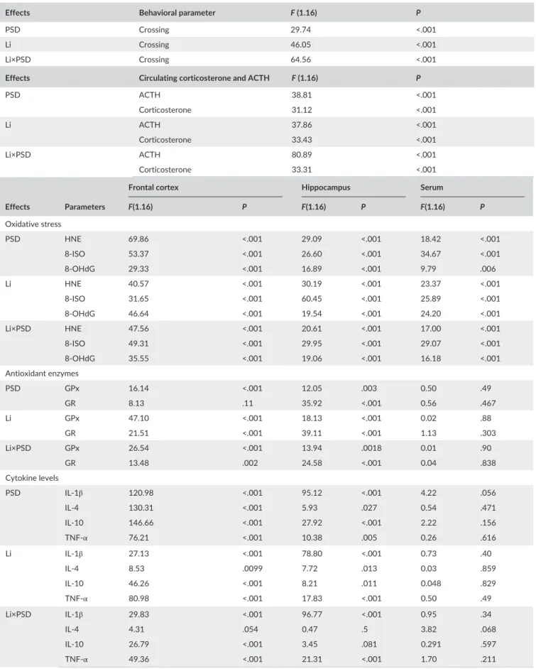
Table 1 Data from two- away ANOVA
Effects Behavioral parameter F (1.16) P
PSD Crossing 29.74 <.001
Li Crossing 46.05 <.001
Li×PSD Crossing 64.56 <.001
Effects Circulating corticosterone and ACTH F (1.16) P
PSD ACTH 38.81 <.001
Corticosterone 31.12 <.001
Li ACTH 37.86 <.001
Corticosterone 33.43 <.001
Li×PSD ACTH 80.89 <.001
Corticosterone 33.31 <.001
Effects Parameters
Frontal cortex Hippocampus Serum
F(1.16) P F(1.16) P F(1.16) P
Oxidative stress
PSD HNE 69.86 <.001 29.09 <.001 18.42 <.001
8- ISO 53.37 <.001 26.60 <.001 34.67 <.001
8- OHdG 29.33 <.001 16.89 <.001 9.79 .006
Li HNE 40.57 <.001 30.19 <.001 23.37 <.001
8- ISO 31.65 <.001 60.45 <.001 25.89 <.001
8- OHdG 46.64 <.001 19.54 <.001 24.20 <.001
Li×PSD HNE 47.56 <.001 20.61 <.001 17.00 <.001
8- ISO 49.31 <.001 29.95 <.001 29.07 <.001
8- OHdG 35.55 <.001 19.06 <.001 16.18 <.001
Antioxidant enzymes
PSD GPx 16.14 <.001 12.05 .003 0.50 .49
GR 8.13 .11 35.92 <.001 0.56 .467
Li GPx 47.10 <.001 18.13 <.001 0.02 .88
GR 21.51 <.001 39.11 <.001 1.13 .303
Li×PSD GPx 26.54 <.001 13.94 .0018 0.01 .90
GR 13.48 .002 24.58 <.001 0.04 .838
Cytokine levels
PSD IL- 1β 120.98 <.001 95.12 <.001 4.22 .056
IL- 4 130.31 <.001 5.93 .027 0.54 .471
IL- 10 146.66 <.001 27.92 <.001 2.22 .156
TNF- α 76.21 <.001 10.38 .005 0.26 .616
Li IL- 1β 27.13 <.001 78.80 <.001 0.73 .40
IL- 4 8.53 .0099 7.72 .013 0.03 .859
IL- 10 46.26 <.001 8.21 .011 0.048 .829
TNF- α 80.98 <.001 17.83 <.001 0.50 .49
Li×PSD IL- 1β 29.83 <.001 96.77 <.001 0.95 .34
IL- 4 4.31 .054 0.47 .5 3.82 .068
IL- 10 26.79 <.001 3.45 .081 0.291 .597
TNF- α 49.36 <.001 21.31 <.001 1.70 .211
2.6.2
|
Brain samples
The mice were killed by decapitation immediately after the OFT. The frontal cortex and hippocampus from the mouse brains were then dis-sected, rapidly frozen, and stored at −70°C until assayed. The sam-ples taken from the frontal cortex and hippocampus of the mice were homogenized in KCl KH2PO4 (12 mM KCl and 0.038 mM KH2PO4,
pH=7.4).
2.7
|
Corticosterone and ACTH circulating levels
Corticosterone levels were determined using enzyme immunoassay (EIA) kits (from Diagnostic Products Corporation, Los Angeles, CA, USA). Serum concentrations of ACTH were determined using
com-mercially available radioimmunoassay kits (from Diagnostic Products
Corporation) for animals.
2.8
|
Evaluation of oxidative stress parameters in the
mouse brains
2.8.1
|
Measures of lipid peroxidation
Two separate markers of lipid peroxidation, 4- HNE (Cell Biolabs, Inc., San Diego, CA, USA; STA- 338) and 8- ISO (Cayman Chemical, Paulinia, Brazil; Item No. 516351), were analyzed following the manufacturers’ instructions. 4- HNE protein adducts to lysine, histidine, or cysteine were quantified by standard sandwich enzyme- linked immunosorb-ent assay (ELISA) using an EIA. 8- ISO was quantified using ACE™ competitive EIAs with 8- ISO- acetylcholinesterase conjugate as a tracer and 8- ISO- specific rabbit anti- serum (Cayman Chemical; Item No. 500431). As 8- ISO and the tracer compete for limited anti- serum binding, the color intensity caused by tracer binding was inversely proportional to the amount of 8- ISO.
2.8.2
|
Nuclear extraction from frontal cortex and
hippocampus
The obtained samples were flash- frozen and stored at −80°C until nuclear proteins were extracted. Tissue samples were subjected to a nuclear extraction protocol with a commercial Nuclear Extraction Kit (Chemicon, Temecula, CA, USA; Item No. 2900). Briefly, samples were homogenized in cytoplasmic lysis buffer containing dithiothrei-tol (DTT) and protease inhibitors. The suspension was kept on ice for
15 minutes and was later centrifuged at 250g for 5 minutes at 4°C.
The supernatant was discarded, and the pellet was resuspended in two volumes of cold cytoplasmic lysis buffer. The suspension was homogenized using a small- gauge needle syringe and centrifuged at
8000g for 20 minutes at 4°C. The resulting pellet contained the
clear portion of the cell lysate. The pellet was resuspended in a nu-clear extraction buffer containing DTT and protease inhibitors, and the suspension was homogenized with a small- gauge needle syringe. The resulting sample was kept in slow agitation for 30−60 minutes in an orbital shaker at 4°C. Later, the nuclear suspension was centrifuged
at 16 000g for 5 minutes at 4°C, and the nuclear extract- containing
supernatant was transferred to a new tube and stored at −80°C until
further analysis.
2.8.3
|
8-OHdG analysis
8- OHdG is produced by oxidative damage of DNA by reactive oxygen and nitrogen species and serves as an established marker of oxidative stress. An 8- hydroxy- 20- deoxy guanosine assay kit purchased from Cell Biolabs (STA- 320) was used. It is a competitive assay that can be used for the quantification of 8- OHdG in serum and nuclear extraction from the frontal cortex and hippocampus. It recognizes both free and DNA- incorporated 8- OHdG. This assay depends on the competition between 8- OHdG and 8- OHdG- acetylcholinesterase (ache) conjugate (8- OHdG tracer) for a limited amount of 8- OHdG monoclonal anti-body. All procedures were carried out in accordance with the manu-facturer’s instructions.
2.9
|
Activity of antioxidant enzymes
2.9.1
|
Glutathione peroxidase (GPx)
GPx activity was measured using the assay kit from Cayman Chemical. Oxidized glutathione is produced via the reduction of hydrogen per-oxide by GPx, and is recycled into its reduced state by GR and oxi-dized nicotinamide adenine dinucleotide phosphate (NADP+). The oxidation of nicotinamide adenine dinucleotide phosphate (NADPH) to NADP+ is followed by a decrease in the absorbance of light at 340 nM. One unit of GPx is defined as the amount of enzyme that will cause the oxidation of 1.0 nmol of NADPH to NADP+ per minute at 25°C.
2.9.2
|
Glutathione redutase (GR)
GR activity was measured using the assay kit from Cayman Chemical. Using this kit, it was possible to measure the rate of oxidation of NADPH to NADP+, which is followed by a decrease in absorbance at 340 nM. One unit of GR is defined as the amount of enzyme that causes the oxidation of 1.0 nmol of NADPH to NADP+ per minute at 25°C.
2.9.3
|
Assessment of IL- 1, IL- 4, IL- 10, and
TNF-
α
levels
The hippocampus and frontal cortex were homogenized in phosphate- bufferid saline extraction solution containing aprotinin (100 mg of tissue per 1 mL). The concentrations of cytokines were determined for the serum, hippocampus, striatum and frontal cortex using com-mercially available ELISAs, following the instructions supplied by the manufacturer (DuoSet kits; R&D Systems, Minneapolis, MN, USA). The results are shown in pg/100 mg of tissue for the hippocampus and frontal cortex. The results are shown in pg/mL of sample for the
2.9.4
|
Protein determination
All biochemical measures were normalized to the protein content with
bovine albumin as standard.36
2.10
|
Statistical analysis
All data are presented as mean±SEM. Differences among experi-mental groups were determined by two- way ANOVA followed
Behavioral test (crossing)
Group Mean±SD
Control+Sal 76.6±6.76757
Control+Li 82.6±11.80254
PSD+Sal 141.4±14.02854*
PSD+Li 70.2±8.92749#
ACTH Corticosterone
Group Mean±SD Mean±SD
Circulating corticosterone and ACTH
Control+Sal 8.38±1.398928 10.5±1.64469
Control+Li 11.34±2.514558 10.48±1.90184
PSD+Sal 24.24±3.537372* 32.86±8.03449*
PSD+Li 8.46±0.95551# 10.1±2.59519#
Oxidative stress parameters
Frontal cortex Hippocampus Serum
Group Mean±SD Mean±SD Mean±SD
HNE levels
Control+Sal 0.00205±0.000723 0.003125±0.000726 0.003655±0.00071 Control+Li 0.002205±0.000486 0.002763±0.001006 0.003341±0.000839 PSD+Sal 0.006535±0.000839* 0.006893±0.001000* 0.007364±0.001569* PSD+Li 0.002635±0.000515# 0.003087±0.000582# 0.003416±0.000467#
8- ISO levels
Control+Sal 11.5138±1.320112 15.041±1.586463 14.4598±3.09785 Control+Li 12.5892±1.606498 12.4114±2.902931 14.8968±2.710521 PSD+Sal 22.55068±2.010965* 27.1804±3.076175* 30.6932±4.12929* PSD+Li 12.8074±1.87088# 12.051±2.382667# 15.611±2.724967#
8- OHdG levels
Control+Sal 2.38388±0.727886 2.452±0.837902 2.8648±1.127493 Control+Li 2.0978±0.848764 2.4324±0.844751 2.4804±0.804611 PSD+Sal 6.1382±0.742536* 5.4846±0.798465* 5.9268±1.012993* PSD+Li 1.9174±0.612942# 2.3408±0.712302# 2.0982±0.849576#
GPx activity
Control+Sal 0.04512±0.005614 0.047076±0.007096 0.04846±0.008583 Control+Li 0.0395±0.006003 0.04492±0.008549 0.0471±0.011301 PSD+Sal 0.075226±0.006658* 0.076758±0.01264* 0.044376±0.008439 PSD+Li 0.035776±0.010185# 0.04384±0.007492# 0.044236±0.014604
GR activity
Control+Sal 0.033097±0.019177 0.025174±0.007829 0.039892±0.010927 Control+Li 0.02786±0.009096 0.021128±0.007743 0.043986±0.007558 PSD+Sal 0.068434±0.007296* 0.05936±0.005645* 0.04247±0.010156 PSD+Li 0.02342±0.009121# 0.02436±0.006466# 0.048548±0.013365
T A B L E 2 Data from two- away ANOVA
by Tukey’s post hoc test. P<.05 was considered statistically significant.
3
|
RESULTS
3.1
|
Behavioral test
The PSD protocol increased crossings (locomotion), whereas the
loco-motor activity was decreased with Li administration. Li in control mice
not subjected to PSD did not alter behavioral measures, indicating that the effects of mood stabilizer on PSD- subjected mice were not associ-ated with sedation (Figure2). Table 1 presents the results of the two- way ANOVA, and means and standard deviations are given in Table 2.
3.2
|
Corticosterone and ACTH circulating levels
Figure3A shows that ACTH and corticosterone levels in the serum of the mice were increased in the PSD group, and the Li treatment pre-vented these PSD- induced HPA- axis alterations. Table 1 presents the
results of the two- way ANOVA, and means and standard deviations
are given in Table 2.
3.3
|
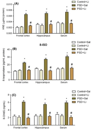
Oxidative stress parameters in the mouse
brains and serum
Figures4A and B show oxidative damage to lipids determined by as-sessing the levels of HNE and 8- ISO in samples of the frontal cortex, hippocampus and serum of the mice used in the study. PSD induced a marked increase of HNE and 8- ISO levels in the frontal cortex, hippocampus and serum. The administration of Li prevented PSD- induced increases of HNE and 8- ISO levels in all samples analyzed. Table 1 presents the results of the two- way ANOVA, and means and
standard deviations are given in Table 2.
Figure4C shows oxidative damage to DNA determined by as-sessing the amount of 8- OHdG in the samples of the frontal cortex, hippocampus and serum of the mice used in the study. OHdG was also significantly different between groups, with significant increases in PSD- subjected mice compared with controls. The administration of Li prevented the PSD- induced increase of OHdG levels in all samples
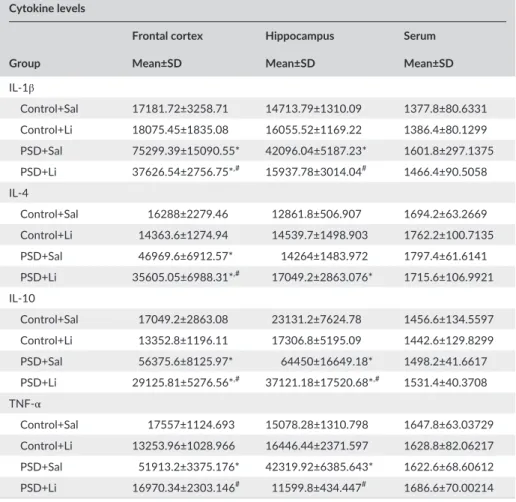
Cytokine levels
Frontal cortex Hippocampus Serum
Group Mean±SD Mean±SD Mean±SD
IL- 1β
Control+Sal 17181.72±3258.71 14713.79±1310.09 1377.8±80.6331 Control+Li 18075.45±1835.08 16055.52±1169.22 1386.4±80.1299 PSD+Sal 75299.39±15090.55* 42096.04±5187.23* 1601.8±297.1375 PSD+Li 37626.54±2756.75*,# 15937.78±3014.04# 1466.4±90.5058
IL- 4
Control+Sal 16288±2279.46 12861.8±506.907 1694.2±63.2669 Control+Li 14363.6±1274.94 14539.7±1498.903 1762.2±100.7135 PSD+Sal 46969.6±6912.57* 14264±1483.972 1797.4±61.6141 PSD+Li 35605.05±6988.31*,# 17049.2±2863.076* 1715.6±106.9921
IL- 10
Control+Sal 17049.2±2863.08 23131.2±7624.78 1456.6±134.5597 Control+Li 13352.8±1196.11 17306.8±5195.09 1442.6±129.8299 PSD+Sal 56375.6±8125.97* 64450±16649.18* 1498.2±41.6617 PSD+Li 29125.81±5276.56*,# 37121.18±17520.68*,# 1531.4±40.3708
TNF- α
Control+Sal 17557±1124.693 15078.28±1310.798 1647.8±63.03729 Control+Li 13253.96±1028.966 16446.44±2371.597 1628.8±82.06217 PSD+Sal 51913.2±3375.176* 42319.92±6385.643* 1622.6±68.60612 PSD+Li 16970.34±2303.146# 11599.8±434.447# 1686.6±70.00214
Mean and standard deviation (SD) are shown. *P<.05 compared to the Control+Sal group.
#P<.05 compared to the ouabain group.
4- HNE, 4- hydroxy- 2- nonenal; 8- OHdG, 8- hydroxy- 2′- deoxyguanosine; ACTH, adrenocorticotropic hor-mone; GPx, glutathione peroxidase; GR, glutathione reductase; IL, interleukin; Li, lithium; PSD, paradoxical sleep deprivation; Sal, saline; TNF- α, tumor necrosis factor alpha.
analyzed. Table 1 presents the results of the two- way ANOVA, and
means and standard deviations are given in Table 2.
3.4
|
Activity of antioxidant enzymes in the mouse
brains and serum
The GPx and GR activities in the hippocampus, frontal cortex and serum of the mice are shown in Figures5A and B, respectively. The PSD protocol increased GPx activity in the hippocampus and frontal cortex of the mice, but not in the serum. The administration of Li pre-vented PSD- induced increases of GPX and GR levels in all brain sam-ples analyzed. Table 1 presents the results of the two- way ANOVA,
and means and standard deviations are given in Table 2.
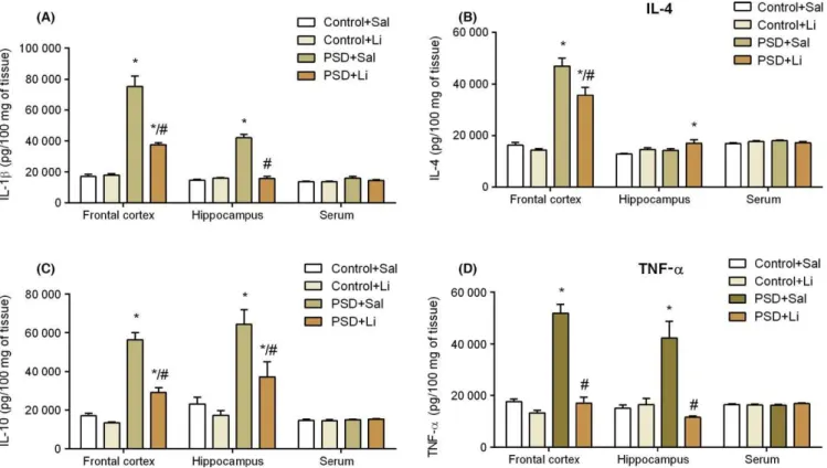
3.5
|
IL- 1
β
, IL- 4, IL- 10, and TNF-
α
levels in the mouse
brains and serum
Figure6 shows the IL- 1β (A), IL- 4 (B), IL- 10 (C), and TNF- α (D) levels in the
frontal cortex, hippocampus and serum of the mice. The PSD protocol increased the IL- 1β levels in the frontal cortex and hippocampus;
how-ever, Li treatment reversed the IL- 1β alteration in the hippocampus. The
Li pretreatment significantly diminished the PSD- induced IL- 1β increase
in the frontal cortex, although it was not able to return the IL- 1β concen
-tration to control levels (Figure6A). PSD increased the IL- 4 levels in the
frontal cortex, but not in the hippocampus. Li pretreatment significantly diminished the IL- 4 increase induced by PSD, although it was not able to return the IL- 4 concentration to control levels (Figure6B). In addition, PSD increased the IL- 10 levels in the frontal cortex and hippocampus (Figure6C), whereas Li treatment reduced this PSD- induced increase. Figure6D shows that PSD increased the IL- 10 levels in the frontal cor-tex and hippocampus; however, Li treatment was able to prevent these
cytokine alterations. No significant alterations in cytokine levels were
observed in the serum. Table 1 presents the results of the two- way ANOVA, and means and standard deviations are given in Table 2.
4
|
DISCUSSION
The present results demonstrate that PSD, an environmental model of mania, induced mania- like behavior, and treatment with Li reversed
F I G U R E 3 Effects of administration of lithium (Li) on the levels of adrenocorticotropic hormone (ACTH) (A) and corticosterone (B) in the serum of animals subjected to the paradoxical sleep deprivation (PSD)- induced animal model (n=8 per group). Data were analyzed by two- way analysis of variance followed by the Duncan test when F
was significant. Values are expressed as mean±SD. *P<.05 compared to the Control+Sal group. #P<.05 compared to the ouabain group. Sal,
saline [Colour figure can be viewed at wileyonlinelibrary.com]
FIGURE 4 Effects of administration of lithium (Li) on the levels of 4- hydroxy- 2- nonenal (HNE) (A), 8- isoprostane (8- ISO) (B) and 8- hydroxy- 2′- deoxyguanosine (8- OHdG) (C) in the frontal cortex, hippocampus and serum of animals subjected to the paradoxical sleep deprivation (PSD)- induced animal model (n=8 per group). Data were analyzed by two- way analysis of variance followed by the Duncan test
when F was significant. Values are expressed as mean±SD. *P<.05
compared to the Control+Sal group. #P<.05 compared to the ouabain
this effect. Likewise, a previous study using the animal model of mania induced by PSD showed the antimanic proprieties of Li.35,37 These re
-sults suggested that alterations in circadian rhythm can lead to mania- like behavior, which represents manic episodes in bipolar patients.34
In fact, the manic episodes are characterized by a marked decrease in the need for sleep. Therefore, PSD may be considered an important tool for the elucidation of the physiopathology and etiology of BD.38
In the present study, the mania- like behavior induced by PSD was accompanied by increases in the circulating levels of corticosterone and ACTH, which indicates that this model of PSD activates the HPA axis. In agreement with the present findings, a recent study
demon-strated an elevated corticosterone concentration in the serum of PSD
rats when compared to controls.39 In addition, and corroborating the
findings of the present study, several studies have demonstrated that
the levels of corticosterone are increased after PSD.40-43 Suchecki and
colleagues 40 showed that PSD induced using different methods in
-creased ACTH and corticosterone secretion. Indeed, insomnia is asso-ciated with increases of ACTH and cortisol secretion, and activation of the HPA axis or administration of glucocorticoids can lead to arousal and sleeplessness.44,45
It has been reported that there is HPA axis dysregulation in bipolar patients.46 A previous clinical study demonstrated increases in cortisol
secretion in bipolar patients, suggesting HPA axis hyperactivity in this
disorder.47 Kim and colleagues 48 found that ACTH- treated animals
exhibited increased locomotor activity. In the same study, Kim and
colleagues 48 suggested that ACTH administration in rats could induce
a putative animal model of mania, with face validity, by mimicking the manic episodes of BD. Therefore, the mania- like behavior induced by PSD in the present study can be explained, at least in part, by an in-crease of ACTH levels also induced by PSD.
It is important to note that, in the present study, administration of Li prevented the increases of ACTH and corticosterone levels in the serum of mice subjected to PSD. On the other hand, previous
stud-ies have demonstrated that Li administration increased the levels of
ACTH and corticosterone in the plasma of rats.49-53 Wood and col -leagues 54 demonstrated that 36 days of Li treatment did not signifi
-cantly alter corticosterone levels. The effects of Li on the HPA axis are therefore somewhat controversial. This discrepancy can be explained
by the fact that the methodologies and durations of the Li treatments
used in the studies were different in each case. It is important to em-phasize that, in the present study, Li alone did not alter the levels of corticosterone or ACTH, its effects being limited to alterations within the HPA axis, which were induced by PDS. It is well described in the
literature that Li acts on the mechanisms of circadian rhythms.55,56 A
previous preclinical study demonstrated that Li delays biochemical cir-cadian rhythms in rats. In that study, the authors found that the levels of prolactin, corticosterone, and aldosterone in plasma showed delays in their circadian rhythms in the Li- treated rats.57 Therefore, it can be
suggested that Li indirectly regulates the levels of corticosterone and
ACTH in PSD- subjected rats, acting on the mechanisms controlling
circadian rhythms.
Some studies have demonstrated that glucocorticoids released
from the adrenal gland in response to stress- induced activation of the HPA axis can induce oxidative stress.15 In the present study, it was
demonstrated that PSD increased oxidative damage to lipids (4- HNE and 8- ISO) and DNA (8- OHdG) in the frontal cortex, hippocampus and serum. Previous studies found that PSD increased lipid oxidative damage parameters, such as lipid hydroperide, 4- HNE and malond-ialdehyde levels in the frontal cortex and hippocampus of mice.58,59
In agreement with the present study, Vollert and colleagues 60 also
demonstrated that an increase in serum corticosterone was accom
-panied by increases in lipid peroxidation markers (8- ISO and malondi-aldehyde) in the hippocampus, cortex and amygdala of rats subjected to PSD. There is also evidence linking the redox state and circadian
rhythm.61,62 Wang and colleagues 61 showed alterations to the redox
systems in mutant mice that had undergone deletion or disruption of their CLOCK genes. The circadian rhythm has a natural oscillation in the redox state which is controlled by a biological clock. This in turn triggers the production of antioxidant enzymes and ROS depending on
the time of day.61 Thus, it can be suggested that PSD, which interrupts
the circadian rhythm oscillation, may be deregulating the redox state, thus generating oxidative stress.
Oxidative damage to lipids and DNA in bipolar patients has also
been observed in both the brain and serum.7,18,63 8- ISO and 4- HNE
were found to be increased in the postmortem prefrontal cortex from
FIGURE 5 Effects of administration of lithium (Li) on the levels of glutathione peroxidase (GPx) (A) and glutathione reductase (GR) (B) in the frontal cortex, hippocampus and serum of animals subjected to the paradoxical sleep deprivation (PSD)- induced animal model (n=8 per group). Data were analyzed by two- way analysis of variance
followed by the Duncan test when F was significant. Values are
expressed as mean±SD. *P<.05 compared to the Control+Sal group.
#P<.05 compared to the ouabain group. Sal, saline [Colour figure can be
patients with BD when compared with controls.18 In addition, clinical
research has also demonstrated elevated levels of lipid peroxidation in the serum of bipolar patients.63 Interestingly, Versace and colleagues 63 demonstrated a relationship between white matter damage and
pe-ripheral lipid peroxidation in bipolar patients. Additionally, a previous study demonstrated that the levels of DNA 8- OHdG were higher in BD patients when compared to healthy controls.7 Together, these studies
suggest the role of lipid peroxidation and DNA oxidation in BD. Given that PSD also induces an increase in the markers of lipid peroxidation and DNA oxidation, PSD- induced mania- like behavior may provide a useful animal model in which to test the hypothesis of the involvement of oxidative stress in BD.
Herein, Li was able to prevent lipid peroxidation and DNA oxida-tion in the brain and serum of mice subjected to PSD. Previous
pre-clinical studies using other animal models of mania have demonstrated
the antioxidant effects of Li. Valvassori and colleagues 64 found that
Li modulated antioxidant enzymes and prevented ouabain- induced mania- like behavior and oxidative damage in the brains of rats that were subjected to this model of mania. Li also reversed the oxidative
damage in the brains of rats subjected to the animal model of mania
induced by amphetamine.65 It was also found that, in an animal model
of sepsis, treatments with Li at doses of 25 and 50 mg/kg decreased the levels of 8- ISO.66 Together with the present results, these studies
suggest that Li has antioxidant properties, protecting the brain against oxidative brain damage.
In the present study, the levels of GPx and GR were increased in the frontal cortex and hippocampus of rats after PSD, and Li prevented these enzyme alterations. In agreement with our data, previous studies have demonstrated that PSD induces GPx and GR alterations in the brains of
mice.59 Another study examined glutathiolation in brain slices taken at
several points across the circadian cycle. It was observed that there was a peak in glutathiolation in the early night, indicating an oxidized state of glutathione, while glutathiolation was lower at midday, indicating a
relatively reduced state.61 Thus, it can be hypothesized that PSD, which
interrupts the oscillation of the circadian rhythm, may be deregulating the glutathiolation cycle and, consequently, altering glutathione activity.
It is important to note that Li prevented the PSD- induced increase in GPx and GR levels in the frontal cortex and hippocampus of mice. Brocardo and colleagues 67 demonstrated that Li modulates GPx and
GR, protecting the brain against oxidative damage induced by ouabain in an animal model of mania. In addition, a previous study showed that Li modulates other antioxidant enzymes, such as superoxide dis-mutase and catalase, and prevents ouabain- induced oxidative damage in the frontal cortex and hippocampus of rats subjected to the model
of mania induced by ouabain.68 In a preclinical study using the animal
model of mania induced by methylphenidate, it was demonstrated that the rats with mania- like behavior showed decreases in their levels of both glutathione and GPx activity, and these alterations were reversed
by Li treatment.69 Together, these data suggest that Li can modulate
antioxidant enzymes, protecting the brain against oxidative damage.
FIGURE 6 Effects of administration of lithium (Li) on the levels of interleukin (IL)- 1β (A), IL- 4 (B), IL- 10 (C) and tumor necrosis factor alpha (TNF- α)
As discussed above, the antioxidant properties of Li have been already described in the literature; however, the present study demonstrated that the neuroprotector effect of Li can be HPA axis- modulated. In fact, it is know that ACTH and cortisol release can lead to an oxidative stress state,15 and these alterations could play a pivotal
role in PSD- induced behavioral changes.40,44 As Li was able to reduce
ACTH and cortisol release induced by PSD, it is possible that, in the present study, the effects of Li on oxidative stress may have been me-diated, at least in part, by ACTH and cortisol modulation.
It is known that oxidative stress is an important etiologic factor in the pathogenesis of chronic inflammatory diseases such as athero-sclerosis, metabolic disorders, and cancer.70-72 There is an increasing
amount of data showing that sleep loss is associated with HPA axis alterations, activation of inflammatory parameters and increases in oxidative stress parameters in humans and in animal models.39,59,73,74
Indeed, the present study showed that HPA axis alterations and oxi-dative stress were accompanied by increased levels of IL- 1β, IL- 4, IL-
10 and TNF- α in the brains of mice subjected to PSD. In agreement
with our study, Ashley and colleagues 39 demonstrated that PSD mice
exhibited increases in IL- 1β and TNF- α pro- inflammatory gene
expres-sion in the hypothalamus, hippocampus and frontal cortex. In a previ-ous study, using the animal model of mania induced by amphetamine, it was demonstrated that IL- 4, IL- 6, IL- 10 and TNF- α were increased
in the brains of rats with mania- like behavior.24 Some clinical studies
have demonstrated that the progressive neuropathological processes in BD are linked to alterations in inflammatory cytokines.25,75 A clinical
study demonstrated that changes observed in the sleep of patients with BD are also related to the elevation of IL- 6.74 Therefore, together,
these studies suggest that sleep deprivation and mania- like behaviors
are associated with inflammatory system alteration.
It is important to note that Li reversed the alterations to cytokines induced by PSD. A previous study from our laboratory demonstrated that Li reversed the alterations to cytokines in the frontal cortex and
striatum of rats subjected to the animal model of mania induced by
amphetamine.24 Interestingly, Li showed anti- inflammatory effects
in an animal model of sepsis.66 The anti- inflammatory effects of Li,
such as the suppression of cyclooxygenase- 2 expression, inhibition of IL- 1β and TNF- α production, and enhancement of IL- 2 and IL- 10
synthesis, have been well described in the literature.76-78 A clinical
study demonstrated that cytokine concentrations in patients in
re-mission treated with Li were no different from those of healthy sub
-jects, suggesting that Li maintenance brings the cytokine status to a
level similar to that in healthy control subjects.79 As demonstrated
previously, Li prevents oxidative damage to lipids and DNA in rats subjected to PSD. Because PSD- induced ROS overproduction can lead to inflammation, Li could act as an antioxidant and indirectly
modulate cytokine levels.
In conclusion, the present data demonstrated that PSD induced HPA axis alterations, and increases in the levels of cytokines and oxi-dative stress parameters. Therefore, it can be suggested that circadian rhythm alterations observed in BD may be related to alterations in the HPA axis, activation of the inflammatory system and oxidative stress;
all these effects were also observed in the animal model induced by
PSD. In addition, Li could reverse the neurochemical alterations in-duced by PSD, suggesting that this mood stabilizer could act on the mechanisms controlling circadian rhythms, and protect the brain and serum against HPA axis alteration and increases in the levels of
oxida-tive stress and cytokines.
ACKNOWLEDGEMENTS
We thank Conselho Nacional de Desenvolvimento Científico e Tecnológico; Fundação de amparo à pesquisa e inovação do estado de Santa Catarina; Instituto Cérebro e Mente; and Universidade do Extremo Sul Catarinense for financial support and Ian Wayne Miles and Paula Abelaira for assistance with language.
DISCLOSURE
The authors declares that there is no conflict of interest regarding this
article.
REFERENCES
1. Kendall T, Morriss R, Mayo-Wilson E, Marcus E, on behalf of the Guideline Development Group of the National Institute for Health and Care Excellence. Assessment and management of bipolar disor-der: summary of updated NICE guidance. BMJ. 2014;349:g5673
2. Miller TH. Bipolar Disorder. Prim Care 2016;43:269-284.
3. American Psychiatric Association. Bipolar disorder. In: American Psychiatric Association, eds. Diagnostic and statistical manual of mental disorders. 5th edition. Washington, DC: American Psychiatric Publishing; 2013:124-125.
4. Geddes JR, Miklowitz DJ. Treatment of bipolar disorder. Lancet
2013;381:1672-1682.
5. Geddes JR, Burgess S, Hawton K, Jamison K, Goodwin GM. Long- term lithium therapy for bipolar disorder: systematic review and meta- analysis of randomized controlled trials. Am J Psychiatry.
2004;161:217-222.
6. Brown NC, Andreazza AC, Young LT. An updated meta- analysis of oxidative stress markers in bipolar disorder. Psychiatry Res.
2014;218:61-68.
7. Soeiro-de-Souza MG, Andreazza AC, Carvalho AF, Machado-Vieira R, Young LT, Moreno RA. Number of manic episodes is as-sociated with elevated DNA oxidation in bipolar I disorder. Int J Neuropsychopharmacol. 2013;16:1505-1512.
8. Kim HK, Andreazza AC, Yeung PY, Isaacs-Trepanier C, Young LT. Oxidation and nitration in dopaminergic areas of the prefrontal cor-tex from patients with bipolar disorder and schizophrenia. J Psychiatry Neurosci. 2014;39:276-285.
9. Girshkin L, Matheson SL, Shepherd AM, Green MJ. Morning cor-tisol levels in schizophrenia and bipolar disorder: a meta- analysis.
Psychoneuroendocrinology. 2014;49:187-206.
10. Luo Y, He H, Zhang M, Huang X, Fan N. Altered serum levels of TNF- α, IL- 6 and IL- 18 in manic, depressive, mixed state of bipolar disorder
pa-tients. Psychiatry Res. 2016;244:19-23.
11. van den Ameele S, van Diermen L, Staels W, et al. The effect of mood- stabilizing drugs on cytokine levels in bipolar disorder: a systematic
review. J Affect Disord. 2016;203:364-373.
12. Muneer A. The Neurobiology of Bipolar Disorder: an Integrated Approach. Chonnam Med J. 2016;52:18-37.
13. Wieck A, Grassi-Oliveira R, do Prado CH, et al. Pro- inflammatory cytokines and soluble receptors in response to acute psychoso-cial stress: differential reactivity in bipolar disorder. Neurosci Lett
14. Ventriglio A, Gentile A, Baldessarini RJ, Bellomo A. Early- life stress and psychiatric disorders: epidemiology, neurobiology and innovative pharmacological targets. Curr Pharm Des. 2015;21:1379-1387. 15. Spiers JG, Chen HJ, Sernia C, Lavidis NA. Activation of the
hypothalamic- pituitary- adrenal stress axis induces cellular oxidative
stress. Front Neurosci 2015;8:456.
16. Cochrane CG. Mechanisms of oxidant injury of cells. Mol Aspects Med.
1991;12:137-147.
17. De Vasconcelos AP, Bouilleret V, Riban V, Wasterlain C, Nehlig A. Role of nitric oxide in cerebral blood flow changes during kainite sei-zures in mice: genetic and pharmacological approaches. Neurobiol Dis.
2005;18:270-281.
18. Andreazza AC, Wang JF, Salmasi F, Shao L, Young LT. Specific subcellu-lar changes in oxidative stress in prefrontal cortex from patients with bipolar disorder. J Neurochem. 2013;127:552-561.
19. Gawryluk JW, Wang JF, Andreazza AC, Shao L, Young LT. Decreased levels of glutathione, the major brain antioxidant, in post- mortem prefrontal cortex from patients with psychiatric disorders. Int J Neuropsychopharmacol. 2011;14:123-130.
20. Andreazza AC, Cassini C, Rosa AR, et al. Serum S100B and antioxidant enzymes in bipolar patients. J Psychiatr Res. 2007;41:523-529. 21. Andreazza AC, Frey BN, Erdtmann B, et al. DNA damage in bipolar
disorder. Psychiatry Res. 2007;153:27-32.
22. Machado-Vieira R, Andreazza AC, Viale CI, et al. Oxidative stress pa-rameters in unmedicated and treated bipolar subjects during initial manic episode: apossible role for lithium antioxidant effects. Neurosci Lett. 2007;421:33-36.
23. Ferrero-Miliani L, Nielsen OH, Andersen PS, Girardin SE. Chronic in-flammation: importance of NOD2 and NALP3 in interleukin- 1beta
generation. Clin Exp Immunol. 2007;147:227-235.
24. Valvassori SS, Tonin PT, Varela RB, et al. Lithium modulates the produc-tion of peripheral and cerebral cytokines in an animal model of mania induced by dextroamphetamine. Bipolar Disord. 2015;17:507-517. 25. Berk M, Kapczinski F, Andreazza AC, et al. Pathways underlying
neuroprogression in bipolar disorder: focus on inflammation, ox-idative stress and neurotrophic factors. Neurosci Biobehav Rev.
2011;35:804-817.
26. Barbosa IG, Rocha NP, Assis F, et al. Monocyte and lymphocyte acti-vation in bipolar disorder: a new piece in the puzzle of immune
dys-function in mood disorders. Int J Neuropsychopharmacol. 2014;18:1-7 27. Munkholm K, Braüner JV, Kessing LV, Vinberg M. Cytokines in bipolar disorder vs. healthy control subjects: a systematic review and meta-
analysis. J Psychiatr Res. 2013;47:1119-1133.
28. Modabbernia A, Taslimi S, Brietzke E, Ashrafi M. Cytokine alterations in bipolar disorder: a meta- analysis of 30 studies. Biol Psychiatry.
2013;74:15-25.
29. Rosenblat JD, Kakar R, Berk M, et al. Anti- inflammatory agents in the treatment of bipolar depression: a systematic review and meta-
analysis. Bipolar Disord. 2016;18:89-101.
30. Gessa GL, Pani L, Fadda P, Fratta W. Sleep deprivation in the rat: an
animal model of mania. Eur Neuropsychopharmacol. 1995;5:89-93. 31. Benedetti F, Fresi F, Maccioni P, Smeraldi E. Behavioural sensitization
to repeated sleep deprivation in a mice model of mania. Behav Brain Res. 2008;187:221-227.
32. Kaplan KA, Harvey AG. Behavioral treatment of insomnia in bipolar
disorder. Am J Psychiatry. 2013;170:716-720.
33. Bunney BG, Bunney WE. Mechanisms of rapid antidepressant effects of sleep deprivation therapy: clock genes and circadian rhythms. Biol Psychiatry. 2013;73:1164-1171.
34. Roybal K, Theobold D, Graham A, et al. Mania- like behavior induced by disruption of CLOCK. PNAS. 2007;104:6406-6411.
35. Tufik S, Andersen ML, Bittencourt LR, Mello MT. Paradoxical sleep deprivation: neurochemical, hormonal and behavioral alter-ations. Evidence from 30 years of research. An Acad Bras Cienc.
2009;81:521-538.
36. Armani F, Andersen ML, Andreatini R, Frussa-Filho R, Tufik S, Galduróz JC. Successful combined therapy with tamoxifen and lithium in a par-adoxical sleep deprivation- induced mania model. CNS Neurosci Ther.
2012;18:119-125.
37. Lowry OH, Rosebrough NJ, Farr AL, Randall RJ. Protein measurement with the Folin phenol reagent. J Biol Chem. 1951;193:265-275. 38. Abrial E, Bétourné A, Etiévant A, et al. Protein kinase C inhibition
res-cues manic- like behaviors and hippocampal cell proliferation deficits in the sleep deprivation model of mania. Int J Neuropsychopharmacol
2014;18:1-11.
39. Berns GS, Nemeroff CB. The neurobiology of bipolar disorder. Am J Med Genet C Semin Med Genet. 2003;123C:76-84.
40. Ashley NT, Sams DW, Brown AC, Dumaine JE. Novel environment influences the effect of paradoxical sleep deprivation upon brain and peripheral cytokine gene expression. Neurosci Lett. 2016;615:
55-59.
41. Suchecki D, Lobo LL, Hipólide DC, Tufik S. Increased ACTH and cor-ticosterone secretion induced by different methods of paradoxical sleep deprivation. J Sleep Res. 1998;7:276-281.
42. Dugovic C, Maccari S, Weibel L, Turek FW, Van Reeth O. High corti-costerone levels in prenatally stressed rats predict persistent para-doxical sleep alterations. J Neurosci. 1999;19:8656-8664.
43. Na JR, Oh DR, Han S, et al. Antistress Effects of Rosa rugosa Thunb. on Total Sleep Deprivation- Induced Anxiety- Like Behavior and Cognitive Dysfunction in Rat: possible Mechanism of Action of 5- HT6 Receptor Antagonist. J Med Food. 2016;19:870-881.
44. Choi JH, Lee SH, Bae JH, et al. Effect of Sleep Deprivation on the Male Reproductive System in Rats. J Korean Med Sci. 2016;31:
1624-1630.
45. Opp MR. Corticotropin- releasing hormone involvement in stressor- induced alterations in sleep and in the regulation of waking. Adv Neuroimmunol. 1995;5:127-143.
46. Chrousos G, Vgontzas AN, Kritikou I. HPA Axis and Sleep. In: De Groot LJ, Chrousos G, Dungan K, Grossman A, Hershman JM, Koch C, Korbonits M, McLachlan R, New M, Purnell J, Rebar R, Singer F, Vinik A, eds. SourceEndotext [Internet]. South Dartmouth (MA): MDText. com, Inc.; 2016:1-47.2000. Jan 18.
47. Manji HK, Lenox RH. The nature of bipolar disorder. J Clin Psychiatry.
2000;61(Supp 13):42-57.
48. Cervantes P, Gelber S, Kin FN, Nair VN, Schwartz G. Circadian secretion of cortisol in bipolar disorder. J Psychiatry Neurosci.
2001;26:411-416.
49. Kim Y, McGee S, Czeczor JK, et al. Nucleus accumbens deep- brain stimulation efficacy in ACTH- pretreated rats: alterations in mitochon-drial function relate to antidepressant- like effects. Transl Psychiatry. 2016;6:e842.
50. Hennessy JW, Smotherman WP, Levine S (1985). Conditioned taste aversion and the pituitary- adrenal system. Behav Biol.
1976;16::413-424. Ann N Y Acad Sci. 443:126-44.
51. Smotherman WP. Glucocorticoid and other hormonal substrates of
conditioned taste aversion. Ann N Y Acad Sci. 1985;443:126-144. 52. Sugawara M, Hashimoto K, Hattori T, Takao T, Suemaru S, Ota
Z. Effects of lithium on the hypothalamo- pituitary- adrenal axis.
Endocrinol Jpn. 1988;35:655-663.
53. Gilmor ML, Skelton KH, Nemeroff CB, Owens MJ. The effects of chronic treatment with the mood stabilizers valproic acid and lithium on corticotropin- releasing factor neuronal systems. J Pharmacol Exp Ther. 2003;305:434-439.
54. Bschor T, Ritter D, Winkelmann P, et al. Lithium monotherapy in-creases ACTH and cortisol response in the DEX/CRH test in unipolar depressed subjects. A study with 30 treatment- naive patients. PLoS ONE. 2011;6:e27613.
55. Wood GE, Young LT, Reagan LP, Chen B, McEwen BS. Stress- induced structural remodeling in hippocampus: prevention by lithium
56. McCarthy MJ, Nievergelt CM, Shekhtman T, Kripke DF, Welsh DK, Kelsoe JR. Functional genetic variation in the Rev- Erbα pathway and lithium response in the treatment of bipolar disorder. Genes Brain Behav. 2011;10:852-861.
57. Moreira J, Geoffroy PA. Lithium and bipolar disorder: impacts
from molecular to behavioural circadian rhythms. Chronobiol Int.
2016;33:351-373.
58. McEachron DL, Kripke DF, Hawkins R, Haus E, Pavlinac D,
Deftos L. Lithium delays biochemical circadian rhythms in rats.
Neuropsychobiology. 1982;8:12-29.
59. Zhang L, Zhang HQ, Liang XY, Zhang HF, Zhang T, Liu FE. Melatonin ameliorates cognitive impairment induced by sleep deprivation in rats: role of oxidative stress, BDNF and CaMKII. Behav Brain Res.
2013;256:72-81.
60. Arent CO, Valvassori SS, Steckert AV, et al. The effects of n- acetylcysteine and/or deferoxamine on manic- like behavior and brain oxidative damage in mice submitted to the paradoxal sleep deprivation model of mania. J Psychiatr Res. 2015;65:71-79. 61. Vollert C, Zagaar M, Hovatta I, et al. Exercise prevents sleep
deprivation- associated anxiety- like behavior in rats: potential role of oxidative stress mechanisms. Behav Brain Res. 2011;224:233-240. 62. Wang TA, Yu YV, Govindaiah G, et al. Circadian rhythm of redox state
regulates excitability in suprachiasmatic nucleus neurons. Science.
2012;337:839-842.
63. Fanjul-Moles ML, López-Riquelme GO. Relationship between Oxidative Stress, Circadian Rhythms, and AMD. Oxid Med Cell Longev. 2016;2016:7420637.
64. Versace A, Andreazza AC, Young LT, et al. Elevated serum measures of lipid peroxidation and abnormal prefrontal white matter in euthymic bipolar adults: toward peripheral biomarkers of bipolar disorder. Mol Psychiatry. 2014;19:200-208.
65. Valvassori SS, Resende WR, Lopes-Borges J, et al. Effects of mood stabilizers on oxidative stress- induced cell death signaling path-ways in the brains of rats subjected to the ouabain- induced animal model of mania: mood stabilizers exert protective effects against ouabain- induced activation of the cell death pathway. J Psychiatr Res.
2015;65:63-70.
66. da-Rosa DD, Valvassori SS, Steckert AV, et al. Effects of lithium and valproate on oxidative stress and behavioral changes induced by ad-ministration of m- AMPH. Psychiatry Res. 2012;198:521-526. 67. Albayrak A, Halici Z, Polat B, et al. Protective effects of lithium: a new look
at an old drug with potential antioxidative and anti- inflammatory effects in an animal model of sepsis. Int Immunopharmacol. 2013;16:35-40. 68. Brocardo PS, Budni J, Pavesi E, et al. Folic acid administration
pre-vents ouabain- induced hyperlocomotion and alterations in oxidative
stress markers in the rat brain. Bipolar Disord. 2010;12:414-424.
69. Jornada LK, Valvassori SS, Steckert AV, et al. Lithium and valproate modulate antioxidant enzymes and prevent ouabain- induced
ox-idative damage in an animal model of mania. J Psychiatr Res.
2011;45:162-168.
70. Arunagiri P, Rajeshwaran K, Shanthakumar J, Tamilselvan T, Balamurugan E. Combination of omega- 3 Fatty acids, lithium, and ar-ipiprazole reduces oxidative stress in brain of mice with mania. Biol Trace Elem Res. 2014;160:409-417.
71. Leitinger N. Oxidized phospholipids as triggers of inflammation in
ath-erosclerosis. Mol Nutr Food Res. 2005;49:1063-1071.
72. Toyokuni S, Okamoto K, Yodoi J, Hiai H. Persistent oxidative stress in
cancer. FEBS Lett 1995;358:1-3.
73. Chen GY, Nuñez G. Sterile inflammation: sensing and reacting to
dam-age. Nat Rev Immunol. 2010;10:826-837.
74. Irwin M, McClintick J, Costlow C, Fortner M, White J, Gillin JC. Partial night sleep deprivation reduces natural killer and cellular immune re-sponses in humans. FASEB J. 1996;10:643-3.
75. Ritter PS, Kretschmer K, Pfennig A, Soltmann B. Disturbed sleep in bipolar disorder is related to an elevation of IL- 6 in peripheral
mono-cytes. Med Hypotheses. 2013;81:1031-1033.
76. Kapczinski F, Dias VV, Kauer-Sant’Anna M, et al. The potential use of biomarkers as an adjunctive tool for staging bipolar disorder. Prog Neuropsychopharmacol Biol Psychiatry. 2009;33:1366-1371. 77. Arena A, Capozza AB, Orlando ME, et al. In vitro effects of lithium
chloride on TNF alpha and IL- 6 production by monocytes from breast cancer patients. J Chemother. 1997;9:219-226.
78. Nahman S, Belmaker RH, Azab AN. Effects of lithium on lipopolysaccharide- induced inflammation in rat primary glia cells.
Innate Immun. 2012;18:447-458.
79. Wang HM, Zhang T, Li Q, Huang JK, Chen RF, Sun XJ. Inhibition of glycogen synthase kinase- 3β by lithium chloride suppresses 6- hydroxydopamine- induced inflammatory response in primary
cul-tured astrocytes. Neurochem Int. 2013;63:345-353.
How to cite this article: Valvassori SS, Resende WR,
Dal-Pont G, et al. Lithium ameliorates sleep deprivation- induced mania- like behavior, hypothalamic- pituitary- adrenal (HPA) axis alterations, oxidative stress and elevations of