HPV detection using primers MY09/MY11
and GP5+/GP6+ in patients with cytologic
and/or colposcopic changes
Detecção de HPV utilizando iniciadores MY09/MY11 e GP5+/GP6+
em pacientes com alterações citológicas e/ou colposcópicas
Emanuella Meneses Venceslau1; Mauro Muniz Bezerra2; Anna Carolina Mota Lopes1; Érick Vieira Souza3; Alexandre Sherlley Casimiro Onofre4; Claudia Moura de Melo5; Verônica de Lourdes Sierpe Jeraldo6; Fabiana Botelho de Miranda Onofre4
First submission on 23/04/12; last submission on 03/12/13; accepted for publication on 16/05/14; published on 20/08/14 1. MSc in Health and Environment-Universidade Tiradentes (UNIT); biomedical Scientist.
2. MSc Health and Environment-Universidade Tiradentes (UNIT); gynecologist. 3. Biomedical scientist.
4. PhD in Theoretical Medicine (Theoretischen Medizin-Doctor rerum medicinalium) – School of Medicine at Rheinisch-Westfalische Technische Hochschule Aachen; Aachen-Germany; adjunct professor at Universidade Federal de Santa Catarina (UFSC).
5. PhD in Parasitology-Universidade Estadual de Campinas (UNICAMP); permanent professor at UNIT. 6. PhD in Parasitology-Universidade de São Paulo (USP); permanent professor at UNIT.
ABSTRACT
Introduction: Cervical cancer is one of the most common diseases among women, and cause considerable morbidity and mortality. Considering that cervical cancer is an important neoplasia in northeastern Brazil, and the prevalence of high-risk human papillomavirus (HPV) is directly associated with it, this work had aimed to correlate the cytological and/or colposcopic indings with HPV infection status, and verify the performance of MY09/MY11 and GP5+/6+ primers for HPV detection. Material and method: Patients in this study were from Penedo-AL, a city with high level of poverty (poverty rate of 60.62%). Out of 70 patients with cytological and/or colposcopic changes, 32 agreed to participate in the study. Results: Regarding cytology, 21 (30%) patients presented atypical squamous cells of undetermined signiicance (ASC-US); 20 (29%), atypical glandular cells of undetermined signiicance (AGUS); 12 (17%), low-grade intraepithelial lesion (LSIL); ive (7%), high-grade intraepithelial lesion (HSIL); and 12 (17%), positive colposcopy. From these, 27 (84%) presented the band gene encoding for human ß-globin. From the 27 patients, eight (30%) were positive for HPV. The results showed that the deoxyribonucleic acid (DNA) of HPV was detected in 15% and 30% by using MY-PCR and GP +-PCR, respectively. Conclusion: This study suggests that more than one type of oligonucleotide primer should be used in clinical samples to increase sensitivity for the detection of HPV.
Key words: cervical cancer; cytology; colposcopy; HPV; PCR.
INTRODUCTION
Cervical cancer is one of the most common diseases among women, and causes considerable morbidity and mortality. According to Globocan (2008), 188 thousand new cases occur annually in Asia, 134 thousand in India, 75 thousand in Africa, and 24 thousand in Brazil. In developed countries, such as the United States of America, the incidence was 11 thousand, and in the European Union was 31 thousand(10).
Cervical cancer mortality worldwide is 275 thousand, and 242 thousand occurred in less developed regions. In Brazil, cervical cancer mortality is still high, with 110 thousand deaths(2). The
differences in incidence and mortality between developed and developing countries may be due to regional differences, prevalence and distribution of major risk factors, practices of detection and/or availability and use of treatment services(1).
Epidemiological and molecular studies have shown close link between human papillomavirus (HPV) and the onset of cervical
cancer, as well as its precursors lesions, implying that the virus presence is a cancer-causing agent(7, 28). Walboomers et al.(26),
using the polymerase chain reaction (PCR) and serological tests, showed that HPV is present in 99.7% of patients with invasive cervical cancer.
Molecular diagnosis of HPV infection is important for virus screening, and is mainly based on methods such as: hybrid capture (CH2)(20, 23), in situ hybridization(2), and PCR(8, 21, 24). These
techniques have vary widely in terms of sensitivity and speciicity, and PCR is the most used today in various areas of molecular diagnostics due to its great ability to detect small fragments of deoxyribonucleic acid (DNA)(4, 11).
The system uses MY09/MY11 and GP5+/GP6+ primers, that amplify the L1 region of viral genome, are more frequently used for HPV detection in clinical and histological studies(12). These primers
are effective for amplifying wide spectrum of HPV genotypes in cells obtained from cervical smears and parafin-embedded tissues(13, 16). The pair of oligonucleotides MY09/MY11 primers
lanks a sequence of approximately 450 pb, while the pair GP5+/ GP6+, a sequence of approximately 150 pb, which is internal to the sequence lanked by MY(22). The pair MY is synthesized from
several degenerate nucleotides in each primer, it is a mixture of 25 primers oligonucleotides able to amplifying > 25 genital HPV types. On the other hand, the pair GP5+/GP6+ consists of a ixed sequence of nucleotide for each oligonucleotide primer. This pair uses low annealing temperature during PCR(18), and may be used
as a single oligonucleotide primer or in PCR “nested”, after the MY oligonucleotide primer ampliication(9).
This study aimed to investigate HPV infection in women with cytological and/or colposcopic abnormalities, which were seen in a poor city of Alagoas, and also verify the performance of MY09/ MY11 and GP5+/GP6+ primers for HPV detection.
MATERIAL AND METHOD
This is a cross-sectional study, performed during May 2009 to March 2011, from epidemiological data on cytological and/or colposcopic abnormalities cases diagnosed at Clinic of Gynecology of Núcleo de Apoio à Saúde da Família (NASF) in Penedo-Alagoas, city with high poverty (60.62%)(14).
The study included women had as cytological result: atypical squamous cells of undetermined signiicance (ASC-US), atypical glandular cells of undetermined signiicance (AGUS), low-grade intraepithelial lesion (LSIL), high-grade intraepithelial lesion (HSIL), and invasive carcinoma. Patients with colposcopic
changes such as acetowhite epithelium (AWE), dotted, mosaic, partial iodine-positive epithelium and iodine-negative epithelium, classiied as positive colposcopy, were also included in this study. Patients’ data were obtained from medical records. Cervical samples were only collected from patients who signed the Informed Consent. This project was approved by Research Ethics Committee of Universidade Tiradentes, Aracaju-SE.
Cervical smears were collected using Ayre spatula, then placed in Falcon tubes, with 25 ml of PBS buffer solution (phosphate-buffered saline, pH 7.4), and stored at -20ºC until DNA extraction.
DNA extraction
DNA extraction from cervical smears was performed using QIAamp DNA Mini Kit (QiagenLtd, Crawley, UK), according to manufacturer’s instructions. Aliquots of 200 μl of samples were digested with 20 µl of K proteinase and 200 μl of AL buffer at 56ºC, for 10 minutes. DNA precipitation was performed by adding 200 μl of ethanol (96%). DNA was eluted in 200 μl of AE buffer and stored at -20ºC until further use.
To determine extracted DNA quality and quantity, each DNA was analyzed by electrophoresis on a 1% agarose gel, stained with
blue green loading dye (LGC) (Figure 1).
FIGURE 1 – Agarose gel (1%) stained with blue green loading dye showing gel electrophoretic proile for DNA integrity
DNA: deoxyribonucleic acid; L: ladder; 1-15: DNA samples.
As an intern control of the reaction, samples with DNA
were subjected to PCR for human β-globin gene ampliication,
Samples that were positive for human ß-globin gene were subjected to DNA viral of HPV research. It was excluded from the analysis only negative samples for ß-globin gene, after reanalyzing samples for conirmation.
DNA amplification
Two different pairs of oligonucleotides primers were used in
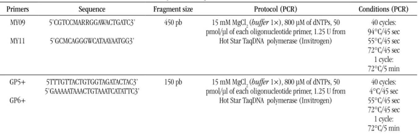
this study to HPV detection: MY09/MY11, GP5+/GP6+ (Figures
3 and 4). PCR reactions for each oligonucleotide primer were
carried out separately, as previously described by Bernard et al.(3)
and De Roda-Husman et al.(6).Table 1 shows the oligonucleotides
primers used in PCR, as well as their ampliication conditions. For each PCR reaction, it was used a positive control (DNA extracted from a condyloma sample) and a negative control (H2O). Result was considered positive for DNA-HPV when one of the MY09/MY11 or GP5+/GP6+ oligonucleotides primers testing detected the viral DNA.
FIGURE 2 – Agarose gel (1,5%) stained with blue green loading dye showing PCR electrophoretic proile using PCO3/PCO4 oligonucleotide primers for human ß-globin gene detection corresponding to ~110 pb
PCR: polymerase chain reaction; L: ladder; C+: positive control; 1-6: positive samples; 7: negative sample; C-: negative control.
FIGURE 3 – Agarose gel (1,5%) stained with blue green loading dye showing PCR electrophoretic proile using MY09/11 oligonucleotide primers for HPV-L1 gene detection corresponding to ~450 pb
PCR: polymerase chain reaction; HPV: human papillomavirus; L: ladder; C+: positive control; 1, 6, 10, 15: positive samples; 2-5, 7-9, 11-14: negative sample; C-: negative control.
FIGURE 4 – Agarose gel (1,5%) stained with blue green loading dye showing PCR electrophoretic proile using GP5+/GP6+ oligonucleotide primers for HPV-L1 gene detection corresponding to ~150 pb
PCR: polymerase chain reaction; HPV: human papillomavirus; L: ladder; C+: positive control; 1-6: positive samples; C-: negative control.
TABLE 1 – Genetic oligonucleotide primers used for DNA of HPV detection
Primers Sequence Fragment size Protocol (PCR) Conditions (PCR) MY09
MY11
5’CGTCCMARRGGAWACTGATC3’
5’GCMCAGGGWCATAAYAATGG3’
450 pb 15 mM MgCl2 (buffer 1×), 800 µM of dNTPs, 50 pmol/µl of each oligonucleotide primer, 1.25 U from
Hot Star TaqDNA polymerase (Invitrogen)
40 cycles: 94°C/45 sec 55°C/45 sec 72°C/45 sec 1 cycle: 72°C/5 min GP5+
GP6+
5TTTGTTACTGTGGTAGATACTAC3’
5’GAAAAATAAACTGTAAATCATATTC3’ 150 pb 15 mM MgCl2
(buffer 1×), 800 µM of dNTPs, 50 pmol/µl of each oligonucleotide primer, 1.25 U from
Hot Star TaqDNA polymerase (Invitrogen)
Statistical analysis
For data statistical analysis we used chi-squared and/or Fisher’s exact test, through SPSS software (Chicago, IL, USA), 16.0
version, considering signiicant variables those that reached p < 0.05 in the two-tailed test.
RESULTS
A sample survey of cytological and/or colposcopic abnormalities cases diagnosed during May 2009 and March 2011 was performed at the Clinic of Gynecology of NASF, in the city of Penedo-AL. The average age of the studied population was 36 years (range: 18-58 years). Regarding cytologic results, 21 patients (30%) showed ASC-US; 20 (29%), AGUS; 12 (17%), LSIL; and ive (7%), HSIL. Twelve patients (17%) showed positive colposcopy (Figure 5 and Table 2).
FIGURE 5 – Results of cytological and/or colposcopic reports from cervical samples of patients admitted in the city of Penedo-AL (2009-2011)
ASC-US: atypical squamous cells of undetermined signiicance; AGUS: atypical glandular cells of undetermined signiicance; LSIL: low-grade squamous intraepithelial lesions; HSIL: high-grade squamous intraepithelial lesions.
TABLE 3 – Percentage of DNA of HPV detection with oligonucleotide primers pairs
DNA of HPV
detection
MY09/MY11 n = 27 (100%)
GP5+/GP6+ n = 27 (100%)
p* value
Negative 23 (85%) 19 (70%) 0.3261
Positive 4 (15%) 8 (30%)
*: Pearson’s chi-square test.
DNA: deoxyribonucleic acid; HPV: human papillomavirus; n: number of collected samples. May 2009 to May 2011
n = 70 (100%)
ASC-US
n = 21 (30%)
AGUS
n = 20 (29%)
LSIL
n = 12 (17%)
HSIL
n = 5 (7%)
Positive colposcopy
n = 12 (17%)
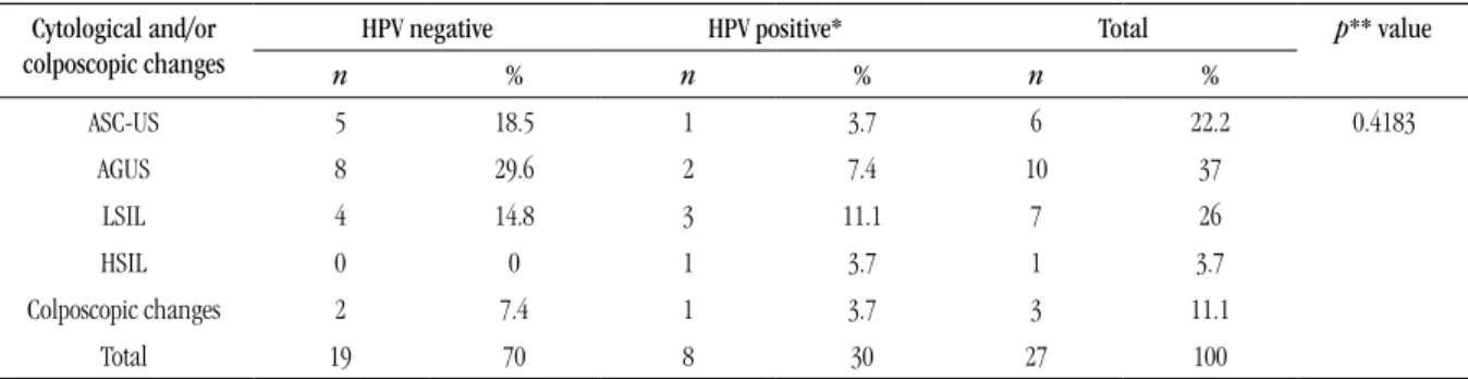
TABLE 2 – Correlation between diagnostic categories to cytologic and colposcopic examinations and HPV molecular detection
Cytological and/or
colposcopic changes
HPV negative HPV positive* Total p** value
n % n % n %
ASC-US 5 18.5 1 3.7 6 22.2 0.4183
AGUS 8 29.6 2 7.4 10 37
LSIL 4 14.8 3 11.1 7 26
HSIL 0 0 1 3.7 1 3.7
Colposcopic changes 2 7.4 1 3.7 3 11.1
Total 19 70 8 30 27 100
*: HPV positive means the sum of the two PCR methods used; **: Fisher’s exact test.
HPV: human papillomavirus; ASC-US: atypical squamous cells of undetermined signiicance; AGUS: atypical glandular cells of undetermined signiicance; LSIL low-grade squamous intraepithelial lesions; HSIL: high-grade squamous intraepithelial lesions; PCR: polymerase chain reaction.
Only one patient has HSIL and a positive sample to HPV, which demonstrate the effectiveness of PCR as standard molecular method. From the seven patients with LSIL, only three were positive for o HPV. The most likely possibility is that the results for the other four samples were false negative.
Results showed that DNA of HPV was detected in 15% and 30%, using MY-PCR and GP+-PCR, respectively. There was no statistical difference between the two oligonucleotides used in PCRs (Table 3).
DISCUSSION
PCR is being increasingly used in clinical laboratories to HPV diagnose. For the L1 region, there is consensus in indicating two oligonucleotides primers systems commonly used: MY09/MY11 and GP5+ /GP6+. Both detect a wide range of HPVs. Degenerated MY09/MY11 oligonucleotide primer uses annealing high temperature (55ºC) and can amplify multiple HPV infections. However, GP5+/GP6+ oligonucleotide primer has lower annealing temperature (42ºC), allowing the best ampliication of single HPV infections, when compared to multiple infections(18).
RESUMO
Introdução: O câncer cervical é uma das doenças mais frequentes entre mulheres, e causa considerável morbidade e mortalidade. Com base nos fatos de que o câncer cervical é uma neoplasia importante no nordeste brasileiro, e que a prevalência do papilomavírus humano (HPV) de alto risco está diretamente associado a ele, este trabalho teve como objetivos correlacionar os achados citológicos e/ou colposcópicos com status de infecção de HPV e veriicar o desempenho dos iniciadores MY09/MY11 e GP5+/GP6+ para detecção do HPV. Material e métodos: Os pacientes deste estudo foram de Penedo-AL, uma cidade com elevado nível de pobreza (índice de pobreza de 60,62%). Do total de 70 pacientes com alterações citológicas e/ou colposcópicas, 32 aceitaram participar do estudo.
Resultados: Com relação a citologia, 21 (30%) pacientes apresentaram células escamosas atípicas de signiicado indeterminado (ASC-US); 20 (29%), células glandulares atípicas de signiicado indeterminado (AGUS); 12 (17%), lesão intraepitelial de baixo grau (LSIL); cinco (7%), lesão intraepitelial de alto grau (HSIL); e 12 (17%), colposcopia positiva. Destas, 27 (84%) apresentaram banda do gene que codiica para ß-globina humana. Das 27 pacientes, oito (30%) apresentaram positividade para o HPV. Os resultados mostraram que o ácido desoxirribonucleico (DNA) do HPV foi detectado em 15% e 30%, usando MY-PCR e GP +-PCR, respectivamente. Conclusão: Este estudo sugere que mais de um tipo de iniciador de oligonucleotídeo deve ser utilizado em amostras clínicas para aumentar a sensibilidade na detecção do HPV.
Unitermos: câncer cervical; citologia; colposcopia; HPV; PCR.
In this study we observed a greater number of positive samples using GP5+/GP6+ oligonucleotides, when compared to MY09/ MY11. From 27 samples, eight (30%) were positive for HPV. DNA-HPV was found in 15% and 30%, using MY e GP+ oligonucleotides primers, respectively. From these, four (15%) were detected by both methods, and eight (30%), only by PCR do GP+. However, there was no statistical difference between the two oligonucleotides used in
PCRs (p = 0.3261), even with twice detection eficiency using GP+
oligonucleotides primers. According to Poljak et al.(17) and Vince
et al.(25), one cannot rule out the possibility of false negative in PCR
due to DNA-HPV low concentration in the clinical sample and the presence of PCR endogenous inhibitors or from sample processing.
In a Zehbe and Wilander study (26), they compared the sensitivity
of these oligonucleotides primers for PCR in cervical biopsies classiied as cervical intraepithelial neoplasia (CIN) I to III. The authors demonstrated that the two oligonucleotides primers systems were equally sensitive, with a correlation of 98%. Using GP5+/GP6+, the authors found HPV positivity of 95% with MY09/MY11 of 94%.
QU et al.(18), compared the detection rates of the same
oligonucleotides primers used in our experiment (MY09/MY11 and GP5+/GP6+) in cervicovaginal washing sample, they found DNA of HPV in 45,5% and 42,8%, respectively, with no statistical difference between the two oligonucleotides used in PCRs, only in the evaluation of HPV types detected by each pair of oligonucleotide, result similar to ours, which also showed no statistical difference.
Remmerbach et al.(19), through oral mucosal smears analysis
of patients suspected of carcinoma lesions, showed that DNA-HPV was detected in 2.8% of sample using PCR with MY09/MY11 oligonucleotides and 35,8% with GP5+/GP6+ oligonucleotides. These authors also reported that HPV detection with GP5+/GP6+ oligonucleotides was higher than MY09/MY11 oligonucleotides,
corroborating the results found in our study. This can be explained by the smaller size of DNA fragment ampliied by GP-PCR system. Thus, this oligonucleotide would have greater eficiency in HPV detection in samples with fragmented DNA. These authors also found a greater HPV detection in biopsy samples, when compared to cervical smear sample, which may indicate the difference between our study and the Zehbe and Wilander study(27).
Iftner and Villa(15) reported that the sensitivity and speciicity
of PCR method may vary depending mainly on the choice of oligonucleotides primers used, the number of base pairs of the product ampliied by PCR, performance of DNA polymerase used in the reaction, DNA-HPV type spectrum ampliied, and the ability to detect different types in the infection. Studies in the literature indicate variables HPV detection rates.
The choice of suitables oligonucleotides to clinical and epidemiological uses should consider the source of clinical material, the size of PCR product, range of DNA-HPV types to be ampliied, oligonucleotides ability to amplify the HPV multiple types and the availability of sequencers systems or speciic probes to identifying the different HPV genotypes(5).
CONCLUSION
MAILING ADDRESS
Emanuella Meneses Venceslau
Rua Construtor Genival Maciel, 182; Atalaia; CEP: 49036-090; Aracaju-SE, Brazil; e-mail: [email protected]
REFERENCES
1. AHMEDIN JEMAL, D. V. M. et al. Global câncer statistics. CA Cancer J Clin, v. 61, p. 69-90, 2011.
2. BAGARELLI, L. B.; OLIANI, A. H. Tipagem e estado físico de papilomavírus humano por hibridização in situ em lesões intra-epiteliais do colo uterino. Rev Bras Ginecol Obstet, v. 26, n. 1, p. 59-64, 2004.
3. BERNARD, H. U. et al. Identiication and assessment of known and novel human papillomaviruses by polymerase chain reaction ampliication, restriction fragment length polymorphisms, nucleotide sequence, and phylogenetic algorithms. J Infect Dis, v. 170, n. 5, p. 1077-85, 1994. 4. CHANG, D. Y. et al. Comparison of detection of human papillomavirus 16 DNA in cervical carcinoma tissues by Southern blot hybridisation and nested polymerase chain reaction. J Med Microbiol, v. 43, n. 6, p. 430-5, 1995.
5. DEMATHE, A. et al. Comparação entre dois métodos de detecção de DNA de papilomavírus humano em carcinoma epidermoide de lábio. J Bras
Patol Med Lab, v. 46, n. 2, p. 85-90, 2010.
6. DE RODA HUSMAN, A. M. et al. The use of general primers GP5 and GP6 elongated at their 3`ends with adjacent highly conserved sequences improves human papillomavirus detection by PCR. J GenVirol, v. 76, n. 4, p. 1057-62, 1995.
7. DE VILLIERS, E. M. et al. Classiication of papillomaviruses. Virology, v. 324, p. 17-27, 2004.
8. DINC, B. et al. Prevalence of human papillomavirus (HPV) and HPV-16 genotyping by real-time PCR in patients with several cervical pathologies.
Braz J Infect Dis,v. 14, n. 1, p. 19-23, 2010.
9. EVANDER, M. et al. Comparison of a one-step and a two-step polymerase
chain reaction with degenerate general primers in a populationbased study of human papillomavirus infection in young Swedish women. JClin
Microbiol, v. 30, p. 987-92, 1992.
10. FERLAY, J. et al. Globocan 2008, Cancer Incidence and Mortality
Worldwide: IARC CancerBase Nº. 10. Lyon, France: International Agency for Research on Câncer; Year. Available at: <http://globocan.iarc. fr/.2010>. Acessed on: Mar. 05, 2011.
11. GRAVITT, P. E. et al. Improved ampliication of genital human
papillomaviruses. J Clin Microbiol, v. 38, n. 1, p. 357-61, 2000. 12. HAWS, A. L. F. et al. Nested PCR with the PGMY09/MY11 and GP5+/6+ primer sets improves detection of HPV DNA in cervical samples. J Virol
Methods, v. 122, n. 1, p. 87-93, 2004.
13. HUBBARD, R. A. Human papillomavirus testing methods. Arch Pathol
Lab Med, v. 127, n. 8, p. 940-5, 2003.
14. IBGE. Censo demográico 2000 − resultados do universo. Available at: <http://www.ibge.gov.br>. Acessed on: Jan. 15, 2011.
15. IFTNER, T.; VILLA, L. L. Human papillomavirus technologies. J Nat
Cancer Inst Monogr, v. 31, p. 80-8, 2003.
16. MANOS, M. M. et al. The use of polymerase chain reaction ampliication for the detection of genital human papillomaviruses.
Cancer Cell, v. 7, p. 209-14, 1989.
17. POLJAK, M. et al. Rapid extraction of DNA from archival clinical specimens: our experiences. Plugers Arch, v. 439, n. 3, p. 42-4, 2000. 18. QU, W. et al. PCR Detection of human papillomavirus: comparision between MY09/MY11 and GP5+/GP6+ primer systems. J Clin Microbiol, v. 35, n. 6, p. 1304-10, 1997.
19. REMMERBACH, T. W. et al. PCR detection of human papillomavirus of the mucosa: comparison between MY09/MY11 and GP5+/6+ primer sets.
J Clin Virol, v. 30, n. 4, p. 302-8, 2004.
20. RODRIGUES, A. D. et al. Comparação das técnicas de captura de hibridos e PCR para a detecção de HPV em amostras clinicas. J Bras Patol
Med Lab, v. 45, n. 6, p. 457-62, 2009.
21. SEAMAN, W. T. et al. Detection and quantitation of HPV in genital and oral tissues and luids by real time PCR. Virol J, v. 7, n. 194, p. 1-17, 2010. 22. SNIJDERS, P. J. et al. The use of general primers in the polymerase chain reaction permits the detection of a broad spectrum of human papillomavirus genotypes. J Gen Virol, v. 71, n. 1, p. 173-81, 1990. 23. SÖDERLUND-STRAND, A. et al0 Comparison between the hybrid
capture II test and a PCR-based human papillomavirus detection method for diagnosis and posttreatment follow-up of cervical intraepithelial neoplasia. J Clin Microbiol, v. 43, n. 7, p. 3260-6, 2005.
24. TSIODRAS, S. et al. Hybrid capture vs. PCR screening of cervical human papilloma virus infections. Cytological and histological associations in 1270 women. BMC Cancer, v. 10, n. 53, p. 1-8, 2010.
25. VINCE, A. et al. DNA extraction from archival giemsa-stained bone-marrow slides: comparison of six rapid methods. Br JHaematol, v. 101, n. 2, p. 349-51, 1998.
26. WALBOOMERS, J. M. et al. Human papillomavirus is a necessary cause of invasive cervical câncer worldwide. J Pathol, v. 189, n. 1, p. 12-9, 1999. 27. ZEHBE, I.; WILANDER, E. Two consensus primer systems and nested polymerase chain reaction for human papillomavirus detection in cervical biopsies: a study of sensitivity. Hum Pathol, v. 27, n. 8, p. 812-5, 1996.