REVISTA PAULISTA
DE PEDIATRIA
1984-1462/© 2015 Sociedade de Pediatria de São Paulo. Published by Elsevier Editora Ltda. All rights reserved. www.rpped.com.br
ORIGINAL ARTICLE
Application of molecular assay for adenovirus
detection among different pediatric patients
Diane Puerari, Clarice Camargo, Sandra Gratura, Aripuanã Sakurada Aranha Watanabe,
Celso Granato, Nancy Cristina Junqueira Bellei*
Universidade Federal de São Paulo (Unifesp), São Paulo, SP, Brazil
Received 17 April 2014; accepted 22 September 2014
KEYWORDS
Respiratory viral infection; Adenovirus and polymerase chain reaction
Abstract
Objective: Adenoviruses play an important role in the etiology of severe acute lower respiratory infection, especially in young children. The aim of the present study was to evaluate the Human Adenovirus (HAdV) detection by different methods (Direct Fluores-cence Assay – DFA and Nested Polymerase Chain Reaction – nested PCR), among samples collected from different groups of pediatric patients.
Methods: Collection of samples was made in children with congenital heart disease (CHD – 123 nasal aspirates collected in the years of 2005, 2007 and 2008) and in community children (CC – 165 nasal aspirates collected in 2008). Children were eligible if they pre-sented acute respiratory infection (ARI) of probable viral etiology, within up to 7 days of symptoms’ onset. All studied samples were evaluated by DFA and nested PCR assay.
Results: Of the 290 samples included during the study period, 43 (14.8%) were positive on at least one test: 17/165 (10.3%) of the CC and 26/125 (20.8%) of the CHD children. The nested PCR detection rates in the community children were 15/165 (9.1%), and for children with CHD, 24/125 (19.2%). Molecular method showed higher detection rates when compared to the DFA test (p<0.001). Univariate analysis showed that children with
congenital heart disease presented a signiicantly higher chance for acquiring the HAdV
(Odds Ratio 2.3; 95% CI: 1.18-4.43).
Conclusions: Based on data obtained in the present evaluation, we suggest that a routine surveillance should be performed in high risk patients by molecular methods, thus
im-proving diagnostic low and eficiency.
© 2015 Sociedade de Pediatria de São Paulo. Published by Elsevier Editora Ltda. All rights reserved.
DOI of original article: http://dx.doi.org/10.1016/j.rpped.2014.09.004 *Corresponding author.
Aplicação de teste molecular para detecção de adenovírus em pacientes pediátricos distintos
Resumo
Objetivo: Os adenovírus desempenham um papel importante na etiologia da infecção aguda grave do trato respiratório inferior, especialmente entre crianças. O objetivo do estudo foi avaliar a detecção do adenovírus humano (HAdV) por diferentes métodos
(imu-noluorescência direta – DFA e reação em cadeia da polimerase nested – nested PCR) em amostras coletadas de diferentes populações de pacientes pediátricos.
Métodos: O material foi coletado de crianças portadoras de doença cardíaca congênita
(DCC – 123 aspirados nasais coletados em 2005, 2007 e 2008) e de crianças da comunidade (CC – 165 aspirados nasais coletados em 2008). As crianças eram consideradas elegíveis se apresentas sem infecção respiratória aguda (IRA) de provável etiologia viral, com até sete dias de início dos sintomas. Todas as amostras coletadas no estudo foram avaliadas por meio de DFA e nested PCR.
Resultados: De 209 amostras incluídas, 43 (14,8%) foram positivas em pelo menos um dos testes feitos: 17/165 (10,3%) das crianças da comunidade e 26/125 (20,8%) das
crian-ças cardiopatas. As taxas de detecção por nested PCR foram 15/165 (9,1%) em crianças
da comunidade e 24/125(19,2%) em crianças cardiopatas. O método molecular mostrou
maiores taxas de detecção quando comparado com a DFA (p<0,001). A análise univariada
mostrou que as crianças portadoras de cardiopatia congênita apresentaram chance signi
-icativamente maior de adquirir HAdV (odds ratio 2,3; IC 95%: 1,18-4,43).
Conclusões: Baseado nos resultados obtidos na presente avaliação, recomenda-se a
vi-gilância de rotina em pacientes de risco (DCC) por métodos moleculares, que melhora o luxo diagnóstico e a eiciência da detecção.
© 2015 Sociedade de Pediatria de São Paulo. Publicado por Elsevier Editora Ltda. Todos os direitos reservados.
PALAVRAS-CHAVE
Infecção respiratória viral;
Adenovírus e reação em cadeia da polimerase
Introduction
Adenoviruses play an important role in the etiology of severe acute lower respiratory infection, especially in young children.1 Human adenoviruses (HAdV) spread rapidly
in closed environments, which often cause epidemic dis-ease in crowded communities. Furthermore, adenovirus infection is difficult to clinically distinguish from other viral or bacterial respiratory infections.2 This combination of
factors indicates that improved clinical microbiological methods are necessary for detection of the acute respira-tory disease caused by adenoviruses.
Prior to the advent of PCR, DFA and viral isolation were the most sensitive methods that were available for respira-tory viruses detection.3,4 However, a significant number of
specimens in patients with clinically compatible viral respi-ratory infection were incorrectly determined to be nega-tive by DFA and viral culture, implying a failure to identify the causative virus in a significant percentage of cases.5-7
Molecular methods demonstrate greater sensitivity when compared to conventional assays for detecting adenovirus in respiratory samples.8,9
In the pediatric population HAdV is the second most
com-mon detected pathogen10 and the virus is responsible for
5%–15% of respiratory disease.10,11 Clinical samples from
children often present higher viral loads when compared to adults patients.12 Brazilian studies described an incidence
ranging from 3% to 7.1%, depending on the technique
used.13-16 Other group at risk to acquired HAdV respiratory
infection was the congenital heart disease children, but there are no published data.
In this context, the aim of the present study was to assess HAdV occurrence by two different methods (Direct Fluorescence Assay – DFA and Nested Polymerase Chain Reaction – nested PCR) in samples collected from different pediatric populations.
Method
This cross sectional study included two different popula-tions assessed from 2005 to 2008:
1. Congenital heart disease children (CHD): during 2005, 2007, and 2008, 123 outpatients with CHD assisted in the Congenital Heart Disease Pediatric Service were includ-ed. During the year of 2006 the congenital heart disease ward underwent a structural and administrative reform, preventing sample collection. Among these patients, 49.7% were male, the mean and median age 3.9 and 2.9 years old, ranging from1 year and 6 month to 11 years old.
For both groups, children were eligible if they presented acute respiratory infection (ARI) of probably viral etiology and preferably up to 7 days of symptoms until sample col-lection. In the community children group, patients were included when parents or guardians sought medical care due to acute respiratory infection. Among CHD group, the inclusion of patients was defined by a physician during a previous routine visit. The CHD population was considered at risk for viral respiratory infection complications; there-fore, samples of children with more than 7 days of symp-toms were collected (ranged from 1 to 60 days). Respiratory symptoms assessed were: coryza, cough, sore throat, or nasal congestion; and systemic symptoms were: fever, headache, malaise, chills, or fatigue. Written consent was obtained from all participants’ legal representative before enrollment.
All nasal samples were maintained at 4 ºC and
transport-ed immtransport-ediately to the laboratory. An aliquot (1 mL) was
separated for molecular analysis and stored at –70 ºC. The remaining specimen volume was evaluated on the same day by direct fluorescent assay (DFA), according to previous study.17 Afterwards, one aliquot from each stored sample
had DNA extracted by QIamp DNA Blood extraction kit (Qiagen, USA), according to manufacturer’s instructions, follow by nested-PCR for detection of all adenovirus sero-types, as previously described.18
All studied samples were evaluated by direct
immunoflu-orescence technique (DFA). The tests were performed using
the kit “Simulfluor Respiratory Screen and Panel” (Chemicon Int., EUA), in accordance with the manufacturer’s instruc-tions.
The primers used for detection target the region of HAdV
Hexon gene. The outer primer pair, hex1deg (5′-GCC SCA RTG GKC WTA CAT GCA CAT C-3′) and hex2deg (5′-CAG CAC
SCC ICG RAT GTC AAA-3’), resulted in a 301 bp product. The
nested primer pair, nehex3deg (5′-GCC CGY GCM ACI GAI ACS TAC TTC-3′) and nehex4deg (5′-CCY ACR GCC AGI GTR WAI CGM RCY TTG TA-3′), produced an amplicon of 171 bp.
For the first amplification reaction, 5 µL of extracted DNA were placed in a tube with a reaction mixture consisting of 2.5 µL of 103 buffer (200mM Tris-HCl, pH 8.4, 500mM KCl),
3.5 µM of MgCl2, 0.5 µM of primers Hex1deg and Hex2deg,
1µL of dNTPs mixture containing 20 mM of each nucleotide, 2,5U of Platinum® TaqDNA Polymerase (Invitrogen, Brazil)
and autoclaved MilliQ water to a final volume of 25 µL. For the second amplification reaction, 2 µL of first reaction amplicon were placed in a tube with a reaction mixture consisting of 2.5 µL of 103 buffer (200 mM Tris-HCl, pH 8.4, 500 mM KCl), 3.5 µM of MgCl2, 0.5 µM of primers Nehex3deg
and Nehex4deg, 1µL of dNTPs mixture containing 20 mM of
each nucleotide, 2,5U of Platinum® TaqDNA Polymerase
(Invitrogen, Brazil) and autoclaved MilliQ water to a final volume of 25 µL. Positive controls (Adenovirus Serotype 3) and redundant negative control (autoclaved MilliQ water) were included in each series. Positive results were consid-ered when amplicon were visualized after 2% agarose gel electrophoresis. The sensibility of the reaction were stan-dardized and showed a detection limit of 10–4 TCID
50/mL.
Association between different categorical variables (response variable: presence or absence of HAdV; indepen-dent variable: community children and congenital heart
disease children) was tested using the chi-square test and
univariate Odds Ratio (SPSS, version 11.5). p<0.05 was con-sidered to be statistically significant. To compare used methods, the Kappa test was conducted (R software version 2.11.1) and the interpretation of the coefficients was per-formed using the classification of Altman.19
The present study was approved by the Institutional Review Board of São Paulo Hospital and the Federal University of São Paulo (1654/09).
Results
Samples from 290 symptomatic patients were analyzed (48.6% (141/290) females and 51.4% (149/290) males), being 56.9% (165/290) from the community and 43.1% (125/290) from CHD children. The time between onset of symptoms and sample collection ranged from 1 to 60 days.
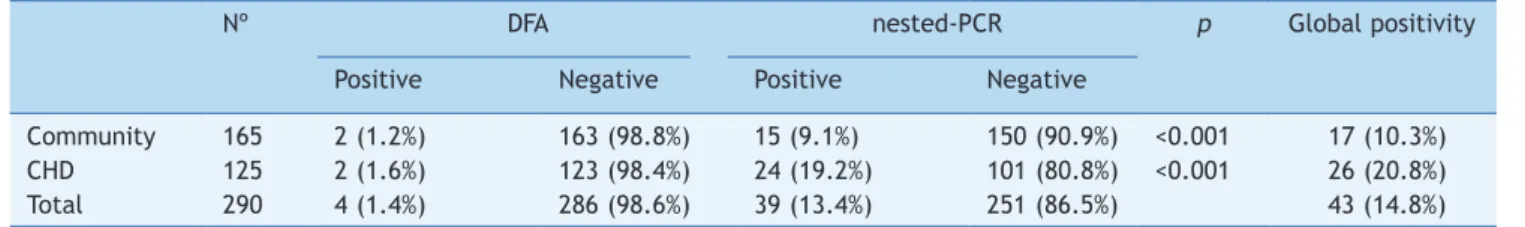
Among the 290 samples included during the study period, 41 (14.1%) were positive for at least one test: 17/165 (10.3%) children from community, 26/125 (20.8%) CHD chil-dren. Table 1 show the differential detection between assays according to different studied populations. Overall, 4/290 (0.7% of samples were DFA positive and 39/290 (13.40%) were nested-PCR positive. Nested-PCR presented a higher detection, statistically significant, among all stud-ied populations when compared to DFA (p<0.001). Comparing the children groups by molecular methods, CHD presented higher detection rate (p=0.02).
Analysis of the length of time between symptoms onset and the sample collection day showed a statistical differ-ence between DFA and nested-PCR (Table 2). Among commu-nity children, the mean ± standard deviation, median and range since symptoms onset were: 3.2±2.72; 3; 1-20 days; and among CHD children the mean ± standard deviation, median and range since symptoms onset were: 6.5±7.1; 5; 1-60 days. Nested-PCR compared to DFA detected a higher
Table 1 HAdV detection by DFA and nested-PCR among different studied populations.
Nº DFA nested-PCR p Global positivity
Positive Negative Positive Negative
Community 165 2 (1.2%) 163 (98.8%) 15 (9.1%) 150 (90.9%) <0.001 17 (10.3%)
CHD 125 2 (1.6%) 123 (98.4%) 24 (19.2%) 101 (80.8%) <0.001 26 (20.8%)
Total 290 4 (1.4%) 286 (98.6%) 39 (13.4%) 251 (86.5%) 43 (14.8%)
number of cases both for samples collected within 5 days of symptoms onset, and >5 days of symptoms onset.
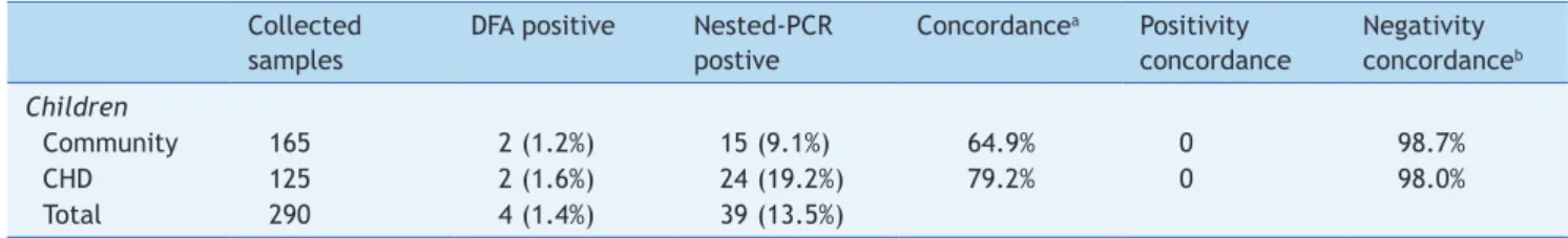
Table 3 shows the agreement between the evaluated tests. The general agreement was low, mainly among com-munity children. The concordance regarding negative results was high for both studied populations.
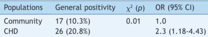
Table 4 shows the result of the univariate Odds Ratio analysis of HAdV occurrence among different studied popu-lations. The response variable was the presence or absence of HAdV, and the independent variable was the studied group: community children and CHD children. CHD children presented approximately two times more chance to
acquired HAdV infection when compared to community
children.
Discussion
Among community children, the adenovirus detection rate was similar to that described in the national literature, with rates ranging from 6% to 7.1%.13-16 A recent Brazilian
study evaluating 1,121 samples collected from hospitalized infants found adenovirus occurrence in 15.8%, which cor-roborates the findings of the present study.10 International
studies report HAdV occurrence ranging from 2%-3.8% among pediatric populations, which is slightly lower when
compared to our results.20-22 Studies with CHD children
demonstrate respiratory viruses circulation, with relative high occurrence rates, showing that these agents must be considered in clinical and diagnostic practice.22 The present
study is the first to describe HAdV respiratory infection in Brazilian children with congenital heart disease. The HAdV detection rate in CHD children was higher (20.8%) when
compared to community children (10.3%). It is noteworthy that, among CHD children sample collection was accom-plished during routine consultation. If the patient present-ed respiratory infection during the schpresent-edulpresent-ed appointment, the sample was collected. Therefore the rate found could be underestimated, because probable cases may have been lost due to lack of active demand from patients. One hypothesis for the higher detection can be related to comorbidities presence among this children.23
When evaluating the two tests used in this study some
important questions for both clinicians and hospital admin -istrators are raised: Which is the best test? Up to which day is ideal to collect the sample? Is there a need to repeat the test? Which is the ideal number of samples for laboratory routine flow? What is the impact of negative and positive tests for high-risk patients? Some of these aspects could be evaluated in the present study.
The analysis of the association between patient´s symp-tom onset and laboratorial test results showed that the molecular method was always more sensible than the DFA, and this difference was more evident after the fifth day of symptoms (p<0.001). Lower detection by DFA was relat-ed to low viral load in respiratory samples.24 The higher
detection rates among patients with more than five days of symptom were expected due to higher nested-PCR sen-sibility.25,26 The success of DFA test depend on some key
factors as good sample collection and experienced
labora-tory staff,10 while nested-PCR, based on nucleic acid
amplification, can detect a very low number of viral par-ticles.10,26
Univariate analysis showed that CHD children have a
higher risk of acquiring HAdV (Odds Ratio: 2.29), when
compared to community children. Utokaparch (2011)27
described that pediatric patients with lower respiratory infection presented higher viral load when compared to patients with upper respiratory infection. Echavarria et al.28 described that children are 2 to 3.5 times more likely
to become infected by HAdV than adults. These data cor-roborates the results of the present study.
One of the study limitations was the lack of another respiratory viruses analysis. Among HAdV negative samples, others virus could be present. As a result of the high sensi-tivity of the molecular methods, viral infections could be detected weeks after the symptoms resolution29,30 but, in
the present study, the included samples were collected from children presenting acute respiratory infection,
Table 2 Time of symptoms onset according to evaluated test. ≤5 days >5 days Positive Negative Positive Negative
% % % %
DFA 3 (1.4) 211 (98.6) 1 (1.3) 75 (98.7) Nested-PCR 24 (11.2) 190 (88.8) 15 (19.7) 61 (80.3)
p <0.001 <0.001
Total 214 76
DFA, direct luorescence assay; PCR, Polymerase Chain Reaction.
Table 3 Agreement assessment between the tests. Collected
samples
DFA positive Nested-PCR postive
Concordancea Positivity
concordance
Negativity concordanceb
Children
Community 165 2 (1.2%) 15 (9.1%) 64.9% 0 98.7%
CHD 125 2 (1.6%) 24 (19.2%) 79.2% 0 98.0%
Total 290 4 (1.4%) 39 (13.5%)
CHD, children with congenital heart disease; PCR, Polymerase Chain Reaction.
a Concordance calculated by method to compare two tests when gold standard method was not known.
decreasing the chance of a past infection detection. Another study limitation was the lack of sample collection during 2006; however the main idea of the study was to understand the occurrence of the virus in the community (community children), and in a population with at high risk of complications after a viral respiratory infection (CHD children). Some studies described an occurrence of adeno-virus during different years, and throughout the year,31,32
with similar occurrence patterns; therefore the collection of samples during different years apparently did not com-promise the purpose of the study.
Three parameters can indicate the validity of the diag-nostic test choice, the cost per sample, the turnaround time (time to provide the result) and the test reliability. In a study published in 2009 Mahony et al.33 compared the cost
of DFA versus molecular technique for diagnosing respirato -ry virus infection, and they described that molecular test was slightly more expensive but, at the same time, was highly cost-effective due to reduction in hospitalization time. Different studies described the turnaround time of DFA and PCR, with both tests presenting the ability to pro-vide results in one day.34,35 Gharabaghi et al.34 compared the
sensitivity and specificity of DFA and commercial molecular assay. This study showed lower sensitivity of DFA test in comparison to molecular testing for all surveyed virus, with adenovirus presenting the lowest sensitivity (38.1%)34
We suggest that routine surveillance should be performed in high-risk patients by molecular methods, improving diag-nostic flow and efficiency. Our results also demonstrated that in samples with more than five days of symptom onset nested-PCR might be the best option.
Funding
CNPq study grant (DP).
Conlicts of interest
The authors declare no conflicts of interest.
References
1. Lu MP, Ma LY, Zheng Q, Dong LL, Chen ZM. Clinical characteristics of adenovirus associated lower respiratory tract infection in children. World J Pediatr. 2013;9:346-9.
2. Horwitz MS. Adenovirus immunoregulatory genes and their cellular targets. Virology. 2001;279:1-8.
3. Bellau-Pujol S, Vabret A, Legrand L, Dina J, Gouarin S, Petitjean-Lecherbonnier J, et al. Development of three multiplex RT-PCR assays for the detection of 12 respiratory RNA viruses. J Virol Methods. 2005;126:53-63.
4. Weinberg A, Brewster L, Clark J, Simoes E, ARIVAC consortium. Evaluation of R-Mix shell vials for the diagnosis of viral respiratory tract infections. J Clin Virol. 2004;30:100-5. 5. Alsaleh AN, Grimwood K, Sloots TP, Whiley DM. A retrospective
performance evaluation of an adenovirus real-time PCR assay. J Med Virol. 2013;86:795-801.
6. Gilbert LL, Dakhama A, Bone BM, Thomas EE, Hegele RG. Diagnosis of viral respiratory tract infections in children by using a reverse transcription-PCR panel. J Clin Microbiol. 1996;34:140-3.
7. Freymuth F, Eugene G, Vabret A, Petitjean J, Gennetay E, Brouard J, et al. Detection of respiratory syncytial virus by reverse transcription-PCR and hybridization with a DNA enzyme immunoassay. J Clin Microbiol. 1995;33:3352-5.
8. Kuypers J, Wright N, Morrow R. Evaluation of quantitative and type-speciic real-time RT-PCR assays for detection of respiratory syncytial virus in respiratory specimens from children. J Clin Virol. 2004;31:123-9.
9. Templeton KE, Scheltinga SA, Beersma MF, Kroes AC, Claas EC. Rapid and sensitive method using multiplex real-time PCR for diagnosis of infections by inluenza a and inluenza B viruses, respiratory syncytial virus, and parainluenza viruses 1, 2, 3, and 4. J Clin Microbiol. 2004;42:1564-9.
10. Ferone EA, Berezin EN, Durigon GS, Finelli C, Felício MC, Storni JG, et al. Clinical and epidemiological aspects related to the detection of adenovirus or respiratory syncytial virus in infants hospitalized for acute lower respiratory tract infection. J Pediatr (Rio J). 2014;90:42-9.
11. Moura FE, Mesquita JR, Portes SA, Ramos EA, Siqueira MM. Antigenic and genomic characterization of adenovirus associated to respiratory infections in children living in Northeast Brazil. Mem Inst Oswaldo Cruz. 2007;102:937-41. 12. Luiz LN, Leite JP, Yokosawa J, Carneiro BM, Pereira Filho E,
Oliveira TF, et al. Molecular characterization of adenoviruses from children presenting with acute respiratory disease in Uberlândia, Minas Gerais Brazil, and detection of an isolate genetically related to feline adenovirus. Mem Inst Oswaldo Cruz. 2010;105:712-6.
13. Walsh EE, Falsey AR, Swinburne IA, Formica MA. Reverse transcription polymerase chain reaction (RT-PCR) for diagnosis of respiratory syncytial virus infection in adults: use of a single-tube ‘‘hanging droplet’’ nested PCR. J Med Virol. 2001;63:259-63.
14. Straliotto SM, Siqueira MM, Muller RL, Fischer GB, Cunha ML, Nestor SM. Viral etiology of acute respiratory infections among children in Porto Alegre, RS Brazil. Rev Soc Bras Med Trop. 2002;35:283-91.
15. Thomazelli LM, Vieira S, Leal AL, Sousa TS, Oliveira DB, Golono MA, et al. Surveillance of eight respiratory viruses in clinical samples of pediatric patients in southeast Brazil. J Pediatr (Rio J). 2007;83:422-8.
16. Bellei N, Carraro E, Perosa AH, Benica D, Granato CF. Inluenza and rhinovirus infecrtions among health-care workers. Respirology. 2007;12:100-3.
17. Allard A, Albinsson B, Wadell G. Rapid typing of human adenovirus by a general PCR combined with restriction endonuclease analysis. J Clin Microbiol. 2001;39:498-505. 18. Abd-Jamil J, Teoh BT, Hassan EH, Roslan N, Abubakar S.
Molecular identiication of adenovirus causing respiratory tract infection in pediatric patients at the University of Malaya Medical Center. BMC Pediatr. 2010;10:46.
19. Altman DG. Practical Statistics for Medical Ressearch. London: Chapman & Hall; 1991.
Table 4 Univariate Odds Ratio analysis of HAdV occurrence among studied populations.
Populations General positivity 2 (p) OR (95% CI)
Community 17 (10.3%) 0.01 1.0
CHD 26 (20.8%) 2.3 (1.18-4.43)
20. Berkley JA, Munywoki P, Ngama M, Kazungu S, Abwao J, Bett A, et al. Viral etiology of severe pneumonia among Kenyan infants and children. JAMA. 2010;303:2051-7.
21. Herrera-Rodríguez DH, De la Hoz F, Mariño C, Ramirez E, López JD, Vélez C. Adenovirus in children under ive years of age Circulation patterns and clinical and epidemiological characteristics in Colombia, 1997-2003. Rev Salud Publica (Bogota). 2007;9:420-9.
22. Medrano Lópes L, García-Guereta L, CIVIC Study Group. Community-acquired respiratory infections in young children with congenital heart diseases in the palivizumab era: the Spanish 4-season civic epidemiologic study. Pediatr Infect Dis J. 2010;29:1077-82.
23. Fujimoto T, Okafuji T, Okafuji T, Ito M, Nukuzuma S, ChikahiraM, et al. Evaluation of a bedside immunochromatographic test for detection of adenovirus in respiratory samples, by comparison to virus isolation, PCR, and real-time PCR. J Clin Microbiol. 2004;42:5489-92.
24. Sanalios RB. Análise comparativa das técnicas de imunoluorescência indireta, isolamento em cultura e reação em cadeia polimerase no diagnóstico de infecções respiratórias por adenovirus e vírus respiratório sincicial. São Paulo: ICB-USP; 2004.
25. Stroparo E, Cruz CR, Debur MC, Vidal LR, Nogueira MB, Almeida SM, et al. Adenovirus respiratory infection: signiicant increase in diagnosis using PCR comparing with antigen detection and culture methods. Rev Inst Med Trop Sao Paulo. 2010;52:317-21. 26. Doing KM, Jerkofsky MA, Dow EG, Jellison JA. Use of luorescent-antibody staining of cytocentrifuge-prepared smears in combination with cell culture for direct detection of respiratory viruses. J Clin Microbiol. 1998;36:2112-4.
27. Utokaparch S, Marchant D, Gosselink JV, McDonough JE, Thomas EE, Hogg JC, et al. The relationship between respiratory viral
loads and diagnosis in children presenting to a pediatric hospital emergency department. Pediatr Infect Dis J. 2011;30:e18-23.
28. Echavarría M. Adenoviruses in immunocompromised hosts. Clin Microbiol Rev. 2008;21:704-15.
29. De Lima CR, Mirandolli TB, Carneiro LC, Tusset C, Romer CM, Andreolla HF, et al. Prolonged respiratory viral shedding in transplant patients. Transpl Infect Dis. 2014;16:165-9.
30. Peltola V, Waris M, Kainulainen L, Kero J, Ruuskanen O. Virus shedding after human rhinovirus infection in children, adults and patients with hypogammaglobulinaemia. Virology. 2013;19:E322-7.
31. Khor CS, Sam IC, Hooi PS, Quek KF, Chan YF. Epidemiology and seasonality of respiratory viral infections in hospitalized children in Kuala Lumpur Malaysia: a retrospective study of 27 years. BMC Pediatr. 2012;12:32.
32. Marcone D, Ellis A, Videla C, Ekstrom J, Ricarte C, Carballal G, et al. Viral etiology of acute respiratory infections in hospitalized and outpatient children in Buenos Aires, Argentina. Pediatr Infect Dis J. 2013;32:e105-10.
33. Mahony JB, Blackhouse G, Babwah J, Smieja M, Buracond S, Chong S, et al. Cost analysis of multiplex PCR testing for diagnosing respiratory virus infections. J Clin Microbiol. 2009;47:2812-7.
34. Gharabaghi F, Hawan A, Drews SJ, Richardson SE. Evaluation of multiple commercial molecular and conventional diagnosticas says for the detection of respiratory viruses in children. Clin Microbiol Infect. 2011;17:1900-6.