212
CASE REPORTSurgical resection of a cutaneous nodule
in the left foot caused by mycobacteria
Exérese cirúrgica de nódulo cutâneo no pé esquerdo causado por uma micobactéria
Neiva Aparecida Grazziotin1; Itamar Luís Gonçalves2; Clara Grazziotin3
First submission on 18/01/13; last submission on 19/03/13; accepted for publication on 03/05/13; published on 20/06/13
1. Master’s in Biological Sciences; professor in Mycology, Microbiology and Parasitology at Universidade Regional Integrada (URI) Erechim; biochemical pharmacist. 2. Graduate student in Pharmacy at URI Erechim; Scientiic Initiation scholarship at URI Erechim.
3. Doctor specialist in Dermatology.
ABSTRACT
Nontuberculous mycobacteria are etiologic agents of opportunistic human infections. Although they usually affect supericial tissues, infections in bones and joints have been described. The contamination is associated with increased environmental exposure. With appropriate therapy, the cases usually progress to complete recovery of the patient. This study reports the case of a patient who developed a cutaneous nodule in her left foot acquired when her skin was punctured by a ish. The anatomopathological examination revealed chronic central suppurative granulomatous dermo-hypodermal inlammation. Furthermore, the screening for resistant acid-fast bacilli was positive.
Key words:nontuberculous mycobacteria; mycobacteriosis; Mycobacterium sp.
J Bras Patol Med Lab, v. 49, n. 3, p. 212-215, junho 2013
INTRODUCTION
Mycobacteria include important pathogens such as
Mycobacterium turbeculosis and M. leprae. Approximately 150
species are indigenous in the human environment(7, 8). Several species
of nontuberculous mycobacterium have been reported in the states of Mato Grosso do Sul and Rio de Janeiro, Brazil(13, 15).
Mycobacterium marinum is among the pathogenic
mycobacteria. It was initially described in 1926 during an investigation into infectious diseases in marine ish(2). It has
worldwide distribution and it is generally associated with cutaneous and bone-articular lesions(9). Moreover, it is accountable
for approximately 150 new cases in the United States annually(5) .
M. marinum is a natural pathogen in ectothermic animals,
including amphibians and ish. In the latter they cause systemic granulomatous disease. Diseases caused by mycobacteria are among the most prevalent in ish(9, 14). It is particularly worrying
the fact that bacteria involved in ish bacteriosis are able to infect humans(4). Human infections caused by M. marinum produce
nodular or ulcerated cutaneous lesions in upper or lower limb extremities(12).
Delayed diagnosis is common and invasion of deeper structures such as synovium, bursae and bone occur in approximately a third of the reported cases(12). A rare case of long bone osteomyelitis
has been described in an immunosuppressed patient(16). The
contamination usually occurs through direct contact with ish or contaminated water, generally in the presence of a pre-existing wound or trauma(1, 4).
The present study describes the clinical progression of a patient who presented a wound caused by a ish. The microscopic exam of the affected area revealed the presence of resistant acid-fast bacilli (RAFB).
CASE REPORT
213
The lesion was caused by trauma during ishing, which occurred in a city in the north region of Rio Grande do Sul, Brazil.
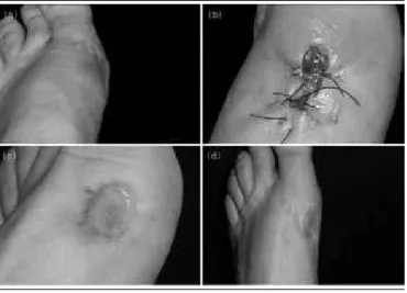
Fifteen days after the initial wound, the patient developed a small asymptomatic papule on the site. Two months afterwards, after trekking with sneakers, the lesion volume increased considerably, presenting a painful swollen erytematous nodule
(Figure 1). Shortly after the onset of these symptoms, a traumatologist raised the diagnostic hypothesis of bursitis and prescribed applying ice to the affected region concomitantly with the use of a non-steroid anti-inlammatory drug orally. The clinical progression is illustrated in Figure 1.
FIGURE 1 – Wound progression
(a) erytematous nodule observed three months after contact with etiologic agent; (b) aspect of the lesion after surgical excision; (c) aspect of the affected area approximately four months after surgical procedure; (d) affected foot ive years after infection.
FIGURE 2 – RAFB screening and anatomopathologic exam
(a) and (b) resistant acid-fast bacilli visualized under optical microscopy at 1,000×. Samples were obtained from the lesion site and underwent Ziehl-Neelsen staining; (c) and (d) suppurative granuloma at 40× and 400×, respectively.
RAFB: resistant acid-fast bacilli.
As the patient did not present any clinical improvement after prescribed treatment, microscopic, bacteriological and anatomopathologic exams were requested. Gram staining allowed to establish that there were no microorganisms in the site, though there were numerous leukocytes. Microscopic and bacteriological exams were negative. Moreover, there were no fungi in the direct exam and culture. The absence of microbiological indings led to RAFB screening through Ziehl-Neelsen staining, yielding positive results (Figure 2A and Figure 2B – Figure 2).
The patient received rifampicin 300 mg, which was administered every 12 hours during 3 days. Subsequently, the patient underwent total surgical excision (Figure 1B). The anatomopathologic results from the surgical specimen revealed chronic central suppurative granulomatous dermo-hypodermal inlammation (Figure 2C and Figure 2D). After surgical resection of the affected area, followed by epithelial grafts and hyperbaric treatment, the patient achieved full recovery with no relapses for ive years and eight months (Figure 1D).
DISCUSSION
Based on the patient’s clinical history, Mycobacterium marinum was considered the most probable infectious agent, which is found in a wide variety of aquatic habitats worldwide. Accordingly, people who work with aquaculture or have aquariums have a higher risk of acquiring this infection.
Most patients with infections caused by M. marinum respond
to antimicrobial treatment, though prolonged treatments are sometimes required for several months(1, 6, 17). A review study assessed
the clinical progression of 63 cases and concluded that the most commonly prescribed antibiotics were clarithromycin, tetracycline and rifampicin(3). The use of clarithromycin is effective for most
patients with cutaneous infections(10). Nonetheless, refractory cases
requiring several antibiotics and surgical procedures have been reported (18) as well as cases in which the infection has led to death(11).
The clinical progression of some cases reported by the literature is shown in the Table below.
In clinical practice, the diagnosis of cutaneous infection by
M. marinum is established on the basis of the clinical history,
lesion features, microbiological exam and molecular techniques. The contact with ish and aquariums plays a major role in the differential diagnosis, hence the need to investigate the patient’s exposure to aquatic habitats. The better understanding of this infection enables an early approach, which reduces the potentially high risk of deeper tissue involvement. Therefore, in this speciic case, the total surgical excision of a unique mycobacterial lesion was the treatment of choice.
Neiva Aparecida Grazziotin; Itamar Luís Gonçalves; Clara Grazziotin
A
C D
214
TABLE– Clinical progression of infections caused by M. marinum
Reference Age Gender Symptoms Contamination Therapy Outcome
1 45 M Draining sinuses in the
chest and arms Swimming in water park R, CL and AM
Improvement after a three-month treatment
6 60 M
Epithelial lesion in the right hand progressing to nodules that reached the
forearm
Cleaning aquarium
containing dead ish R and CL
Cured four months after
diagnosis
6 23 M Nodule on the dorsal
surface of the right hand
Skin lesion cleaning
aquarium R, E and CL
Cured three months after
diagnosis
11 67 M
Painful subcutaneous nodules on the right arm
and leg
Aquarium with tropical
ish and turtle R, E, CL and CP Died after 111 days
17 16 F Slow healing nodular lesion on the left leg Contact with barnacle (marine crustacean) E and CL Full recovery in four
months M: male; F: female; R: rifampicin, E: ethambutol, CL: clarithromycin, CP: ciproloxacin, AM: amikacin.
REFERENCES
1. AFZAL, A. et al. Mycobacterium marinum infection: a case report.
JPAD, v. 19, p. 48-51, 2009.
2. ARONSON, J. D. Spontaneous tuberculosis in salt water ish. J Infect Dis,
v. 39, p. 315-20, 1926.
3. AUBRY, A. et al. Sixty-three cases of Mycobacterium marinum
infection: clinical features, treatment, and antibiotic susceptibility of causative isolates. Arch Intern Med, v. 162, n. 15, p. 1746-52, 2002.
4. BOWSER, P. R. Fish diseases: mycobacteriosis of ish. NRAC Publication, v. 202, p. 1-3, 2002.
5. DOBOS, K. M. et al. Emergence of a unique group of necrotizing mycobacterial diseases. Emerg Infect Dis, v. 5, n. 3, p. 367-78, 1999.
6. EL AMRANI, M. H. et al. Upper extremity Mycobacterium marinum
infection. Orthop Traumatol Surg Res, v. 96, n. 6, p. 706-11, 2010. 7. EUZÉBY, J. P. List of bacterial names with standing in nomenclature: a folder available on the internet. Int J Syst Bacteriol, v. 47, n. 2, p. 590-2, 1997.
RESUMO
Micobactérias não tuberculosas são agentes causadores de infecções oportunistas no homem. Embora, geralmente, afetem tecidos supericiais, infecções em ossos e articulações têm sido descritas. O contágio está associado ao aumento da exposição do homem ao meio ambiente. Diante da terapêutica adequada, os casos normalmente evoluem com recuperação total do indivíduo. Este estudo descreve o caso clínico de um paciente que apresentou nódulo cutâneo no pé esquerdo após traumatismo com um peixe. O exame anatomopatológico revelou inlamação crônica granulomatosa centralmente supurativa dermo-hipodérmica e a pesquisa de bacilos álcool-ácido resistentes (BAAR) foi positiva.
Unitermos: micobactérias não turbeculosa; micobacteriose; Mycobacterium sp.
8. FALKINHAM, J. Nontuberculous mycobacteria in the environment. Clin
Chest Med, v. 23, n. 3, p. 529-51, 2002.
9. FARD, S. M. H. et al. Mycobacterium marinum as a cause of skin chronic granulomatous in the hand. Casp J Intern Med, v. 2, n. 1,
p. 198-200, 2011.
10. FENG, Y. et al. Outbreak of a cutaneous Mycobacterium marinum
infection in Jiangsu Haian, China. Diagn Microbiol Infect Dis, v. 71, n. 3, p. 267-72, 2011.
11. IMAKADO, S.; KOJIMA, Y.; MORIMOTO. S. Disseminated
Mycobacterium marinum infection in a patient with diabetic
nephropathy. Diabetes Res Clin Pract, v. 83, n. 2, p. 35-6, 2009.
12. LAHEY, T. Invasive Mycobacterium marinum infections. Emerg
Infect Dis, v. 9, n. 11, p. 1496-8, 2003.
13. MORAES, P. R. S. et al. Identiication of non-tuberculous mycobacteria from the Central Public Health Laboratory from Mato Grosso do Sul and analysis of clinical relevance. Braz J Microbiol, v. 39, n. 2, p. 268-72, 2008.
14. ROMANO, L. A.; SAMPAIO, L. A.; TESSER, M. B. Micobacteriose por
Mycobacterium marinum em linguado Paralichthys orbignyanus e
215
MAILING ADDRESSNeiva Aparecida Grazziotin
Avenida Sete de Setembro, 1.621; Fátima; CEP: 99700-000; Erechim-RS, Brazil; e-mail: neivagra@uri.com.br..
em barber goby Elacatinus igaro: diagnóstico histopatológico e
imuno-histoquímico. Pesq Vet Bras, v. 32, n. 3, p. 254-8, 2012.
15. SENNA, S. G. et al. Identiicação de micobactérias não tuberculosas isoladas de sítios estéreis em pacientes em um hospital universitário na cidade do Rio de Janeiro. J Bras Pneumol, v. 37, n. 4, p. 521-6, 2011.
16. SIVAN, M. et al. Ostéomyélite à Mycobacterium marinum d’un
os long Mycobacterium marinum osteomyelitis of a long bone. Rev
Rhum, v. 75, n. 9, p. 875-8, 2008.
17. TEBRUEGGE, M. et al. Mycobacterium marinum infection following kayaking injury. Int J Infect Dis, v. 14, n. 3, p. 305-6, 2010.
18. VAN SEYMORTIER, P.; VERELLEN, K.; DE JONGE, I. Mycobacterium
marinum causing tenosynovitis. Fish tank inger. Acta Orthop Belg,
