Fisioter. Mov., Curitiba, v. 28, n. 1, p. 169-186, Jan./Mar. 2015 Licenciado sob uma Licença Creative Commons DOI: http://dx.doi.org.10.1590/0103-5150.028.001.AR02
[T]
Assessment of the strength of the trunk and upper limb muscles
in stroke subjects with portable dynamometry: a literature review
[I]
Avaliação da força muscular pós-AVE pela dinamometria
portátil: uma revisão da literatura
[A]
Júlia Caetano Martins, Luci Fuscaldi Teixeira-Salmela, Larissa Tavares Aguiar, Lucas Araújo Castro e Souza, Eliza Maria Lara, Christina Danielli Coelho de Morais Faria*
Universidade Federal de Minas Gerais (UFMG), Belo Horizonte, MG, Brazil
[R] Abstract
Introduction: Clinical measurements of strength in stroke subjects are usually performed and portable dyna-mometers are one of the most employed instruments. Objective: To verify the standardization procedures of the methods used to assess the strength of the trunk and upper limb muscles with portable dynamometers in stroke subjects, as well as to assess the psychometric properties which were already investigated. Materials and methods: An extensive search was performed on the MEDLINE, SciELO, LILACS, and PEDro databases, by combining specific key words, followed by active manual searches by two independent researchers. Results and discussion: Fifty-eight studies were included: three related to the trunk and 55 to the upper limb mus-cles, including handgrip and pinch strength assessments. The most investigated muscular groups were hand-grip, elbow flexors/extensors, wrist extensors, and lateral pinch. Nine studies reported adequate reliability
* JCM: MSc, e-mail: [email protected] LFTS: Ph.D, e-mail: [email protected]
170
levels and the seated position was employed in the majority of the studies which assessed trunk, handgrip, and pinch strength, while the supine position was used for the other muscular groups. The number of trials most used was three, while the reported contractions and rest times were variable. Final considerations: Most studies reported the positioning and/or the data collection protocols; however, there was no consensus on the standardization procedures. The only investigated psychometric property was reliability. Few studies evaluated the trunk muscles and other psychometric properties.
[P]
Keywords: Dynamometer. Trunk. Upper limbs. Reliability. Validity. [B]
Resumo
Introdução: A mensuração da força muscular em indivíduos acometidos pelo Acidente Vascular Encefálico (AVE) é comumente realizada na clínica, sendo os dinamômetros portáteis os instrumentos mais utilizados para tanto. Objetivo: Verificar se há uma padronização dos métodos utilizados para avaliação da força mus -cular de tronco e membros superiores (MMSS) com o uso de dinamômetros portáteis em indivíduos pós-AVE,
bem como verificar quais propriedades de medida já foram investigadas. Materiais e métodos: As buscas foram realizadas nas bases de dados MEDLINE, SciELO, LILACS e PEDro com combinação de termos específi
-cos, seguidas de busca manual ativa. A seleção dos estudos e a extração das informações foram realizadas por dois examinadores independentes. Resultados e discussão: Foram incluídos 58 estudos (três de tronco e 55
de MMSS, incluindo preensão manual e pinça). Os grupos musculares mais avaliados foram preensão manual, flexores de cotovelo, extensores de punho, extensores de cotovelo e pinça lateral. Nove estudos reportaram confiabilidade adequada do método. A maioria dos estudos que avaliaram os músculos de tronco, de preensão manual e de pinça utilizou a postura sentada, enquanto o decúbito dorsal foi mais utilizado na avaliação dos demais músculos. O número de repetições mais utilizado foi três, já o tempo de contração e o período de re -pouso variaram entre os estudos. Considerações finais: A maioria dos estudos relatou o posicionamento e/ou
o protocolo de coleta, porém não houve uma padronização. A única propriedade de medida investigada foi a confiabilidade. Poucos estudos avaliaram os músculos de tronco e as outras propriedades de medida. [K]
Palavras-chave: Dinamômetro. Tronco. Membros superiors. Confiabilidade. Validade.
Introduction
Stroke is an important cause of disabilities. Every year, thousands of working-age adults become par-tially or totally disabled by this health condition (1), which results in emotional distresses for the patients and their families and socio-economic impact on the health systems (2). Stroke subjects may demonstrate several impairments, being the motor ones the most common (3-5) and those that affect the performance of daily life activities (6).
Among the observed motor impairments, muscu-lar weakness has shown significant associations with activity limitations (3, 7, 8) and social participation restriction (7, 8). Specifically, weakness of the upper limb (UL) (9-13) and trunk (14, 15) muscles, which are involved in the performance of many basic, instru-mental, work, and leisure activities, lead to important
functional limitations. About 70% of the subjects with paresis of the UL muscles have some degree of functional limitation (13, 16, 17). Moreover, after the onset of the hemiparesis, stroke subjects demonstrate difficulties in moving and controlling their trunk (18), which affect their balance, transfer, gait performance, and independence in many daily activities (15). Thus, the strength of the UL and trunk muscles strength become an important outcome to be evaluated and considered within the clinical decision-making pro-cess for the rehabilitation of stroke subjects.
171
objective and sensitive measures to detect chang-es in strength, such as the portable and isokinetic dynamometers (19). Portable devices, such as the handheld, handgrip, and pinch dynamometers, are more easily applied within clinical settings, when compared to the isokinetic dynamometer (21).
The portable dynamometers, which record the maximal isometric force generated during an iso-metric contraction (22), have been used to assess the strength of the trunk (14, 23), UL (24-27), and handgrip (9, 12, 28) and pinch (29-31) muscles of subjects with stroke. They are practical devices that can be placed between the examiner's hand and the muscle group to be tested, similar to the MMT assess-ment (32) or used with the subject exerting force directly on the equipment, in the case of handgrip and pinch assessments (33-36). Furthermore, they provide quantitative measures of strength, which has an important advantage, compared to the MMT assessment (37).
Studies have reported several factors that could influence the measures obtained with portable dy -namometers (21, 33), such as positioning of the subjects and the device, number of repetitions, con-traction and rest time, prior demonstration and fa-miliarization with the procedures, and supply of ver-bal or visual encouragement. Other factors should also be considered when selecting these devices for the assessment of strength in subjects with stroke, such as unilateral or bilateral assessments and the measurement properties already established for this specific population. Before the portable dynamom -eters be appropriately employed for the measure-ment of the strength of the UL and trunk muscles of subjects with stroke, it is necessary to standardize the assessment procedures and to ensure that they show appropriate psychometric properties. Within this context, the aims of this study were to investi-gate whether there were standardized protocols for the use of the portable dynamometers for the assess-ment of the strength of the UL and trunk muscles, including handgrip and pinch strength, in subjects with stroke, and verify the investigated psychomet-ric properties. Based upon the results of this review, it will be possible to determine the most commonly used protocols and the psychometric properties, to allow a scientifically-based clinical decision making regarding the use of portable dynamometry for the assessment of the UL and trunk muscles in subjects with stroke.
Methods
Initially, electronic searches were performed in MEDLINE (via PUBMED), SciELO, LILACS, and PEDro databases. The MEDLINE search strategy followed guidelines developed by the Cochrane group (38), which was adjusted for the other databases, using descriptors related to UL, trunk, and handheld dyna-mometry. The search terms used for the UL included words related to handgrip and pinch, were: upper limb, upper extremity, hand grip, palmar grip, grip, grasp, hand strength, pinch, hand, and palmar. For the trunk, the search terms included word related to back, trunk, abdomen, and thorax. Finally, for the dynamometry, the following words were used: Dynamometer, pinch gauge, pinch strength, Preston pinch gauge, Jamar, handheld dynamometer, and muscle strength.
To be included, the studies should clearly report in the methodology section that the strength of the trunk or UL muscles, including handgrip or pinch strength, with portable dynamometers was assessed in subjects with stroke. There were no restrictions regarding the language of publication and all studies published until November 2011 were included.
The selection of the studies was performed by two independent examiners, following three steps, as recommended and commonly used (39-41). The first step consisted of reading the titles and excluding the that clearly did not meet the established criteria (39-41). Then, the selected abstracts were analyzed and those that did not meet the inclusion criteria were also excluded (39-41). The last step consisted of reading the full papers. An active manual search from all selected studies was also performed, following the same previously described criteria and procedures.
Results
172
days to 30 years. The studies which assessed pinch strength included 536 subjects of both sexes, who had ages ranging from 16 to 94 years and the time since the onset of the stroke ranging from two days to 23 years. The studies which evaluated other UL muscles included 468 individuals of both sexes, who had ages ranging from 17 to 89 years and the mean time since the onset of stroke ranging from two days to 30 years.
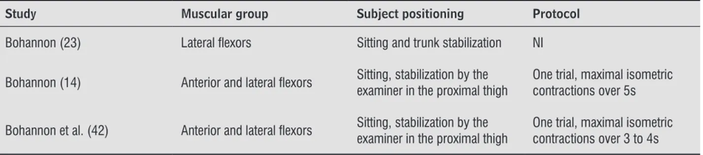
Regarding the trunk muscles (Table 1), the ante-rior flexors were evaluated in two studies (66.7%) (14, 42) and the lateral flexors in all three included studies (100%) (14, 23, 42). In all studies, the seated position was used and two reported the data collec-tion protocols, describing the number of trials and the duration of the isometric contractions (14, 42). One study (33.3%) performed unilateral assessment of the lateral flexors (23) and two (66.7%) bilateral (14, 42).
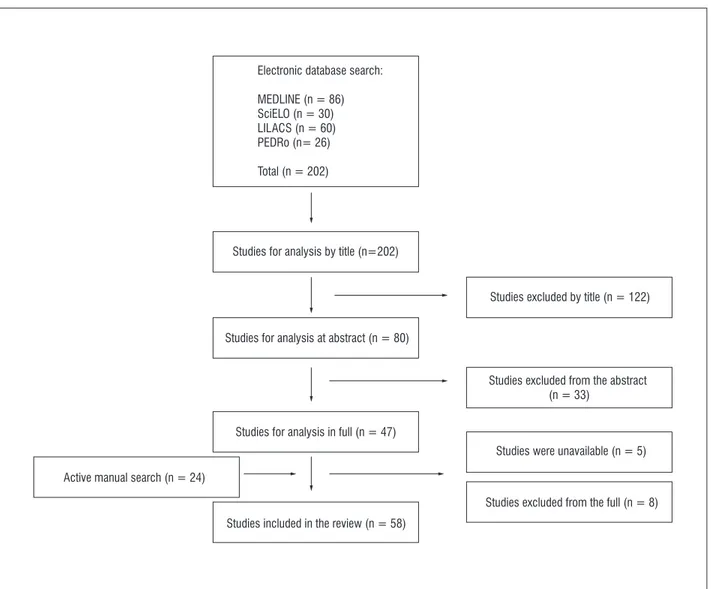
34 studies, 24 others were included. Therefore, a total of 58 studies fulfilled all eligibility criteria and were included in this review (Figure 1).
Amongst the 58 included studies, three evaluated the strength of the trunk (5.17%) (14, 23, 42), while 55 (94.83%) analyzed the strength of the UL muscles, including handgrip and pinch strength. Out of the 55, 41 assessed handgrip (4, 9, 12, 28, 31, 43-78), 15 pinch (29-31, 47, 54, 55, 57, 58, 61, 71, 76, 78-81), and 17 the strength of other UL muscles (24-27, 46-48, 55, 59, 63, 70, 76, 82-86). The studies that measured the strength of the trunk muscles included 59 individuals of both sexes, who had ages ranging from 27 to 87 years and were at the acute stages (three to 27 days post-stroke). Those that assessed handgrip strength included 1,408 individuals of both sexes, who had ages ranging from 16 to 93 years and the time since the onset of stroke ranging from two
Figure 1 - Flow chart of the selection of the studies
Electronic database search:
MEDLINE (n = 86) SciELO (n = 30) LILACS (n = 60) PEDRo (n= 26)
Total (n = 202)
Studies for analysis by title (n=202)
Studies for analysis at abstract (n = 80)
Studies for analysis in full (n = 47)
Studies included in the review (n = 58)
Studies excluded by title (n = 122)
Studies excluded from the abstract (n = 33)
Studies were unavailable (n = 5)
173
Table 1 - Data extraction of the studies which assessed the strength of the trunk muscles in subjects with stroke with portable dynamometers
Study Muscular group Subject positioning Protocol
Bohannon (23) Lateral lexors Sitting and trunk stabilization NI
Bohannon (14) Anterior and lateral lexors Sitting, stabilization by the examiner in the proximal thigh One trial, maximal isometric contractions over 5s
Bohannon et al. (42) Anterior and lateral lexors Sitting, stabilization by the examiner in the proximal thigh
One trial, maximal isometric contractions over 3 to 4s
Note: NI = not informed.
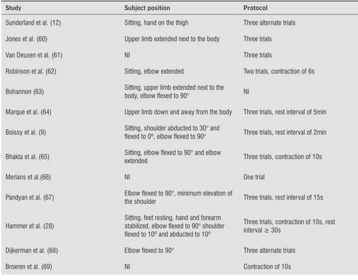
From the 41 studies that evaluated handgrip strength, 24 (58.54%) provided detailed informa-tion regarding the subjects’ posiinforma-tioning or the data collection protocols (Table 2). Since 17 studies did not provide this information (43-59), they were not included in the table. As can be seen in Table 2, the seated position was employed in 11 studies (73.33%) (9, 12, 28, 31, 62, 63, 65, 70, 72, 76, 77). Twenty-two studies reported the number of trials and the major-ity of them performed three trials (72.73%) (9, 12, 28, 31, 60, 61, 64, 65, 67, 68, 71-73, 75, 76, 78). The duration of the maximal isometric contractions was reported by five studies, and 10 seconds was the time most commonly used (60%) (28, 65, 69). The resting time was reported in seven studies with alternated measurements between the UL being the most em-ployed method (42.9%) (12, 68, 74). Eighteen stud-ies (69.2%) repoted bilateral measures of handgrip strength (4, 9, 12, 28, 47, 48, 52, 54-56, 59, 61, 62, 68, 72, 74, 76, 77), while eight (30.8%) only assessed the paretic limb (31, 45, 58, 64, 65, 70, 71, 73).
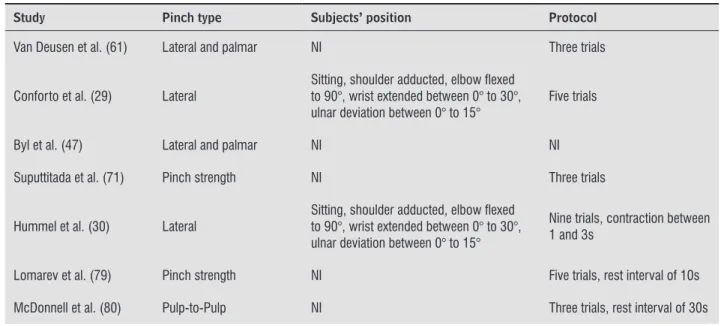
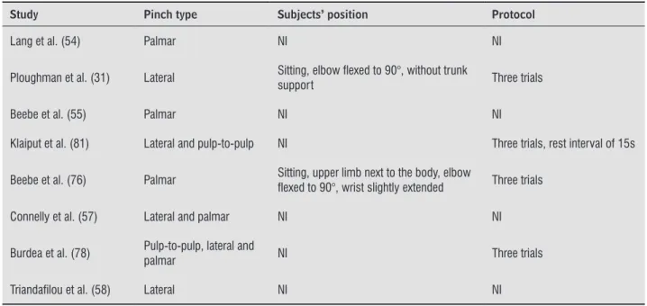
From the 15 studies that assessed pinch strength, lateral pinch was evaluated by nine (29-31, 47, 57, 58, 61, 78, 81), the palmar pinch by seven (47, 54, 55, 57, 61, 76, 78), pulp-to-pulp pinch by two (78, 80) and tip-to-tip pinch by one (81). Two studies did not specify the type of pinch that was measured (71, 79). As observed in Table 3, five studies did not provide in -formation regarding the subjects’ positioning or data collection protocols (47, 54, 55, 57, 58). The seated position was adopted by all four studies that provided information regarding the participants’ positioning (29-31, 76). Ten studies reported the number of tri-als, and seven used three trials (70%) (31, 61, 71, 76, 78, 80, 81). The duration of the contractions was reported by only one study (30), and varied from 1
to 3 seconds. Three studies reported different rest-ing times between the trials: 10s (79), 15s (81), and 30s (80). Six studies (50%) assessed bilateral pinch strength (47, 54, 55, 61, 76, 80) and six (50%) only the paretic side (29-31, 58, 71, 81).
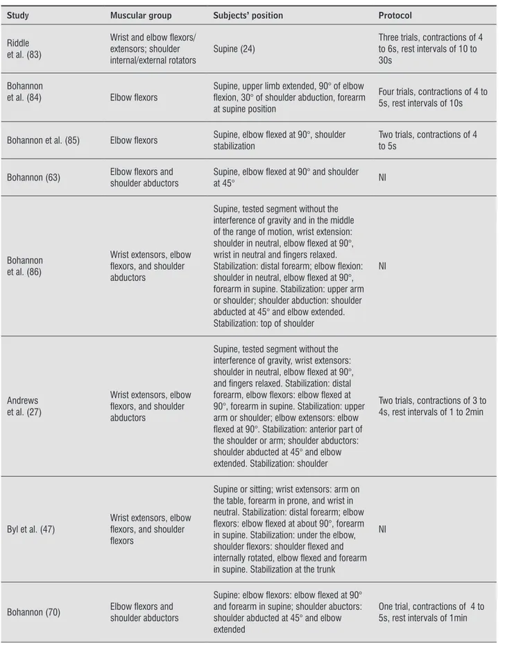
The main evaluated other muscular groups of the UL (except for handgrip and pinch strength) were the wrist flexors: eight studies (24, 46, 48, 55, 59, 76, 82, 83); wrist extensors: 13 studies (24-27, 46-48, 55, 59, 76, 82, 83, 86); elbow flexors: 17 studies (24-27, 46-48, 55, 59, 63, 70, 76, 82-86); elbow extensors: ten studies (24, 25, 27, 46, 48, 55, 59, 76, 82, 83); shoul -der flexors: nine studies (24, 25, 46-48, 55, 59, 76, 82); shoulder extensors: eight studies (24, 25, 46, 48, 55, 59, 76, 82); internal shoulder rotators: five studies (24, 25, 59, 82, 83); external shoulder rotators: six studies (24-26, 59, 82, 83); and shoulder abductors: nine studies (24, 25, 27, 46, 48, 63, 70, 82, 86). Other muscular groups, such as the shoulder adductors (24, 82) and flexors (55, 76) and extensors of the index finger (55, 76) were evaluated in two studies.
174
UL muscles reported that the device was positioned perpendicular to the evaluated segment in its distal region, and stabilization was provided to the proximal region (24-27, 55, 63, 70, 76, 82-85).
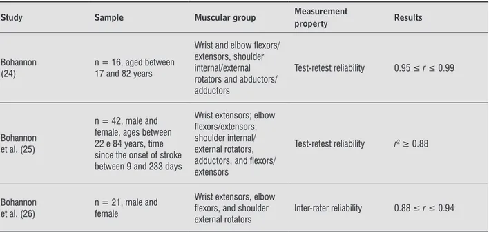
Out of the 58 included studies, nine reported the psychometric properties of the portable dynamom-eters. In these studies, the strength of the trunk (42) and some UL muscles (24-26, 63, 83-85), including handgrip (9, 28, 63), were evaluated, as shown in Table 5. All studies assessed the reliability, five re -ported test-retest (9, 24, 25, 28, 63), one intra-rater (85), two inter-rater (26, 42), and one intra- and inter-session reliabilities (83). All studies that inves-tigated the psychometric properties of the dynamom-eter, except one (28), reported the magnitudes of the correlation coefficient values above 0.80, indicating excellent reliability (87).
describe the subjects’ positioning or data collection protocol were not included. Most studies, 11 (73.3%), performed bilateral measures (25, 27, 46-48, 55, 59, 76, 82, 83, 86) and four (26.7%) unilateral (26, 70, 84, 85), three of the paretic hand.
Regarding the positioning of the dynamometers, two studies on trunk assessment reported that the device was placed in the lower portion of the jugu-lar notch for the anterior trunk flexors, and in the lateral lower portion of the acromion for the lateral trunk flexors (14, 42). For the assessment of handgrip strength, the device was positioned between the palm of the hand and the fingers (12, 65) with its handle on the second position (70, 77). For the evaluation of pinch strength, the end portion of the device was placed between the thumb and the finger involved in the assessed pinch (29, 30, 81). Studies of other
Table 2 - Data extraction of the 24 studies which assessed handgrip strength in subjects with stroke with portable dyna-mometers and provided information regarding the subject’ positioning or the data collection protocol
Study Subject position Protocol
Sunderland et al. (12) Sitting, hand on the thigh Three alternate trials
Jones et al. (60) Upper limb extended next to the body Three trials
Van Deusen et al. (61) NI Three trials
Robinson et al. (62) Sitting, elbow extended Two trials, contraction of 6s
Bohannon (63) Sitting, upper limb extended next to the body, elbow lexed to 90° NI
Marque et al. (64) Upper limb down and away from the body Three trials, rest interval of 5min
Boissy et al. (9) Sitting, shoulder abducted to 30° and lexed to 0º, elbow lexed to 90° Three trials, rest interval of 2min
Bhakta et al. (65) Sitting, elbow lexed to 90° and elbow extended Three trials, contraction of 10s
Merians et al.(66) NI One trial
Pandyan et al. (67) Elbow lexed to 90°, minimum elevation of the shoulder Three trials, rest interval of 15s
Hammer et al. (28) Sitting, feet resting, hand and forearm stabilized, elbow lexed to 90° shoulder lexed to 10º and abducted to 10º
Three trials, contraction of 10s, rest interval ≥ 30s
Dijkerman et al. (68) Elbow lexed to 90° Three alternate trials
Broeren et al. (69) NI Contraction of 10s
175
Table 2 - Data extraction of the 24 studies which assessed handgrip strength in subjects with stroke with portable dyna-mometers and provided information regarding the subject’ positioning or the data collection protocol
Study Subject position Protocol
Bohannon (70)
Sitting, shoulder adducted, elbow lexed to 90°, wrist extended between 0° to 30°, ulnar deviation between 0° to 15°
One trial
Suputtitada et al. (71) NI Three trials
Kamper et al. (4) NI Two trials
Wolf et al. (72) Sitting, elbow lexed to 90° Three trials, contraction of 3s
Restemeyer et al. (73) NI Three trials
Ploughman et al. (31) Sitting, elbow lexed to 90°, without trunk support Three trials
Gosselin et al. (74) NI Two alternate trials
Kang et al. (75) NI Three trials
Beebe et al. (76) Sitting, upper limb next to the body, elbow lexed to 90°, wrist slightly extended Three trials
Bohannon (77) Sitting, shoulder adduced, elbow lexed to 90° One trial
Burdea et al. (78) NI Three trials
Note: NI = not informed. The assessment of strength was performed with isometric contractions in all studies.
(Conclusion)
Table 3 - Data extraction of the 15 studies which assessed pinch strength in subjects with stroke with portable dynamometers
Study Pinch type Subjects’ position Protocol
Van Deusen et al. (61) Lateral and palmar NI Three trials
Conforto et al. (29) Lateral
Sitting, shoulder adducted, elbow lexed to 90°, wrist extended between 0° to 30°, ulnar deviation between 0° to 15°
Five trials
Byl et al. (47) Lateral and palmar NI NI
Suputtitada et al. (71) Pinch strength NI Three trials
Hummel et al. (30) Lateral
Sitting, shoulder adducted, elbow lexed to 90°, wrist extended between 0° to 30°, ulnar deviation between 0° to 15°
Nine trials, contraction between 1 and 3s
Lomarev et al. (79) Pinch strength NI Five trials, rest interval of 10s
176
Table 3 - Data extraction of the 15 studies which assessed pinch strength in subjects with stroke with portable dynamometers
Study Pinch type Subjects’ position Protocol
Lang et al. (54) Palmar NI NI
Ploughman et al. (31) Lateral Sitting, elbow lexed to 90°, without trunk
support Three trials
Beebe et al. (55) Palmar NI NI
Klaiput et al. (81) Lateral and pulp-to-pulp NI Three trials, rest interval of 15s
Beebe et al. (76) Palmar Sitting, upper limb next to the body, elbow
lexed to 90°, wrist slightly extended Three trials
Connelly et al. (57) Lateral and palmar NI NI
Burdea et al. (78) Pulp-to-pulp, lateral and palmar NI Three trials
Triandailou et al. (58) Lateral NI NI
Note: NI = not informed. The assessment of muscular strength was performed with isometric contraction for all studies.
(Conclusion)
Table 4 - Data extraction of the 14 studies which assessed the upper limb strength in subjects with stroke with portable dynamometers and provided information regarding the subject’s positioning or the data collection protocol
Study Muscular group Subjects’ position Protocol
Bohannon (24)
Wrist and elbow lexors/ extensors;
shoulder internal/external rotators, abductors/ adductors
Supine, wrist and elbow lexion/extension, shoulder internal/external rotation: upper limb next to the body and elbow lexed at 90°; shoulder abduction/adduction: elbow extended and shoulder abducted at 45°. Manual stabilization next to the evaluated segment
Three trials, contractions of 4 to 5s, rest intervals between 10 and 30s
Bohannon et al. (82)
Wrist and elbow lexors/ extensors; shoulder internal/external rotators, abductors/adductors, lexors/extensors
Supine, tested segment without the interference of gravity and in the middle of
range of motion Same as above
Bohannon et al. (25)
Wrist extensors; elbow lexors/extensors; shoulder internal/ external rotators, abductors, and lexors/extensors
Supine, tested segment without the interference of gravity and in the middle of range of motion
Same as above
Bohannon et al. (26)
Wrist extensors; elbow lexors, and shoulder external rotators
Supine, tested segment without the interference of gravity and the middle of range of motion, described by Bohannon (24)
One trial, contractions of 4 to 5s
177
Table 4 - Data extraction of the 14 studies which assessed the upper limb strength in subjects with stroke with portable dynamometers and provided information regarding the subject’s positioning or the data collection protocol
Study Muscular group Subjects’ position Protocol
Riddle et al. (83)
Wrist and elbow lexors/ extensors; shoulder internal/external rotators
Supine (24) Three trials, contractions of 4 to 6s, rest intervals of 10 to 30s
Bohannon
et al. (84) Elbow lexors Supine, upper limb extended, 90° of elbow lexion, 30° of shoulder abduction, forearm at supine position
Four trials, contractions of 4 to 5s, rest intervals of 10s
Bohannon et al. (85) Elbow lexors Supine, elbow lexed at 90°, shoulder stabilization Two trials, contractions of 4 to 5s
Bohannon (63) Elbow lexors and shoulder abductors Supine, elbow lexed at 90° and shoulder at 45° NI
Bohannon et al. (86)
Wrist extensors, elbow lexors, and shoulder abductors
Supine, tested segment without the interference of gravity and in the middle of the range of motion, wrist extension: shoulder in neutral, elbow lexed at 90°, wrist in neutral and ingers relaxed. Stabilization: distal forearm; elbow lexion: shoulder in neutral, elbow lexed at 90°, forearm in supine. Stabilization: upper arm or shoulder; shoulder abduction: shoulder abducted at 45° and elbow extended. Stabilization: top of shoulder
NI
Andrews et al. (27)
Wrist extensors, elbow lexors, and shoulder abductors
Supine, tested segment without the interference of gravity, wrist extensors: shoulder in neutral, elbow lexed at 90°, and ingers relaxed. Stabilization: distal forearm, elbow lexors: elbow lexed at 90°, forearm in supine. Stabilization: upper arm or shoulder; elbow extensors: elbow lexed at 90°. Stabilization: anterior part of the shoulder or arm; shoulder abductors: shoulder abducted at 45° and elbow extended. Stabilization: shoulder
Two trials, contractions of 3 to 4s, rest intervals of 1 to 2min
Byl et al. (47)
Wrist extensors, elbow lexors, and shoulder lexors
Supine or sitting; wrist extensors: arm on the table, forearm in prone, and wrist in neutral. Stabilization: distal forearm; elbow lexors: elbow lexed at about 90°, forearm in supine. Stabilization: under the elbow, shoulder lexors: shoulder lexed and internally rotated, elbow lexed and forearm in supine. Stabilization at the trunk
NI
Bohannon (70) Elbow lexors and shoulder abductors
Supine: elbow lexors: elbow lexed at 90° and forearm in supine; shoulder abuctors: shoulder abducted at 45° and elbow extended
One trial, contractions of 4 to 5s, rest intervals of 1min
178
Table 4 - Data extraction of the 14 studies which assessed the upper limb strength in subjects with stroke with portable dynamometers and provided information regarding the subject’s positioning or the data collection protocol
Study Muscular group Subjects’ position Protocol
Beebe
et al. (55) Index inger, wrist, elbow, and shoulder lexors and extensors
Supine, tested segment without the interference of gravity; foreinger lexors/ extensors: NI; wrist lexors: NI; wrist extensors: shoulder in neutral, elbow lexed at 90°, and ingers relaxed. Stabilization: distal forearm; elbow lexors: elbow lexed at 90°, forearm in supine. Stabilization: anterior part of the shoulder or arm; shoulder lexors: shoulder lexed at 90° and elbow extended. Stabilization: axillary region, shoulder extensors: shoulder lexed at 90°, elbow lexed. Stabilization: shoulder
NI
Beebe et al. (76)
Index inger, wrist, elbow, and shoulder lexors and
extensors Same as above NI
Note: NI = not informed. The assessment of strength was performed with isometric contractions in all studies.
(Conclusion)
Table 5 - Results of the nine studies which assessed the measurement properties of the portable dynamometers
Study Sample Muscular group Measurement
property Results
Bohannon (24)
n = 16, aged between 17 and 82 years
Wrist and elbow lexors/ extensors, shoulder internal/external rotators and abductors/ adductors
Test-retest reliability 0.95 ≤ r ≤ 0.99
Bohannon et al. (25)
n = 42, male and female, ages between 22 e 84 years, time since the onset of stroke between 9 and 233 days
Wrist extensors; elbow lexors/extensors; shoulder internal/ external rotators, adductors, and lexors/ extensors
Test-retest reliability r2 ≥ 0.88
Bohannon
et al. (26) n = 21, male and female
Wrist extensors, elbow lexors, and shoulder external rotators
Inter-rater reliability 0.88 ≤ r ≤ 0.94 (To be continued) Few studies reported the use of visual or verbal
feedback to motivate the participants during the per-formance of maximal isometric contractions: only two studies that evaluated the strength of the UL muscles (26, 84) reported some stimulus. The dem-onstration and familiarization with the procedures
179
Table 5 - Results of the nine studies which assessed the measurement properties of the portable dynamometers
Study Sample Muscular group Measurement
property Results
Riddle et al. (83)
n = 31, mean age of 54.6 ± 18.1 years, time since the onset of stroke between 5 and 150 days
Wrist lexors and extensors, elbow lexors and extensors, and shoulder internal and external rotators
Test-retest and inter-rater reliabilities
Test-retest 0.91 ≤ r ≤ 0.99; ICC: 0.93-0.98 Inter-rater 0.92 ≤ r ≤ 0.98; ICC: 0.90-0.98
Bohannon
et al. (85) n = 23 Elbow lexors Inter-rater reliability ICC = 0.99
Bohannon (63)
n = 10, male and female, ages between 46 and 81 years, time since the onset of stroke between 2 and 10 days
Elbow lexors, shoulder abductors, and grip
strength Test-retest reliability 0.95 ≤ rs ≤ 0.96
Bohannon et al. (42)
n = 11 male and female, mean age of 67.4 ± 10.2 years, time since the onset of stroke of 14.2 ± 11.5 days
Anterior and lateral trunk
lexors Inter-rater reliability ICC = 0.80-0.82
Boissy et al. (9)
n = 15, male and female, ages between
29 and 65 years Grip Test-retest reliability ICC = 0.91
Hammer et al. (28)
n = 18, male and female, ages between 38 and 63 years, time since the onset of stroke between 2 and 25 weeks
Grip Test-retest reliability CR = 48.2 N
Note: r = Pearson correlation coeficients; r2 = coeficients of determination; ICC = Intra-class correlation coeficient; rs = Spearman
correlation coeficients; CR = reproducibility coeficient. The assessment of strength was performed with isometric contractions in all
studies.
(Conclusion)
Discussion
The aim of this study was to investigate whether there were standardized protocols for the use of por-table dynamometers for the assessment of strength of the trunk and UL muscles, including handgrip and pinch strength in subjects with stroke, as well as to verify which measurement properties were investigat-ed. The majority of the studies assessed handgrip, fol-lowed by elbow flexors, wrist extensors, elbow exten -sors and lateral pinch strength. In addition, adults and elderly subjects at the acute, sub-acute, and chronic
phases after stroke were included, thus covering a large sample variability. Most studies described the positioning of the subjects and/or the data collection protocols, however, without standardized procedures. The only investigated measurement property was reli-ability, with excellent results in most studies.
180
obtained, and are commonly used within clinical set-tings, where the measures of the same professional are compared before and after an intervention, for example. Since the results indicated reliable mea-sures when they are performed by the same examiner, the changes observed in measures performed by the same examiner before and after an intervention, for example, can be attributed to changes obtained with the performed intervention (87).
Most of the studies which investigated reliabil-ity, calculated the Pearson correlation coefficients to correlate the measurements obtained in differ-ent sessions (defined by the authors as intra-rater or test-retest reliability) or by different examiners (inter-rater reliability). However, this statistical test only evaluates the degree of associations between the measures, without considering the levels of agree-ment and, therefore, it is not considered the most adequate method for the assessment of reliability (83, 87). On the other hand, intra-class correlation coeffi -cients (ICCs) are mostly recommended to assess reli-ability, since they reflect both the associations and the agreement between two or more measures (83, 87). All four studies that used ICCS, reported coefficients 0.80, which are indicative of excellent reliability.
Another important issue to be considered is that the terminology used in the studies to specify the types of similar reliability varied: test-retest, intra-, inter-session, and intra-rater reliability. Test-retest reliability is used to determine whether an instru-ment or test provides consistent measures, keeping all other measurement conditions as constant, as possible (87). In the case of portable dynamometry assessment, in which the resistance exerted by the examiners is critical, it is necessary to guarantee that their measures are reliable. As pointed out by Portney and Watkins (87), “in a test-retest situation, when a rater’s skill is relevant to the accuracy of the test, intra-rater reliability and test-retest reliability are essentially the same estimate. The effects of rater and the test cannot be separated out”.
The results of this review found that validity was not investigated for portable dynamometer with stroke subjects. Despite the fact that portable dynamometers are devices with adequate face validity for the mea -surement of strength, studies were found that com-pared the measurements provided by the portable dynamometers with those obtained with isokinetic dy-namometers, which are considered the gold standard for the assessment of strength (37). These studies, subjects with portable dynamometry, but they did not
include subjects at the chronic phase nor evaluated the strength of the trunk extensor and rotator muscles.
Although weakness of the trunk muscles were al-ready identified in stroke (14, 42, 88, 89), possibly the strength of the trunk muscles has been poorly evalu-ated, because the weakness is most remarkable in the upper and lower limb muscles, especially those contra-lateral to the side of the brain injury (14, 42). The nerve supply of the trunk muscles provided by both cerebral hemispheres (90), which may justify less remarkable impairment of this segment, compared to limbs (15). Moreover, according to Bohannon (14), the recovery of the strength of the trunk muscles follows the time of the onset of stroke (14), and therefore, impairments of the trunk muscles are most evident at the acute and sub-acute phases after stroke (15). In addition, according to Bohannon (14), the greatest recovery of strength after stroke was found for the anterior trunk flexors, which is usually the most affected muscular group. Possibly, these are the reasons that the stud-ies that assessed the strength of the trunk muscles included subjects at the acute phases and the assess-ment of anterior trunk flexors. Within this context, it is important to note that subjects at the chronic phases also demonstrate weakness of the trunk muscles, which is associated with functional limitations (15, 91). Furthermore, this weakness is observed not only on the anterior trunk flexors, but also on the extensors and rotators (88, 89).
All trunk muscles play an important role in sup-porting the body during antigravity postures and in stabilizing the proximal body during functional move-ments of the limbs (92). Adequate function of these muscles is crucial for balance, transfers, gait, and other functional activities (15), providing stability and mobility for the performance of daily tasks (93). Therefore, the assessment of the strength of the trunk muscles is essential (15, 94) for all subjects affected by stroke, because they have significant impairments of these muscles (15, 42).
181
position, while the supine position was further used to evaluate the muscles of the other UL muscles. Most studies that evaluated the strength of other UL muscles placed the limb in a position to avoid the influence of the gravity. The MMT, which is the most common method for the assessment of strength within clinical settings usually follows the position recommended by Kendall et al. (98). Only one study (47) cited the same position described by Kendall et al. (98) and did not avoid the influence of gravity to test the strength of the UL muscles. For the assess-ment of the trunk and UL muscles, the equipassess-ment was positioned perpendicular to the evaluated segment and in the case of the UL, in the distal extremity.
The contraction time, which was most used for the UL muscles varied from 4 to 5 seconds (14, 24-26, 70, 82, 84, 85); for the handgrip strength, it was about 10 seconds (28, 65, 69). Only one study regarding pinch strength described contraction time of 1 to 3 seconds (30), and for the trunk muscles, this time ranged from 3 to 5 seconds (14, 42). The time of maximum effort was also quite varied. However, most of the studies included in this review used 4 to 5 seconds, whose values can be used as references.
The rest interval also varied between the studies. The most widely used for the UL muscles was 10 to 30 seconds (24, 25, 82, 83) and for the handgrip strength was the alternate method (12, 68, 74). Mathiowetz (99) reported that it is not really necessary to extend the rest interval, because the differences between measurements with different rest interval are small. Trossman et al. (100) investigated the effect of rest interval between five trials and did not found signifi -cant differences between rest intervals of 60s, 30s, and 15s. Therefore, rest intervals of 15s seem to be sufficient to avoid effects of fatigue.
The scoring method most commonly used to ana-lyze the maximal isometric strength in stroke subjects was the mean of three trials (9, 12, 24, 25, 60, 61, 65, 82, 83). Variations of the scoring were reported in healthy subjects, for example, the use of only one trial, the best value of two or three trials (101). Coldham et al. (101) evaluated handgrip strength in healthy sub-jects and in subsub-jects who had undergone orthopedic surgery, and reported that the use of only one trial of maximum strength was appropriate, less painful, and as reliable as the mean or the best value of three trials. Similar studies in subjects with stroke are needed to determine if the mean of three trials is the best scor-ing method. However, none of the studies included in which evaluated various muscular groups and
sub-jects with different health conditions, reported good concurrent criterion-related validity for the portable dynamometry. However, they did not assessed the strength of the UL and trunk muscles nor stroke sub-jects (37). Considering that the subsub-jects’ characteris-tics could influence the measurements obtained with these devices, such as difficulty in understanding the commands (2) and recruiting motor units for the generation of strength (95), it becomes necessary to investigate the concurrent criterion-related validity of the portable dynamometry for the assessment of these muscular groups with this population.
Amongst the muscular groups commonly evaluated with portable dynamometers in stroke subjects, the measurement properties of the pinch strength were not investigated. According to Araújo et al. (35), pinch strength measures are related to dexterity and accuracy of the movements. Faria-Fortiniet al.(7) found that impairments of the lateral pinch strength in subjects with stroke were associated with deficits in functional activities. Thus, the measurement properties of the por-table dynamometers for the assessment of strength in this population should be investigated. To recommend the use of an instrument in a given population, such as stroke subjects, for the assessment of a specific muscu -lar group, it is necessary that its measurement proper-ties be established, considering the context of interest, such as the population and/or muscular groups, for example. The validity and reliability of a method and/ or a measurement instrument is not guaranteed if they are used within contexts, which are different from those for which they were developed (87, 96).
Most of the studies performed bilateral measures of the strength of the UL, including handgrip strength. The loss of strength of the paretic side is a common impairment in stroke subjects. However, weakness is also commonly observed on the non-paretic side (60, 86). Due to the decrease in overall strength in subjects affected by stroke, it is necessary that these measures are obtained bilaterally (86, 97).
182
2. World Health Organization. Neurological disorders: public health challenges. Geneva: WHO Library Cateloguin-in-Publication Data; 2006.
3. Ada L, Dorsch S, Canning CG. Strengthening interven-tions increase strength and improve activity after stroke: a systematic review. Aust J Physiother. 2006; 52(4):241-8.
4. Kamper D, Fischer H, Cruz E, Rymer W. Weakness is the primary contributor to finger impairment in chronic stroke. Arch Phys Med Rehabil. 2006;87(9):1262-9.
5. Bohannon RW. Muscle strength and muscle training after stroke. J Rehabil Med. 2007;39(1):14-20.
6. Abe I. Prevalência de acidente vascular cerebral em área de exclusão social na cidade de São Paulo, Brasil: utilizando questionário validado para sintomas [tese]. São Paulo: Universidade de São Paulo; 2010.
7. Faria-Fortini I, Michaelsen S, Cassiano J, Teixeira-Salmela L. Upper extremity function in stroke subjects: relationships between the International Classification of Functioning, Disability, and Health Domains. J Hand Ther. 2011;24(3):257-65.
8. Kwakkel G, Kollen B. Predicting improvement in the upper paretic limb after stroke: a longitudinal prospective study. Restor Neurol Neurosci. 2007; 25(5-6):453-60.
9. Boissy P, Bourbonnais D, Carlotti MM, Gravel D, Arse-nault BA. Maximal grip force in chronic stroke subjects and its relationship to global upper extremity func-tion. Clin Rehabil. 1999;13(4):354-62.
10. Mercier C, Bourbounais D. Relative shoulder flexor and handgrip strength is related to upper limb func-tion after stroke. Clin Rehabil. 2004;18(2):215-21.
11. Nascimento L. Desempenho muscular isocinético do complexo do ombro de indivíduos com hemiparesia crônica [dissertação]. Belo Horizonte: Universidade Federal de Minas Gerais; 2011.
12. Sunderland A, Tinson D, Bradley L, Hewer R. Arm func-tion after stroke. An evaluafunc-tion of grip strength as a measure of recovery and a prognostic indicator. J Neu -rol Neurosurg Psychiatry. 1989;52(11):1267-72.
13. Harris JE, Eng JJ. Paretic upper-limb strength best ex-plains arm activity in people with stroke. Phys Ther. 2007;87(1):88-97.
this review compared different ways of scoring the measures provided by portable dynamometer (mean of two or three trials, or the value of a single trial).
Few studies reported procedures of demonstra-tion (24-26, 28, 81, 82) and familiarizademonstra-tion with the devices and/or with the data collection protocol (27, 28, 62) or provided stimulation for motivating the participants (26, 84) during data collection. These factors may influence the measurements of strength obtained with portable dynamometry. Consistent in-structions for performing a standardized protocol could minimize the errors and promote better quality of the measures (33, 102). Considering stroke sub-jects, who show difficulties in achieving contractions, especially on the paretic side (70, 83) and in under-standing (2), procedures related to demonstration, familiarization, and encouragement are essential to obtain adequate measures of strength.
Final considerations
Portable dynamometry has been used for the as-sessment of most muscular groups of the UL in stroke subjects, including handgrip and pinch strength, with large and varied samples. However, the same was not observed for the muscles of the trunk. Most studies provided some information regarding the subjects’ positioning and/or data collection protocol, however, without any standardization. Few studies investi-gated the measurement properties of the portable dynamometer and only reliability was reported, with adequate results in most of the studies. Few studies have reported procedures related to familiarization and/or motivation. No studies were found which in -vestigated the reliability of portable dynamometer for the assessment of pinch strength, neither its validity in subjects with stroke. Thus, there are still important gaps that limit adequate scientific foundation for the clinical decision making regarding the use of portable dynamometer for the assessment of the strength of the UL and trunk muscles in individuals with stroke.
References
183
25. Bohannon RW, Smith MB. Assessment of strength defi -cits in eight paretic upper extremity muscle groups of stroke patients with hemiplegia. Phys Ther. 1987; 67(4):522-5.
26. Bohannon RW, Andrews AW. Interrater reliabil-ity of hand-held dynamometry. Phys Ther. 1987; 67(6):931-3.
27. Andrews AW, Bohannon RW. Short-term recovery of limb muscle strength after acute stroke. Arch Phys Med Rehabil. 2003;84(1):125-30.
28. Hammer A, Lindmark B. Test-retest intra-rater reli-ability of grip force in patients with stroke. J Rehabil Med. 2003;35(4):189-94.
29. Conforto A, Kaelin-Lang A, Cohen L. Increase in hand muscle strength of stroke patients after somatosen-sory stimulation. Ann Neurol. 2002;51(1):122-5.
30. Hummel F, Voller B, Celnik P, Floel A, Giraux P, Gerloff C, et al. Effects of brain polarization on re-action times and pinch force in chronic stroke. BMC Neurosci. 2006;7:73.
31. Ploughman M, Shears J, Hutchings L, Osmond M. Con-straint-induced movement therapy for severe upper-extremity impairment after stroke in an outpatient rehabilitation setting: a case report. Physiother Can. 2008;60(2):161-70.
32. Andrews AW, Thomas MW, Bohannon RW. Norma -tive values for isometric muscle force measurements obtained with hand-held dynamometers. Phys Ther. 1996;76(3):248-59.
33. Figueiredo IM, Sampaio RF, Mancini MC, Silva F, Souza MAP. Teste de força de preensão utilizando o dinamô-metro Jamar. Acta Fisiatr. 2007;14(2):104-10.
34. Bohannon RW. Adequacy of hand-grip dynamometry for characterizing upper limb strength after stroke. Isokinet Exerc Sci. 2004;12(4):263-5.
35. Araújo MP, Araújo PMP, Caporrino FA, Faloppa F, Albertoni WM. Estudo populacional das forças das pinças polpa-apolpa, trípode e lateral. Rev Bras Ortop. 2002;37(11-12):496-504.
36. Gonçalves GH, Gomes DA, Teixeira MDM, Shimano SGN, Shimano AC, Fonseca MCR. Força de preensão palmar e pinça digital em diferentes grupos de pilo-tos da Academia da Força Aérea brasileira. Fisioter Pesqui. 2010;17(2):141-6.
14. Bohannon RW. Recovery and correlates of trunk muscle strength after stroke. Int J Rehabil Res. 1995; 18(2):162-7.
15. Karatas M, Çetin N, Bayramoglu M, Dilek A. Trunk muscle strength in relation to balance and functional disability in unihemispheric stroke patients. Am J Phys Med Rehabil. 2004;83(2):81-7.
16. Oullette MM, LeBrasseur NK, Bean JF, Philips E, Stein J, Frontera WR, et al. High-intensity resistance train-ing improves muscle strength, self-reported function, and disability in long-term stroke survivors. Stroke. 2004;35(6):1404-9.
17. Moraes G, Nascimento L, Glória A, Teixeira-Salmela L, Paiva C, Lopes T, et al. A influência do fortalecimento muscular no desempenho motor do membro supe-rior parético de indivíduos acometidos por Acidente Vascular Encefálico. Acta Fisiatr. 2008;15(4):245-8.
18. Lima N, Rodrigues S, Fillipo T, Oliveira R, Oberg T, Cacho E. Versão brasileira da Escala de Comprome-timento do Tronco: um estudo de validade em sujei-tos pós-acidente vascular encefálico. Fisioter Pesqui. 2008;15(3):248-53.
19. Durfee W, Iaizzo P. Rehabilitation and muscle testing. In: Webster J, editor. Encyclopedia of medical devices and instrumentation. 2. ed. Minnesota: Wiley Online Library; 2006.
20. Wadsworth CT, Krishnan R, Sear M, Harrold J, Nielsen DH. Intrarater reliability of manual muscle testing and hand-held dynametric muscle testing. Phys Ther. 1987;67(9):1342-7.
21. Sisto S, Dyson-Hudson T. Dynamometry testing in spi-nal cord injury. J Rehabil Res Dev. 2007;44(1):123-36.
22. Morris S, Dodd K, Morris M. Reliability of dynamom-etry to quantify isometric strength following trau -matic brain injury. Brain Inj. 2008;22(13-14):1030-7.
23. Bohannon RW. Interrelationships of trunk and ex-tremity muscle strengths and body awareness follow-ing unilateral brain lesions. Percept Mot Skills. 1991; 73(3 Pt 1):1016-8.
184
48. McCombe Waller S, Whitall J. Hand dominance and side of stroke affect rehabilitation in chronic stroke. Clin Rehabil. 2005;19(5):544-51.
49. Desrosiers J, Bourbounais D, Bravo G, Roy P, Guay M. Performance of the ‘unaffected’ upper extremity of elderly stroke patients. Stroke. 1996;27(9):1564-70.
50. Levy C, Nichols D, Schmalbrock P, Keller P, Chakeres D. Functional MRI evidence of cortical reorganization in upper-limb stroke hemiplegia treated with constraint-induced movement therapy. Am J Phys Med Rehabil. 2001;80(1):4-12.
51. Jack D, Boian R, Merians A, Tremaine M, Burdea G, Adamovich S, et al. Virtual reality-enhanced stroke rehabilitation. IEEE Trans Neural Syst Rehabil Eng. 2001;9(3):308-18.
52. McAniff C, Bohannon RW. Validity of grip strength dynamometry in acute rehabilitation. J PhysTher Sci. 2002;14(1):41-6.
53. Duncan P, Studenski S, Richards L, Gollub S, Lai SM, Reker D, et al. Randomized clinical trial of thera-peutic exercise in subacute stroke. Stroke. 2003; 34(9):2173-80.
54. Lang CE, Beebe J. Relating movement control at 9 upper extremity segments to loss of hand function in people with chronic hemiparesis. Neurorehabil Neural Repair. 2007;21(3):279-91.
55. Beebe J, Lang CE. Absence of a proximal to distal gra-dient of motor deficits in the upper extremity early after stroke. Clin Neurophysiol. 2008;119(9):2074-85.
56. Trickbroom G, Byrnes M, Archer S, Mastaglia F. Motor outcome after subcortical stroke: MEPs correlate with hand strength but not dexterity. Clin Neurophysiol. 2002;113(12):2025-9.
57. Connelly L, Jia Y, Toro M, Stoykov M, Kenyon R, Kamper DG. A pneumatic glove and immersive virtual reality environment for hand rehabilitative training after stroke. IEEE Trans Neural Syst Rehabil Eng. 2010; 18(5):551-9.
58. Triandafilou K, Ochoa J, Kang X, Fischer HC, Stoykov ME, Kamper DG. Transient impact of prolonged versus repetitive stretch on hand motor control in chronic stroke. Top Stroke Rehabil. 2011;18(4):316-24. 37. Stark T, Walker B, Phillips J, Fejer R, Beck R.
Hand-held dynamometry correlation with the gold standard isokinetic dynamometry: a systematic review. PM R. 2011;3(5):472-9.
38. Wu H, Tang J, Lin X, Lau J, Leung P, Woo J, et al. Acu -puncture for stroke rehabilitation. Cochrane Database Syst Rev. 2006;(3):CD004131.
39. Moher D, Liberati A, Tetzlaff J, Altman DG; PRISMA Group. Preferred reporting items for systematic re-views and meta-analyses: the PRISMA statement. Int J Surg. 2010;8(5):336-41.
40. Puga VOO, Lopes AD, Costa LOP. Avaliação das adap-tações transculturais e propriedades de medida de questionários relacionados às disfunções do ombro em língua portuguesa: uma revisão sistemática. Rev Bras Fisioter. 2012;16(2):85-93.
41. Faria CDCM, Saliba VA, Teixeira-Salmela LF. Musculo-skeletal biomechanics in sit-to-stand and stand-to-sit activities with stroke subjects: a systematic review. Fisioter Mov. 2010; 23(1):35-52.
42. Bohannon RW, Cassidy D, Walsh S. Trunk muscle strength is impaired multidirectionally after stroke. Clin Rehabil. 1995;9(1):47-51.
43. Wade DT, Langton-Hewer R, Wood VA, Skilbeck C, Ismail H. The hemiplegic arm after stroke: measure-ment and recovery. J Neurol Neurosurg Psychiatry. 1983;46(6):521-4.
44. Kraft G, Fitts S, Hammond M. Techniques to improve function of the arm and hand in chronic hemiplegia. Arch Phys Med Rehabil. 1992;73(3):220-7.
45. Taub E, Miller N, Novack T, Cook III E, Fleming W, Nepomuceno C, et al. Technique to improve chronic motor deficit after stroke. Arch Phys Med Rehabil. 1993;74(4):347-54.
46. Whitall J, McCombe Waller S, Silver K, Macko R. Repeti-tive bilateral arm training with rhythmic auditory cue-ing improves motor function in chronic hemiparetic stroke. Stroke. 2000;31(10):2390-5.
185
70. Bohannon RW. Adequacy of simple measures for char -acterizing impairment in upper limb strength follow-ing stroke. Percept Mot Skills. 2004;99(3 Pt 1):813-7.
71. Suputtitada A, Suwanwela N, Tumvitee S. Effec -tiveness of constraint-induced movement thera-py in chronic stroke patients. J Med Assoc Thai. 2004;87(12):1482-90.
72. Wolf SL, Winstein C, Miller J, Taub E, Uswatte G, Morris D, et al. Effect of constraint-induced movement therapy on upper extremity function 3 to 9 months after stroke: the EXCITE randomized clinical trial. JAMA. 2006;296(17):2095-104.
73. Restemeyer C, Weiller C, Liepert J. No effect of a le -vodopa single dose on motor performance and motor excitability in chronic stroke. A double-blind placebo-controlled cross-over pilot study. Restor Neurol Neu -rosci. 2007;25(2):143-50.
74. Gosselin S, Desrosiers J, Corriveau H, Hébert R, Rochette A, Provencher V, et al. Outcomes during and af-ter inpatient rehabilitation: comparison between adults and older adults. J Rehabil Med. 2008;40(1):55-60.
75. Kang H, Sok S, Kang J. Effects of meridian acupres-sure for stroke patients in Korea. J Clin Nurs. 2009; 18(15):2145-52.
76. Beebe J, Lang CE. Relationships and responsiveness of six upper extremity function tests during the first six months of recovery after stroke. J Neurol Phys Ther. 2009;33(2):96-103.
77. Bohannon RW. Grip strength impairments among older adults receiving physical therapy in a home-care setting. Percept Mot Skills. 2010;111(3):761-4.
78. Burdea G, Cioi D, Martin J, Fensterheim D, Holenski M. The Rutgers Arm II rehabilitation system – a fea-sibility study. IEEE Trans Neural Syst Rehabil Eng. 2010;18(5):505-14.
79. Lomarev M, Kim D, Richardson S, Voller B, Hallett M. Safety study of high-frequency transcranial magnetic stimulation in patients with chronic stroke. Clin Neu -rophysiol. 2007;118(9):2072-5.
80. McDonnell M, Hillier S, Miles T, Thompson P, Ridding M. Influence of combined afferent stimulation and task-specific training following stroke: a pilot random -ized controlled trial. Neurorehabil Neural Repair. 2007;21(5):435-43.
59. Stoykov M, Lewis G, Corcos D. Comparison of bilateral and unilateral training for upper extremity hemipa-resis in stroke. Neurorehabil Neural Repair. 2009; 23(9):945-53.
60. Jones R, Donaldson I, Parkin P. Impairment and re-covery of ipsilateral sensory-motor function follow-ing unilateral cerebral infarction. Brain. 1989;112(Pt 1):113-32.
61. van Deusen J, Shalik L, Harlowe D. Construct valida-tion of an acute care occupavalida-tional therapy cerebral vascular accident assessment tool. Can J Occup Ther. 1990;57(3):155-9.
62. Robinson L, Fitts S, Kraft G. Laterality of performance in fingertapping rate and grip strength by hemi -sphere of stroke and gender. Arch Phys Med Rehabil. 1990;71(9):695-8.
63. Bohannon RW. Consistency of paretic upper extremity motor performance soon after stroke. J Phys Ther Sci. 1995;7(2):49-51.
64. Marque P, Felez A, Puel M, Demonet J, Guiraud-Chau -meil B, Roques C, et al. Impairment and recovery of left motor function in patients with right hemiple-gia. J Neurol Neurosurg Psychiatry. 1997;62(1):77-81.
65. Bhakta B, Cozens J, Chamberlain M, Bamford J. Quanti-fying associated reactions in the paretic arm in stroke and their relationship to spasticity. Clin Rehabil. 2001; 15(2):195-206.
66. Merians A, Jack D, Boian R, Tremaine M, Burdea G, Adamovich S, et al. Virtual reality–augmented reha-bilitation for patients following stroke. Phys Ther. 2002;82(9):898-915.
67. Pandyan A, Cameron M, Powell J, Stott D, Granat M. Contractures in the post-stroke wrist: a pilot study of its time course of development and its association with upper limb recovery. Clin Rehabil. 2003;17(1):88-95.
68. Dijkerman H, Ietswaart M, Johnston M, MacWalter R. Does motor imagery training improve hand function in chronic stroke patients? A pilot study. Clin Rehabil. 2004;18(5):538-49.
186
93. Aguiar P, Rocha T, Oliveira E. Escalas de controle de tronco como prognóstico funcional em pacientes após acidente vascular encefálico. Acta Fisiatr. 2008; 15(3):160-4.
94. Wang C, Hsueh I, Sheu CF, Hsieh CL. Discriminative, predictive, and evaluative properties of a trunk con-trol measure in patients with stroke. Phys Ther. 2005; 85(9):887-94.
95. Ng SSM, Shepherd RB. Weakness in patients with stroke: implications for strength training in neuro-rehabilitation. Phys Ther Rev. 2000;5(4):227-38.
96. Streiner DL, Norman GR. Health measurment scales: a pratical guide do their development and use. 4. ed. New York: Oxford; 2008.
97. Andrews AW, Bohannon RW. Distribution of muscle strength impairments following stroke. Clin Rehabil. 2000;14(1):79-87.
98. Kendall FP, McCreary EK, Provance PG, Rodgers MM, Romani WA. Músculos: provas e funções. 5. ed. Baru-eri: Manole; 2007.
99. Mathiowetz V. Effects of three trials on grip and pinch strength measurements. J Hand Ther. 1990;3(4):195-8.
100. Trossman P, Li P. The effect of the duration of intertrial rest periods on isometric grip strength performance in young adults. Occup Ther J Res. 1989;9(6):362-78.
101. Coldham F, Lewis J, Lee H. The reliability of one vs. three grip trials in symptomatic and asymptomatic subjects. J Hand Ther. 2006;19(3):318-26.
102. Innes E. Handgrip strength testing: a review of the literature. Aust OccupTher J. 1999;46(3):120-4.
Received: 10/23/2013 Recebido: 23/10/2013
Approved: 04/09/2014
Aprovado: 09/04/2014
81. Klaiput A, Kitisomprayoonkul W. Increased pinch strength in acute and subacute stroke patients after simultaneous median and ulnar sensory stimulation. Neurorehabil Neural Repair. 2009;23(4):351-6.
82. Bohannon RW, Smith MB. Upper extremity strength deficits in hemiplegic stroke patients: relationship be -tween admission and discharge assessment and time since onset. Arch Phys Med Rehabil. 1987;68(3):155-7.
83. Riddle D, Finucane S, Rothstein J, Walker M. Intra-session and intercession reliability of hand-held dy-namometer measurements taken on brain-damaged patients. Phys Ther. 1989;69(3):182-94.
84. Bohannon RW, Andrews AW. Influence of head-neck rotation on static elbow flexion force of paretic side in patients with hemiparesis. Phys Ther. 1989; 69(2):135-7.
85. Bohannon RW, Warren M, Cogman K. Motor variables correlated with the hand-to-mouth maneuver in stroke patients. Arch Phys Med Rehabil. 1991;72(9):682-4.
86. Bohannon RW, Andrews AW. Limb muscle strength is impaired bilaterally after stroke. J Phys Ther Sci. 1995;7(1):1-7.
87. Portney LG, Watkins MP. Foundations of clinical re-search: applications to practice. 2. ed. New Jersey: Prentice-Hall; 2000.
88. Tanaka S, Hachisuska K, Ogata H. Muscle strength of trunk flexion-extension in post-stroke hemiplegic pa -tients. Am J Phys Med Rehabil. 1998;77(4):288-290.
89. Tanaka S, Hachisuska K, Ogata H. Trunk rotatory mus-cle performance in post-stroke hemiplegic patients. Am J Phys Med Rehabil. 1997;76(5):366-369.
90. Taoka M, Toda T, Iwamura Y: Representation of the midline trunk, bilateral arms, and shoulders in the monkey postcentral somatosensory cortex. Exp Brain Res. 1998;123(3):315-22.
91. Castellassi CS, Ribeiro EAF, Fonseca VC, Beinotti F, Oberg TD, Lima NMFV. Confiabilidade da versão brasileira da escala de deficiências de tronco em hemiparéticos. Fisioter Mov. 2009;22(2):189-99.