J of Evolution of Med and Dent Sci/ eISSN- 2278-4802, pISSN- 2278-4748/ Vol. 3/ Issue 22/June 02, 2014 Page 6158
AN ETIOLOGICAL ANALYSIS OF CHILDHOOD PROPTOSIS
M. Loganathan1, M. Radhakrishnan2
HOW TO CITE THIS ARTICLE:
M. Loganathan, M. Radhakrishnan. An Etiological Analysis of Childhood Proptosis . Journal of Evolution of Medical and Dental Sciences 2014; Vol. 3, Issue 22, June 02; Page: 6158-6162,
DOI: 10.14260/jemds/2014/2717
ABSTRACT: AIMS: To analyse the various causes for proptosis in Children. SETTINGS AND DESIGN:
Prospective analytical study. METHODS AND MATERIAL: A prospective analysis of 50 cases of proptosis in children less than 15 years. Detailed Ocular and Systemic history, examination and relevant investigations were done in necessary cases. Also other related specialties opinion obtained whenever indicated for diagnosis and treatment. Statistical analysis used: Descriptive analysis.
RESULTS: Out of 50 cases 32 were axial and 18 were eccentric proptosis. Incidence of proptosis in male was 60% and female 40%. Etiology of proptosis due to inflammation 19 cases, neoplasm 15 cases and others 8. CONCLUSIONS: Orbital cellulitis is the single most common cause for proptosis in children. Among malignancy secondaries are common than the primary tumors in the orbit. Orbital X-ray, B-Scan, CT Scan and MRI were helpful in diagnosis, treatment and follow up.
KEYWORDS: Early diagnosis, Timely intervention, Save Vision and Life.
INTRODUCTION: Proptosis is the common presenting symptom of wide variety of diseases affecting the structure in and around the orbit. The exact clinical diagnosis of the cause for proptosis is not easy, owing to the inaccessibility of the contents of the orbit.
A lesion in the intraconal region produces axial proptosis, whereas lesion in the extraconal region produces eccentric proptosis. Eccentric proptosis may be due to a lesion within the orbit itself or due to the lesion in the neighboring structures like cranial cavity, paranasal sinuses etc.
A clinical study of etiology of childhood proptosis1 requires the recording of a careful ocular and systemic history, in which a thorough knowledge of all the possibilities that can lead on to proptosis in a patient belonging to a particular age group is essential.
J of Evolution of Med and Dent Sci/ eISSN- 2278-4802, pISSN- 2278-4748/ Vol. 3/ Issue 22/June 02, 2014 Page 6159 The services of Neurosurgeon, Otorhinolaryngologist (ENT) and Physician are sought with thorough examination. The conclusion can be arrived clinically depending on the age of the patient, associated symptoms, signs, duration, direction and presentation of the proptosis. Some diseases are excluded by the absence of their typical manifestations.
A final etiological diagnosis of proptosis can be possible in certain cases only after investigations like Peripheral smear,2 X-ray, B-Scan, Computerized Tomography (CT) Scan, Magnetic Resonance Imaging (MRI) and Histopathological examinations.
MATERIALS & METHODS: A prospective analysis of 50 cases of proptosis in children was done. All the patients were evaluated as follows. Detailed history with reference to the duration of the illness, mode of onset, laterality, 3 progression and associated symptoms like fever, pain etc. Birth trauma, prior medical or surgical treatment and family history of proptosis were also noted.
Complete ocular examinations including visual acuity, examination of orbit, eye lids, anterior and posterior segments were done. Slit lamp biomicroscopy, ophthalmoscopy, Hertel’s exophthalmometry, 4 fields, color vision, forced-duction test, refraction, intra ocular pressure and examination of proptosis were also done.
Examination of other relevant system and laboratory investigations like routine hemogram, culture and sensitivity etc., to find out inflammatory and hematological causes of proptosis. Orbital X-ray, B-Scan and CT scan5 were done to aid the etiological diagnosis.
Patients were also referred to ENT, Hematology, Neurology, Endocrinology, Oncology and Radiology department for the expert opinion regarding the diagnosis and management whenever indicated.
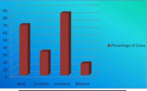
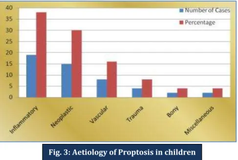
RESULTS: Incidence of proptosis in male was 60% and female 40%. Etiology of proptosis due to Inflammatory cause 38%(19 cases), Neoplasm 30%(15 cases), vascular 16%(8 cases), trauma 8%(4 cases) and others 8%(4 cases). Out of 50 cases 32 were axial and 18 were eccentric proptosis.
J of Evolution of Med and Dent Sci/ eISSN- 2278-4802, pISSN- 2278-4748/ Vol. 3/ Issue 22/June 02, 2014 Page 6160 A bilateral orbital mass lesion often signifies a neoplastic process or underlying systemic disease that is likely to be a malignant.6
The common etiology for proptosis in children is inflammatory (38%) and neoplastic (30%). Orbital cellulitis is the single most common cause for proptosis in children. Leukemic infiltration is the commonest cause for proptosis due to secondary orbital infiltration.
Plain X-ray, B-Scan and CT scan were very useful for diagnosis and to plan the mode of treatment. However confirmation of diagnosis especially in tumors requires FNAC7 or biopsy for tissue diagnosis.8
Fig. 3: Aetiology of Proptosis in children
J of Evolution of Med and Dent Sci/ eISSN- 2278-4802, pISSN- 2278-4748/ Vol. 3/ Issue 22/June 02, 2014 Page 6161
DISCUSSION: Proptosis in children differ from adults, for example malignant tumors in children can present as inflammatory lesions but not in adults.9 Thorough understanding of anatomy of orbit and surrounding structures with systematic approach is necessary to arrive at a correct diagnosis and the management of proptosis in children.
Bilateral hemorrhagic proptosis often signifies a neoplastic process, which often reveals underlying systemic malignancies like leukemias.
Henderson’s review of 4 tumours10 shows female: male ratio is 24:26, this study show similar comparable results of 20:30.
Inflammatory and neoplastic lesions are the leading cause for proptosis in children. It shows similar results of previous studies by Albert and Jakobeic of 257 cases of childhood proptosis11
Incidence of proptosis due to primary intraocular malignancy like retinoblastoma is decreased, due to availability of modern equipments for early diagnosis and treatment.
Among the proptosis due to tumors, malignancies were common, of these majority were due to secondaries from leukemia and Neuroblastoma.12
The exact early etiological diagnosis of proptosis in children and timely referral to appropriate expert orbital surgeon and others like Neurosurgeon, Oncologist is mandatory to save the vision and life of these patients.
J of Evolution of Med and Dent Sci/ eISSN- 2278-4802, pISSN- 2278-4748/ Vol. 3/ Issue 22/June 02, 2014 Page 6162
REFERENCES:
1. Sindhu K, Downie S, Ghabrial R, Martin F. Aetiology of childhood proptosis. J Paediatr Child health 1998; 34:374-6.
2. Sethi A, Ghose S, Gujral S, Jain P, Kumar R. Childhood proptosis: The invaluable but overlooked peripheral blood smear. Indian J Ophthalmology 2001; 49:121-3.
3. Oakhill A, Willshaw H, Mann JR. Unilateral proptosis. Arch Dis Child 1981; 56:549-51.
4. Tamsin J, Sleep, Ruth M. Interinstrument Variability in Hertel Type Exophthalmometers. J Ophth Plastic Reconstructive Surgery 2002; 18:254-7.
5. Rauniyar RK, Thakursk, Panda A. CT in the diagnosis of isolated cysteicercal Infestation of Extra Ocular Muscle. J Clin Radiology 2003; 58:154-6.
6. Bakhshi Sameer, Singh Preetpaul, Chawla Nikhil. Malignant Childhood proptosis. Study of 104 cases. J Paediatr Hematology Oncology 2008; 30:73-76.
7. Das DK, Das J, Bhatt NC, Chachra KL, Natarajan R. Orbital lesions. Diagnosis by fine needle aspiration cytology. Acta Cytol 1994; 38:158-64.
8. Shields JA, Bakewell B, Augsburger JJ, Donoso LA, Bernardino V. Space – occupying orbital masses in children. A review of 250 consecutive biopsies. J Ophthalmology 1986; 93; 379-84. 9. Jerry A, Shields, Carol L, Shields. Rhabdomyosarcoma review for the Ophthalmologist. Sur of
Ophth 2003; 48:1.
10.Garrity AG, Henderson WJ. Vascular hamartomas hyperplasia neoplasm. In, Garrity AG, Henderson WJ, Cameron DJ (ed). Henderson’s Orbital tumours, 4th edition. USA, Lippincott Williams & Wilkins, 2007; 210-215.
11.Dallow LR, Pratt GS, Green PJ. Approach to Orbital disorders and Frequency of Disease occurrence. In, Albert MD, Jakobiec AF (ed). Principle and Practice of Ophthalmology, 2nd edition. USA, W B Saunders Company, 2000; 3057-3067.
12.
Stefanyszyn MA, Robinson DH, Robert BP. Disorders of orbit. In, Nelson LB(ed). Harlyes Paediatric Ophthalmology, 4th edition. USA, WB Saunders company, 1998; 371-396.AUTHORS:
1. M. Loganathan 2. M. Radhakrishnan
PARTICULARS OF CONTRIBUTORS:
1. Associate Professor, Department of Ophthalmology, Sri Venkateshwara Medical College Hospital and Research Centre, Pondy. 2. Professor, Department of Ophthalmology, Sri
Ramachandra Medical College, Chennai.
NAME ADDRESS EMAIL ID OF THE CORRESPONDING AUTHOR:
Dr. M. Loganathan, Associate Professor,
Department of Ophthalmology,
Sri Venkateshwara Medical College Hospital and Research Centre,
13-A Pondy- Villupuram Main Road, Ariyur, Pondicherry – 605102. Email: log_smith@yahoo.com