Establishment of a Developmental
Compartment Requires Interactions between
Three Synergistic
Cis
-regulatory Modules
Dimitri Bieli1, Oguz Kanca1, David Requena2, Fisun Hamaratoglu3, Daryl Gohl4¤,
Paul Schedl4, Markus Affolter1, Matthew Slattery5, Martin Müller1, Carlos Estella2
*
1Biozentrum, University of Basel, Basel, Switzerland,2Departamento de Biología Molecular and Centro de Biología Molecular Severo Ochoa, Universidad Autónoma de Madrid (UAM), Madrid, Spain,3Center for Integrative Genomics, University of Lausanne, Lausanne, Switzerland,4Department of Molecular Biology, Princeton University, Princeton, New Jersey, United States of America,5Department of Biomedical Sciences, University of Minnesota Medical School. Duluth, Minnesota, United States of America
¤ Current address: University of Minnesota Genomics Center, Minneapolis, Minnesota, United States of America
Abstract
The subdivision of cell populations in compartments is a key event during animal develop-ment. InDrosophila, the geneapterous (ap)divides the wing imaginal disc in dorsalvs ven-tral cell lineages and is required for wing formation.apfunction as a dorsal selector gene has been extensively studied. However, the regulation of its expression during wing devel-opment is poorly understood. In this study, we analyzedaptranscriptional regulation at the endogenous locus and identified threecis-regulatory modules (CRMs) essential for wing development. Only when the three CRMs are combined, robustapexpression is obtained. In addition, we genetically and molecularly analyzed the trans-factors that regulate these CRMs. Our results propose a three-step mechanism for the cell lineage compartment expression ofapthat includes initial activation, positive autoregulation and Trithorax-medi-ated maintenance through separable CRMs.
Author Summary
The separation of cell populations into distinct functional units is essential for both verte-brate and inverteverte-brate animal development. A classical paradigm for this phenomenon is the establishment of developmental compartments duringDrosophilawing development. These compartments depend on the restricted expression of two selector genes,engrailed
in the posterior compartment andapterous(ap) in the dorsal compartment. Yet, despite the central role these genes and their restricted expression patterns play inDrosophila
development, we still do not understand how these patterns are established or maintained. Here, by dissecting the regulatory sequences required forapexpression, we solve this problem for this critical selector gene. We used a combination of experimental approaches to identify and functionally characterize thecis-regulatory modules (CRMs) that regulate OPEN ACCESS
Citation:Bieli D, Kanca O, Requena D, Hamaratoglu F, Gohl D, Schedl P, et al. (2015) Establishment of a Developmental Compartment Requires Interactions between Three SynergisticCis-regulatory Modules. PLoS Genet 11(10): e1005376. doi:10.1371/journal. pgen.1005376
Editor:Claude Desplan, New York University, UNITED STATES
Received:May 7, 2015
Accepted:June 19, 2015
Published:October 15, 2015
Copyright:© 2015 Bieli et al. This is an open access article distributed under the terms of theCreative Commons Attribution License, which permits unrestricted use, distribution, and reproduction in any medium, provided the original author and source are credited.
Data Availability Statement:All relevant data are within the paper and its Supporting Information files.
Funding:This study was supported by a grant from the MINECO to CE (No. BFU2012-34353) and grants from the Kantons Basel-Stadt and Basel-Land, and the Swiss National Science Foundation to MA. The funders had no role in study design, data collection and analysis, decision to publish, or preparation of the manuscript.
apexpression duringDrosophilawing development. For these analyses we implement a novel technique allowing us to study the function of these CRMsin vivo, at the nativeap
locus. We found threeapCRMs crucial for wing development: the Early (apE) and the D/ V (apDV) enhancers and theapPRE (apP). Only when all three regulatory elements are combined is a uniform and completeapexpression domain generated. In summary, our results indicate thatapis regulated in time and space by a three-step mechanism that gen-erates a lineage compartment by integrating input from separate CRMs for the initiation, refinement and maintenance of its expression.
Introduction
Animal development requires the segregation of cell populations using both lineage and non-lineage boundaries. These cell boundaries act as signaling centers that organize the growth and patterning of specific tissues (reviewed in [1]). A paradigmatic example is the subdivision of theDrosophilawing disc into anterior-posterior (A/P) and dorsal-ventral (D/V) compart-ments. At the compartment boundaries, ligands encoded bydecapentaplegic(dpp) andwingless
(wg) are secreted and activate signaling pathways that orchestrate wing development [2–8]. The generation of the A/P and D/V compartments is directed by specific transcription factors, the selector genesengrailed(en) andapterous(ap), respectively, that define the identity of the cells using a binary code (on or off) [9–15]. Once the compartmental fates have been assigned, the cells in whichenandapare expressed as well as their descendants maintain that“ deter-mined”state. Unlike the A/P wing division, which is established during embryonic develop-ment, the D/V boundary is defined in the wing disc during the second larval stage by the expression ofap[16].apencodes a LIM-type homeodomain transcription factor and its activ-ity depends on the formation of a complex with the LIM-domain binding protein Chip [17–
19]. Sinceapfunction is crucial to initiate the signaling center at the D/V boundary [20,21],ap
null mutants completely lack the wing [16].
Due to its key role in wing disc development,apfunction has been studied extensively. However, the transcriptional regulation ofapis poorly understood. How a sharp border ofap -expressing and non--expressing cells is generatedde novoduring the growth phase of the imagi-nal disc, and how the expression ofapis maintained and restricted to the dorsal compartment are critical unanswered aspects of wing development.
The spatial and temporal regulation of gene expression is mediated by the binding of tran-scription factors to discrete DNA sequences namedcis-regulatory modules (CRMs). CRMs can be located up to hundreds of kilobases away from their target promoters. Synergistic interac-tions between CRMs may be required to faithfully regulate gene transcription (reviewed in [22]). Several CRMs have been identified controllingapexpression in different tissues, such as in muscle progenitors and in the embryonic nervous system [23,24]. A wing disc specific enhancer, named apC, has been reported to drive expression in the dorsal wing disc [24]. How-ever, it has been demonstrated that this element is not sufficient for properapregulation in the wing [25].apexpression is initially activated in future dorsal cells by theDrosophilaEpidermal Growth Factor Receptor (EGFR) pathway through the secreted neuregulin-like signaling pro-tein Vein (Vn) [26,27]. However, it is still unknown howapexpression is regulated after this initial EGFR-mediated activation. This is particularly critical in a highly proliferating tissue such as the wing imaginal disc.
(PcG and TrxG). These proteins either repress (PcG) or activate (TrxG) the expression of their target genes throughcis-regulatory sequences called Polycomb Response Elements (PREs) (reviewed in [28,29]). It has been suggested thatapexpression is repressed by PcG protein complexes in ventral wing disc cells [30].
In this study, we have analyzed the regulation ofapat the endogenous locus and identified threeapCRMs crucial for wing development: the Early (apE) and the D/V (apDV) enhancers and theapPRE (apP). Importantly, we analyzed these CRMs in the endogenous locus using a novelin siturescue system. We find that only when the three regulatory elements are com-bined, a uniform and completeapexpression domain is observed. Our results indicate thatap
is regulated by a three-step mechanism that generates a lineage compartment through the integration of input from separate CRMs for the initiation, refinement and maintenance of its expression.
Results
Genetic characterization of the
apterous
promoter region
apis expressed in multiple tissues during embryonic and larval stages. Four different tran-scripts starting from three different promoters have been annotated which give rise to three unique polypeptides (FlyBase). We have generated a series of deletions to identify whichap
non-coding sequences are required forapexpression in the wing imaginal disc (Fig 1A–1C; see
Materials and Methodsfor information about each allele). Unless otherwise stated, hemizygous phenotypes resulting from these deletions were analyzed overapDG3, a large deletion removing the bulk of theaplocus [25], and were classified as amorphs or hypomorphs depending on their severity.apamorphs were defined by the absence of wing tissue in discs and adults.
The shortest deletion in our collection with anapnull phenotype isapt11b(Fig 1CandS1D Fig). It specifically deletes the transcription start site (TSS) of transcriptsap-RAandap-RC. Ourin silicoanalysis indicates that this TSS is not controlled by a TATA-box promoter, but rather contains an Initiator (Inr) and a Downstream Promoter Element (DPE) (for review see [31]). In addition,apt11bremoves a PRE located around 100 bp upstream of theap-RA/ap-RC
TSS. This PRE was defined by several chromatin immunoprecipitation studies with various anti-PcG antibodies [30,32–34]. The putative PRE core as defined by Oktabaet al[30] is indi-cated inFig 1C. Two other small deletions with the same distal break point asapt11bwere iso-lated:apc2.73candapc1.2b. They leave the TSS, Inr and DPE intact, but remove the PRE core. In hemizygous flies, small wing stumps are often formed (Fig 1GandS1E Fig). In addition the wing pouches of 3rdinstar wing discs are larger than in amorphic mutants (compareFig 1G” with 1F”). Small amounts of Ap can only be detected in the presumptive hinge and notum (arrows inFig 1G’andS1E Fig). The Wg stripe along the compartment boundary is absent (Fig 1G”andS1E Fig). Hence,apc2.73candapc1.2bbehave as strong hypomorphic alleles. A dramatic improvement of the adult wing phenotype is observed for deletionapc1.60cwhich is a mere 113 bp shorter thanapc1.2b(Fig 1CandS1F Fig). Note that it keeps the TSS, Inr, DPE as well as the PRE core in place. A weak phenotype becomes apparent in hemizygous condition: similar to other weakaploss-of-function alleles, most wing margins have notches. Unexpectedly, this phenotype is brought about by partial ectopicapexpression in the ventral pouch compartment which correlates with gaps in the Wg stripe along the compartment boundary (S1F Fig).
Deletions affecting the intergenic spacer separating
ap
and
l(2)09851
identify two regions important for
ap
function
As a next step, we generated alleles that retain an intact PRE/promoter region, but remove upstream non-coding regions ofap. In the mutantapDG1, 27 kb of the upstream non-coding region are deleted (Fig 1B) [35]. Hemizygous flies of this genotype can be considered as amorphic mutants, since no wing tissue was formed despite weak residualapexpression in the notum (Fig 1E’). Removing proximal upstream regions (apDG6,apDG11, andapDG12) also resulted in amorphic phenotypes (Fig 1B and 1F). These deletions remove the previously iden-tifiedapwing enhancer apC [24,25]. The distal part of the interval defined byapDG1was deleted inapDG14. Hemizygous flies form wing stumps of almost normal length but wing mar-gin formation is severely impaired (Fig 1H). In the corresponding wing discs,apexpression in the wing pouch is reduced and the Wg stripe along the D/V boundary is critically perturbed (Fig 1H’and 1H”). The large size of the apDG14deficiency precludes a precise localization of an enhancer element within the ~16 kb interval. However, a few small deletions extending only proximal to e01573 allowed us to narrow down its approximate distal end: one of them,ap11.1, deletes 1654 bp (Fig 1B; see alsoMaterials and Methods). It can be maintained as a homozy-gous stock and wings look wild-type. Its weak hypomorphic nature is revealed in hemizyhomozy-gous
ap11.1flies: all wings have notches along the margin (Fig 1I). Their origin can be traced to gaps in the Wg stripe along the D/V compartment boundary due to reduced Ap levels in the pouch (Fig 1I’and 1I”).
Two conserved regions harbor essential wing enhancer elements
To further characterize the intergenic spacer betweenapandl(2)09851, we engineered and vali-dated a system which allowed us to investigate the role of given DNA stretches at theaplocus [25]. Briefly, we deleted the 27 kb (apDG1) upstream region ofap, and replaced it by an attP site juxtaposing the promoter/PRE (apattPΔEnh;Fig 2B’). In this amorphic situation, we were able to bring back sub-fragments of the previously deleted regulatory regions byFC31-integrase
The number in between the break points indicates the approximate length of the deletion. Phenotypically, the deletions can be divided into amorphic (in red) or hypomorphic (in blue) wing alleles when hemizygous over apDG3. Please note the different scales of the maps depicted in B and C.(B)Deletions that affect the coding sequence all lead to a no wing phenotype (apDG16,apDG3,ap12.1,apDG8,apDG2,apc1.78a, andapc2.58c).
Deletions in the upstream noncoding region betweenapMMandl(2)09851either lead to amorphic (apDG1,
apDG15,apDG6,apDG11, andapDG12) or hypomorphic wing phenotypes (apDG14andap11.1).(C)Blow up of the
appromoter region specific for transcriptsap-RAandap-RC. The PRE core is depicted by a yellow box. apt11b, a deletion which removes the TSS as well as the PRE core, results in a no wing phenotype. The two deletionsapc2.73candapc1.2bleave the TSS intact but both remove the PRE core and both yield strong
hypomorphic wing phenotypes. The weak hypomorphic alleleapc1.60cleaves TSS and PRE core untouched.
(D-I)Wings and 3rdinstar wing discs of representativeapwing mutants stained for Wg (red) and Ap (green).
(D)Wing and notum of a hemizygous +/apDG3fly. Almost 100% of the wings look normal [25](D’)Ap staining
in the wing disc demarcates the dorsal compartment.(D”)Wg staining: the inner ring outlines the wing pouch (white arrow) and the stripe traversing it corresponds to the D/V compartment boundary (white arrowhead).(E and F)All wing tissue is lost in amophic wing mutants (apDG1/apDG3andapDG12/apDG3).(E
’and F’)Only weak Ap staining is detectable in the notum (white arrow).(E”and F”)The wing pouch is completely lost and the inner Wg ring is reduced to a dot.(G)In strong hypomorphic conditions (apc1.2b/apDG3), only small wing
and haltere stumps form (black arrow).(G’)Low Ap protein levels are detected (white arrow) mainly in the hinge region.(G”)The size of the wing pouch is drastically reduced and no Wg stripe along the D/V boundary is visible.(H)Hypomorphic mutants (apDG14/apDG3) developed considerably more wing tissue with no or little
wing margin or hinge.(H’)Compared to control discs, weaker Ap staining is observed in the pouch region.
(H”)The size of the wing pouch is comparable to wild type while the D/V Wg stripe is disrupted.(I)In weak hypomorphic mutants (ap11.1/apDG3) notching of the wing blade is prominent.(I
’)Compared to control discs, apexpression is mostly compromised in the pouch region.(I”)Pouch size is similar to wild type and the Wg D/V stripe is locally disrupted. All scale bars are 50μm.
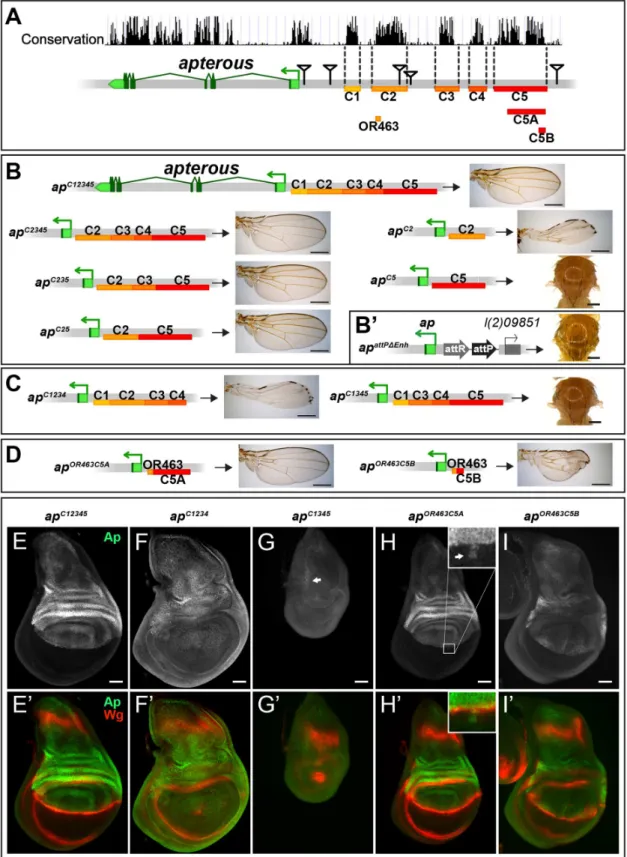
Fig 2. Analysis of theapwing enhancer region. (A)Conservation of theaplocus (data from UCSC genome browser) and subdivision of the 27 kb intergenic region betweenapandl(2)09851into 5 conserved blocks (C1–C5) is shown. OR463, C5A and C5B are subfragments of C2 and C5, respectively. Black triangles mark the locations of the transposon used for the generation of the deletions.(B)Six different constructs consisting of variable combinations of conserved blocks and the corresponding hemizygous wing phenotypes are depicted. When all 5 conserved regions are present (apC12345), a normal sized
mediated insertion and test their ability to rescue wing development. Again, all the newly gener-ated alleles were tested in hemizygous condition. According to sequence conservation and his-tone H3 lysine 4 trimethylation (H3K4me3) patterns, which have been reported to correlate with active promoters and enhancers [36], we have divided the upstream non-coding region of
apinto 5 blocks (C1–5,Fig 2A). Combining all 5 conserved blocks and reintroducing them into
apattPΔEnh(apC12345), fully rescued wing formation (Fig 2B) as well as the Ap and Wg pattern in wing imaginal discs (compare Figs2Ewith1D). Deleting the conserved blocks that showed no H3K4me3 mark (C1 and C4), had no consequence on wing phenotype (Fig 2B). Next, we deleted conserved regions with a methylation mark. Deleting C3 had no influence on wing for-mation (apC25,Fig 2B). In contrast, upon removal of C5, wing development was critically dis-turbed (apC2,Fig 2B). Long wing stumps with defective wing margin and hinge were formed that resembled the hypomorphicapDG14mutant, which completely lacks the conserved C5 block (compare Figs2Band1H). In flies containing only C5, no wing tissue was formed (apC5).
We then tested whether the C2 and C5 fragments were also necessary when all the other conserved elements were present. Removing C5 only (apC1234) had the same effect as maintain-ing C2 alone, since long wmaintain-ing stumps with little margin and hmaintain-inge were formed (compareFig 2C with 2B). Wing discs of this genotype showed drastically reducedapexpression in the pouch region and in most cases lost the Wg stripe along the D/V boundary (Fig 2F). As expected, when only C2 was removed (apC1345, seeFig 2C), no wing tissue was formed, Ap pro-tein was only weakly detected in the notum and the Wg pattern was equivalent to amophicap
wing mutants (Fig 2G).
We also investigated a possible role of the positions of C2 and C5 relative to each other in
apC52andapC15342flies. Both alleles yield wild-type wings in hemizygous flies, indicating that their order on the chromosome is not important (S2A Fig).
Next, we aimed at defining the minimal CRMs which were able to direct wing development. We have recently found that shorter sub-fragments of C2 retain its wing disc specific activity [25]. The combination of a 463 bp fragment of C2 (OR463) and 3.8 kb of C5 (C5A) in
apOR463C5Afully rescued wing development (Fig 2D and 2H). Replacing C5A by C5B, a 600 bp subfragment of C5A, indicated that it lacks certain regulatory input (Fig 2D). The expression ofapinapOR463C5Bwing discs was restricted to the dorsal compartment, but reduced compared toapC12345(compareFig 2I with 2E). Nevertheless, apart from small disruptions at the D/V boundary,wgexpression appeared almost normal (Fig 2I’).
Finally, to investigate whether additional wing-specific CRMs reside within the intronic sequences, we replaced the coding sequences with anapcDNA lacking most intronic sequences (apcDNAint2.3). This allele produces normal wings (S2B and S2C Fig). Thus, we conclude that no essential wing CRMs are present in the intronic regions ofap. In agreement with this notion, fragments taken from intronic sequences (see below andS2D Fig) failed to drive reporter gene expression in the wing disc. Note that the cDNA used for the construction ofapcDNAint2.3 corre-sponds to theap-RA/ap-RCtranscripts.
results in hypomorphic wings (apC2). C5 alone (apC5) is an amophic allele, as no wings are formed.(B’)apattPΔEnh: the docking site of thein siturescue
system for the evaluation of DNA fragments originating from the 27 kb intergenic spacer is shown. An attP site located ~400 bp upstream of theapTSS juxtaposes the promoter/PRE region. As inapDG1, the 27 kb intergenic region is deleted.(C)Removing C2 or C5 in the context ofapC12345(apC1234and
apC1345) leads to the same phenotype as each element alone (apC2orapC5, respectively).
(D)Enhancer bashing of C2 and C5 regions. OR463 and C5A in combination are the shortest fragments that still result in a normal wing (apOR463C5A). C5B, a sub-fragment of C5A, in combination with OR463 does not fully
rescue wing formation (apOR463C5B). Wing size is reduced, but all margin structures are formed.(E-I)Third instar wing discs of different genotypes stained for
Ap (green) and Wg (red).(E-E’)apC12345: Ap and Wg pattern is indistinguishable from wild type.(F-F’)apC1234: a significant reduction of Ap levels in the wing pouch is observed. The Wg stripe along the D/V border is almost completely lost.(G-G’)apC1345: scattered cells with little Ap protein are detectable in the notum (see arrow). Wing pouch is reduced to a small dot ofwgexpression.(H-H’)apOR463C5A: Ap and Wg patterns are similar to wild type. Ap protein can
sometimes be detected in some cells of the ventral part of the disc (arrow in inset).(I-I’)apOR463C5B: although protein levels are reduced, the Ap pattern is
close to wild type. Nevertheless, the appearance of the Wg stripe along the D/V border is not as smooth as in wild type. All scale bars are 50μm.
Combining the results from the two complementaryin vivoapproaches (deletion analysis and thein siturescue system), we have defined three distinct regions which are absolutely required for the correctapexpression in the wing disc: a region next to theapTSS which contains a PRE and two enhancers with distinct regulatory input located in homology blocks C2 and C5.
Identification of
ap cis
-regulatory modules active in the wing imaginal
disc
In parallel to theapdeletion andin siturescue strategies, we performed an unbiased search for
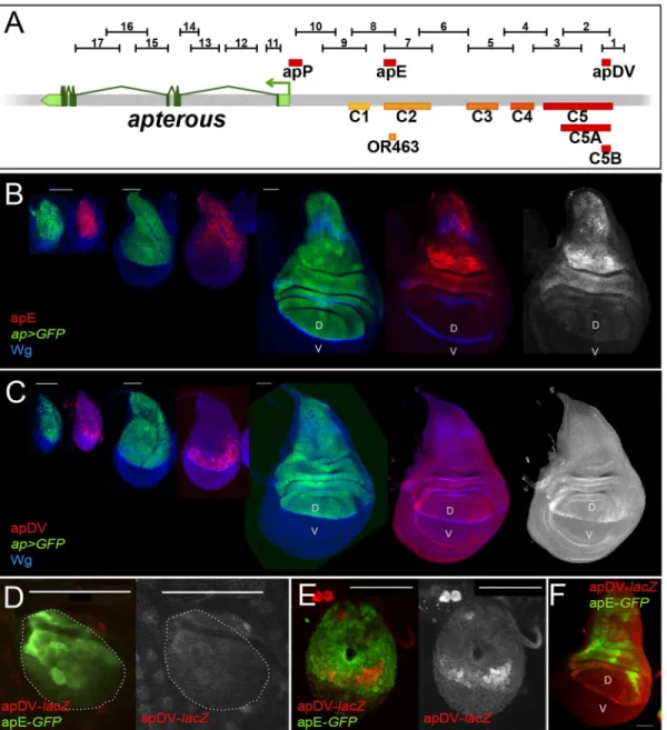
apCRMs active in the wing imaginal disc. Using the Fly Light database [37] and self-made con-structs (seeMaterials and Methods), we screened theapgenomic region for DNA fragments that activate theGal4gene in anap-like expression pattern (Fig 3A). We found that 4 of the 17 lines tested partially recapitulatedap-like expression pattern in third instar wing imaginal disc (S3 Fig). Interestingly, lines 1 and 2 were active in a similar pattern in the wing pouch and hinge but were not active in the notum, while lines 7 and 8 showed identical expression pattern in the notum and hinge with low levels in the dorsal wing pouch (S3 Fig). Subsequently, we cloned the overlapping sequences between lines 1–2 and 7–8 in reporter constructs and com-pared their activity withapexpression as well as with each other during wing imaginal disc development (Fig 3B–3F; seeMaterials and Methods). apE (Early), the first element to be acti-vated in early to mid-second instar imaginal discs, drove expression in allap-expressing cells (Fig 3B and 3D). The other element, named apDV (Dorso-Ventral), was activated a few hours later in dorsal cells close to the D/V boundary (Fig 3C and 3E). As the wing imaginal disc developed, the activity of apE became mainly restricted to the notum and hinge with low expression remaining in the wing pouch (Fig 3B). In contrast, apDV was always restricted to dorsal wing pouch cells close to the D/V boundary, with some cells expressing the reporter in the dorsal wing hinge (Fig 3C).
In line with our previous results, apE and apDV are located within the C2 and C5 regulatory fragments identified with thein siturescue system, respectively, and overlap with OR463 and C5B (Fig 3A). Moreover, in a reporter gene construct, C2 and C5 reproduced the same expres-sion pattern described for apE and apDV, respectively (S3A Fig).
It should be noted that none of the singleap-CRMs identified, apDV or apE, nor the combi-nation of them, apDV-lacZ+apE-GFP, was able to completely reproduce the endogenousap
expression pattern, suggesting that additional elements are necessary (Fig 3Fand see below).
The EGFR pathway transiently regulates the apE element
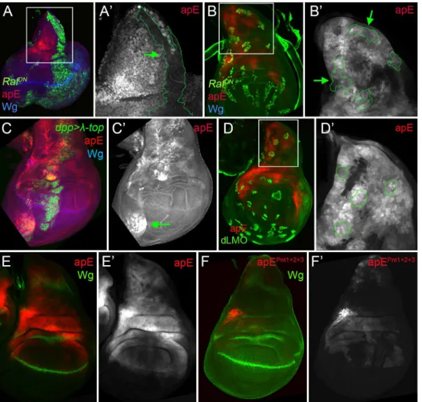
The initialapexpression in the wing disc is activated by the EGFR signalling pathway at early stages of wing development (from early to mid-second instar), while its later expression is EGFR-independent [26,27]. Since the apE element was active in the entireapexpression domain in early wing discs, we tested whether this CRM is regulated by the EGFR pathway. Clones of cells expressing a dominant-negative form of the pathway effector Raf (RafDN) gener-ated early in larval development (24–48 hrs after egg laying, AEL) were unable to activate apE (Fig 4A), while no effect was observed in clones generated later (72–96h AEL,Fig 4B). The same temporal EGFR-dependency of apE was found when the pathway was reduced in the entire wing disc using a temperature-sensitive EGFR allele (S4 Fig).
on apE activity (Fig 4D). Taken together, these results suggest that apE is activated by the EGFR pathway and that other factors regulate its expression afterwards.
To understand how the EGFR pathway regulates apE activity, we searched for putative binding sites of the ETS transcription factor Pnt [39]. Two highly conserved and one less Fig 3. Activity patterns of apE and apDV enhancers. (A)Schematic representation of theapgenomic region is shown as a grey bar.aptranscriptap-RAis shown in green. In the upper part of the panel, horizontal bars represent the DNA elements for which Gal4 drivers were generated by the Janelia Farm consortium except for line 6 (seeMaterials and Methods) (http://flweb.janelia.org/cgi-bin/flew.cgi). Red bars represent regulatory elements apP, apE and apDV. At the bottom of the panel, 8 fragments tested with theap in siturescue system are indicated.(B-C)Pairs of wing imaginal discs isolated from second, early and late third instar larvae are shown (from left to right). They were stained for GFP (ap-Gal4>UAS-GFP, green) and Wg (blue) or apE-lacZ(red) in (B) or apDV-lacZ(red) in (C). Note thatap>GFPrepresents the completeappattern to which those of apE-lacZand apDV-lacZare compared.(B)In early discs, apE is active in all the cells that expressap. Later, its activity is restricted to a subset ofap-expressing cells mainly in the notum and hinge region. Expression in the wing pouch is very low.(C)apDV is active in dorsal-distal cells in early discs. Later, its activity is restricted to dorsal wing pouch cells close to the D/V boundary.(D-F)Early second(D),mid-second(E)and third instar imaginal discs(F)stained for apDV-lacZ(red) and apE-GFP(green) are shown.(D)apE is activated earlier than apDV in proximal wing disc cells.(E)apDV is activated in dorsal-distal cells that already have apE activity.(F)In third instar imaginal discs, apE and apDV occupy complementary territories. apDV is restricted to dorsal wing cells close to the D/V border. apE remains mainly active in the hinge and notum. All scale bars are 50μm. D, dorsal and V, ventral.
conserved sites were identified. When all these sites were mutated simultaneously, the activity of apE was strongly reduced (compareFig 4E with 4F). Altogether, these results suggest that apE is initially activated by the EGFR pathway and that this activation requires Pnt function.
Ap and Vg/Sd regulate the apDV CRM
While the apE element was activated in allap-expressing cells in early second instar wing discs, the apDV element was induced only later and restricted to a subset of apE-positive cells (the wing pouch cells). Therefore, we tested whether the Ap protein itself is needed in an autoregu-latory fashion for the restricted activity of the apDV element in the dorsal compartment. Fig 4. apE is regulated by the EGFR pathway. (A-B)Third instar wing imaginal disc with clones expressing a dominant negative version of Raf (RafDN)
induced at different time points of larval development are marked by GFP (green). Discs were also stained for Wg (blue) and apE-lacZactivity (red).(A)Early inducedRafDNclones (24–48hrs after egg laying, AEL) are unable to activate apE.(A´)Close-up of the disc in(A)with the clone outlined in green (green
arrow). Note that apE is not activated within the clone.(B)Late inducedRafDNclones (72–96hrs AEL) have no effect on apE activity.(B´)Close-up of the disc
in(B)with clones outlined in green (green arrows).(C)dpp-Gal4; UAS-GFP, UAS-EGFRλtop4.2(
λ-top, green) wing disc stained for apE-lacZ(red) and Wg (blue). Ectopic activation of the EGFR pathway induces apE in ventral pleural cells.(C´)Single channel for apE. Green arrow points to ectopic apE activity.
(D)Gain of function clones of the Ap activity repressor dLMO (green) has no effect on apE activity (red).(D´)Close-up of the disc in(D)with clones outlined in green.(E-F)Wing imaginal discs stained for Wg (green) and apE (E, red) and apEpnt1+2+3(F, red) activity. Note that apE activity is strongly reduced after mutating the three identified Pnt binding sites (apEpnt1+2+3). All constructs have been inserted in the same genomic location. Images were obtained keeping the confocal settings constant.
dLMO expressing clones cell-autonomously repressed the apDV element, while forced expres-sion ofapin the ventral compartment cells ectopically activated it (Fig 5A and 5B). This sug-gests that Ap restricts the dorsal activity of apDV. Althoughapis expressed in all dorsal wing disc cells, apDV is only active in dorsal wing cells close to the D/V boundary, which suggests
Fig 5. apDV is regulated by Ap and Sd/Vg. (A)In dLMO expressing clones (green), apDV-lacZactivity (red) is repressed. Wg (blue) is non-autonomously activated in cells surrounding the clones. Single channels are displayed for apDV-lacZ(A’)and Wg(A”). Green arrows point to dLMO expressing clones.(B)
dpp-Gal4; UAS-GFP, UAS-ap(green): upon ectopic Ap induction bydpp-Gal4, apDV-lacZ(red) and Wg (blue) are induced in ventral compartment cells. Single channels are displayed for apDV-lacZ(B’)and Wg(B”). Green arrow points to ectopic apDV andwgexpression.(C)dpp-Gal4; UAS-GFP, UAS-vg-RNAi: RNAi-induced knockdown of Vg indppdomain (green). Wg is in blue. Note that apDV-lacZexpression (red) is strongly downregulated in the central part of the pouch.(C’)Single channel display of apDV-lacZ. Green arrow points to discontinuity in the apDV pattern.(D)dpp-Gal4; UAS-GFP, UAS-vg: ectopicvgexpression induces apDV (red) along thedppdomain (green), but only in the dorsal compartment. Note thatwgis not induced upon the ectopic expression ofvg. Single channels are displayed for apDV-lacZ(D’; green arrow points to ectopic apDV-lacZ) and Wg(D”).(E-F)dpp-Gal4; UAS-GFP(E)and dpp-Gal4; UAS-GFP, UAS-vgleg discs(F):dpp-Gal4; UAS-GFP(green), apDV-lacZ(red) and Wg (blue) patterns are shown. Ectopicvgexpression induces apDV activity in the distal domain of the leg disc (white arrow inF).(G)ChIP experiments with anti-Sd and anti-Ap antibodies. Quantifications of apP, apE and apDV DNA in immunoprecipitates demonstrate that Sd and Ap are preferentially bound to the apDV regulatory region. Representative enrichment values are shown for a single experiment that was conducted in triplicate.(H)DNA sequences of variousDrosophilaespecies surrounding the identified Ap (red shade) and Sd (green shade) binding sites are shown. Note that Ap1 site is deleted inap11.1flies.(I-K)Wing imaginal discs stained for Wg (green) and apDV (I, red),
apDVAp1+2(J, red) and apDVSd1+2(K, red) activity. Mutation of the Ap sites(J)or Sd sites(K)results in loss of apDV activity.(I
’-K’)Single channel pictures are depicted for each apE wild type and mutant condition. All constructs have been inserted in the same genomic location and images were obtained keeping the confocal settings constant.
additional input into this element. Therefore, we tested whether apDV activity is controlled by
wgorvestigial(vg) [40], two key genes required for wing development. Downregulation or ectopic activation of the Wg pathway did not significantly affect apDV-lacZexpression (S4 Fig). However, knockdown ofvgin thedppdomain eliminated apDV-lacZexpression (Fig 5C). Additionally, ectopic expression ofvgstrongly activated apDV in the dorsal compartment (Fig 5D). Remarkably, while apDV is not activated in the leg disc, forced expression ofvgin this disc induced its activity in the distal domain of the leg, where a ring of endogenousap expres-sion has been described (Fig 5E and 5F)[16,41].
As a next step, we tested whether Ap and Scalloped proteins (Sd), the transcriptional com-panion of Vg, directly bind to the apDV CRM. Using Ap and Sd chromatin immunoprecipita-tion (ChIP), we found that Ap and Sd were significantly enriched at the apDV regulatory region in comparison to apE or apP (Fig 5G). Moreover, we identified two conserved consen-sus-binding sites for Sd as well as for Ap in the apDV region. Mutation of these sites completely eliminated apDV activity (Fig 5H–5K). Intriguingly, loss of one of these Ap binding sites likely contributes to the wing defects seen in theap11.1mutant described previously (seeFig 1I).
Taken together, these results suggest that Ap and Vg/Sd directly regulate apDV in the wing pouch, with an Ap autoregulatory input restricting its activity to the dorsal compartment.
Synergistic effect of apE and apDV with the
ap
promoter directs
ap
expression in the wing disc
We have identified twoapCRMs (apE and apDV) that, when combined in a reporter assay, partially recapitulatedapexpression in the wing disc (seeFig 3F), suggesting that other CRMs are needed for full expression. Since PRE-containing sequences are necessary for correctap
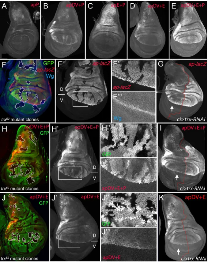
expression and proper wing development (Fig 1C), we tested if a region around theapTSS including the PRE, named apPRE (apP), had an impact on the activity of the distalapCRMs (Fig 3A). On its own, the apP drove weak expression in the wing disc in a pattern not related to the characteristicapexpression (Fig 6A). When placed together in a reporter construct with either apDV or apE (resulting in apDV+P-lacZor apE+P-lacZ), the activity of the resulting reporter gene construct was the sum of both elements and did not reproduce faithfully theap
expression pattern (Fig 6B and 6C). Interestingly, when the three CRMs were placed together, the expression of the apDV+E+P-lacZin third instar wing discs was more accurate than the expression of the previous CRMs combinations or the apDV+E-lacZand more precisely repro-duced the expression pattern ofap(compareFig 6D with 6E).
Therefore, we tested whether theseapCRM combinations placed next toapcDNA were suf-ficient to rescue wing development in anapmutant background. As expected, apE+P-apcDNA
was partially able to rescue wing growth, but completely lacks the D/V margin, whereas an apDV+P-apcDNAtransgene, that lacks the apE enhancer, did not rescue wing formation (S5 Fig). Interestingly, the apDV+E+P-apcDNAtransgene rescued wing formation inapmutants. Although the rescue was not fully wild type, a clear wing margin was observed in wing discs and adult wings (S5 Fig).
In summary, we have identified threeapCRMs that, only when combined, can accurately reproduce the endogenousapexpression pattern in the wing imaginal disc.
Trx maintains robust
ap
expression via the apP element
Fig 6. apP mediatesapexpression maintenance and depends on Trx input (A-E) Third instar wing imaginal discs stained withα-βGal antibody to visualizelacZactivity. (A)apP activity is weak and not related to the endogenousapexpression pattern.(B)apDV+P activity is the sum of both elements.
(C)The combination of apE+P leads to stronger and more homogeneouslacZexpression in the notum and hinge. Note that expression levels remain low in the dorsal wing pouch.(D)apDV+E activity is the sum of apDV and apE and does not reproduce the completeapexpression pattern.(E)Only the
immediately 5’to theapTSS (aprK568). We found that cells devoid oftrxfunction show reduced
ap-lacZexpression (Fig 6F). To analyze this result in more detail, we reducedtrxmRNA levels in the anterior wing disc compartment (ci-Gal4>trx-RNAi) and compared the levels ofap-lacZ
expression with the posterior control compartment (Fig 6G). Consistent withtrxmutant clones,ap-lacZexpression was strongly reduced in the anterior compartment, although the reduction was more prominent in the notum and in the dorsal wing pouch close to the hinge.
To genetically confirm that theapPRE (apP) functions as a Trithorax response element (TRE), we eliminated or downregulated Trx activity and analyzed the expression of the apDV +E+P-lacZreporter construct. IntrxE2mutant clones, apDV+E+P-lacZlevels were strongly reduced, as it was the case forap-lacZ(Fig 6H). In contrast, the same construct without theap
promoter, apDV+E-lacZ, was not altered in thesetrxmutant clones (Fig 6J). Accordingly, reducing the levels of Trx in the anterior compartment cells (ci>trx-RNAi) did not affect
expression of apDV+E-lacZ(compareFig 6K with 6I). Interestingly, the expression pattern of apDV+E+P-lacZwas strongly reduced upon Trx downregulation and resembled the pattern of wild type apDV+E-lacZ(the same construct without the apP, compareFig 6G and 6I with 6K). Altogether, our results suggest that theappromoter region behaves as a PRE/TRE providing the information required to maintainapexpression.
Direct and continuous contact of the apDV and apE CRMs together with
the apP element for
ap
maintenance
Classical transvection experiments usually deal with chromosomes harboring genes lacking either a functional promoter region or a functional enhancer. For combinations of members of the two groups, intragenic complementation can be observed, i.e. the corresponding phenotype is much less severe than seen in allelic combinations involving only one or the other group [42,43]; reviewed in [44]. We have previously reported that transvection is at work atap[35]. For example,apDG12/apDG1flies have no wings because both alleles delete wing enhancer apE. The same phenotype is observed inapt11b/apDG8flies because both alleles remove the promoter region as well as the 5’end ofap. In contrast, the wing phenotype ofapt11b/apDG1flies is much improved (S7B Fig). Models for transvection posit that the apE and apDV enhancers on chro-mosomeapt11bcan activate the transcription machinery of the functionalapgene on chromo-someapDG1. However, theapt11b/apDG1wings are consistently less well formed than those obtained fromapDG3/+flies (seeS7 Fig). These observations suggest that the apP region on the one hand and the two enhancers on the other interact more efficiently if they are locatedin cis. In our study, we have shown that the twoapwing enhancers are clearly separable units: (1) they lie ~10 kb apart and (2) the activity of apE is essential for auto-regulatory activation of apDV. From these premises, one would nota prioriexpect that the two enhancers must bein cisfor full function. However, several allelic combinations containing only one or the other enhancer element (apE or apDV) generated discs and adult wings with defects at the D/V boundary: similar results were obtained for genotypesapC1234/apC1345,apDG14/apDG12,apC2/
clones were generated 48–72hrs AEL and are marked by the absence of GFP (in each disc, several of them are outlined in white). Discs were stained for Wg (blue) andap-lacZ(red,F), apDV+E+P-lacZ(red,H) and apDV+E-lacZ(red,J).(F’,H’,I’)single channel image oflacZstaining.(G, I, and K)ci-Gal4; UAS-trx-RNAi: RNAi-induced knockdown of Trx-activity in the anterior wing disc compartment. Imaginal disc were stained forβ-Gal protein. White arrow points to anterior wing compartment.(G)ap-lacZ: enhancer trapaprK568,(I)apDV+E+P-lacZand(K)apDV+E-lacZ.(F, F’)trxE2mutant clones show downregulation of apexpression.(F”andF”’)Close-up ofap-lacZandwgexpression shown inF’.(G)Knockdown of Trx in the anterior compartment downregulatesap-lacZ expression. Note that the reduction ofap-lacZis stronger in the notum and the wing pouch close to the hinge.(H and H’)trxE2mutant clones show
downregulation of apDV+E+P-lacZexpression. (H”andH”’) Close-up of GFP and apDV+E+P activity inH’.(I)Knockdown of Trx in the anterior
compartment (arrow) downregulates apDV+E+P-lacZexpression. Asap-LacZin(G), apDV+E+P activity is reduced in a spatial dependent manner.(J and J’)trxE2mutant clones show no effect on apDV+E-lacZexpression. (J”andJ”’) Close-up of GFP and apDV+E activity inJ’.(K)Reducing Trx in the anterior
compartment has no effect on apDV+E-lacZexpression. D, dorsal and V, ventral.
apDG12or when asu(Hw)insulator element was inserted between apDV and apE inapf00451/
apDG3animals (Fig 7AandS7 Fig). Our transvection studies suggest that all three CRMs need to be incisto fully rescue wing development.
To better understand how the synergy between the three regulatory elements is achieved, we used chromosome conformation capture (3C) [45], which allowed us to testin vivowhether there is direct physical contact between the apE and apDV CRMs with the apP. Indeed, as seen inFig 7B, we found that in whole third instar larvae, apP preferentially contacted the apE and apDV elements, and did so more frequently when compared to sequences outside theap geno-mic locus. This suggests that the distal apE and apDV regions are in close physical proximity to apPin vivo.
Next, we tested whether apDV and apE CRMs are required either continuously or only tran-siently to directapexpression during wing disc development. To distinguish between these two possibilities, we generated an apDV+E+P-lacZconstruct, in which the apDV+E is flanked by FRT sequences (FRT-apDV+E-FRT+P-lacZ,Fig 7C). This allowed us to remove the apDV+E cassette at different time points of wing development using Flp-mediated recombination [46,47] (seeMaterials and Methods). Deletion of apDV+E early in development in the poste-rior compartment completely abolished reporter expression compared to anteposte-rior control cells (compareFig 7D and 7E). Deletion of the apDV+E at later stages also strongly decreased reporter gene expression (Fig 7F).
In summary, these experiments suggest that there is a direct contact between the apE and apDV with theappromoter and that these three elements need to bein cisthroughout wing disc development to confer optimalapexpression.
Discussion
The selector geneapencodes for a transcription factor that confers dorsal identity in the wing imaginal disc. A precise border ofap-expressing and non-expressing cells is absolutely neces-sary for wing growth and pattern formation. Although the role ofapas a dorsal selector gene has been extensively studied, how its specific spatial expression pattern is brought about during wing development has remained unclear. In this work, we have used complementary strategies to identify and molecularly characterize the endogenous CRMs that regulateapexpression during wing development.
ap cis
-regulatory logic for Dorso-Ventral identity in the wing imaginal disc
Our genetic andcis-regulatory analysis provides information about the logic ofapexpression during wing development. We propose thatapexpression is controlled by at least three CRMs that act in combination (Fig 7G). The first element, apE is the earliest to be activated in proxi-mal wing disc cells via the EGFR pathway; its expression subsequently weakens in the wing pouch. Deletion of this early enhancer (e.g.apDG12orapC1345) completely abolishes wing for-mation. The asymmetry ofapexpression to the proximal domain of the wing disc is probably due to the localized activation of the EGFR pathway by its ligand Vn and a distal repression by Wg signaling [26,48–50]. We have genetically and molecularly confirmed the initial activation of the apE by the EGFR pathway; however, other inputs are required for the continuous activa-tion of this CRM in later wing discs.
Fig 7. Evidence for genetic and physical interaction between apDV, apE and apP. (A)At the top of the panel, the genetic constitution ofapC1234/apC1345 flies is shown. Note that apE and apDV are present intransand that apP (not indicated) is present on both chromosomes.(A’)Ap (green) in the wing disc is uneven leading to derepression ofwgin cells with no Ap (red).(A”)Wings ofapC1234/apC1345flies frequently show wing patterning defects and outgrowths.
expression by triggering Notch signaling at the D/V boundary [20,21,48,51,52]. Thus, the (direct) input of Vg/Sd on apDV can be regarded as an indirect positive autoregulation, which delimits the spatial domain where apDV can be actived. Consequently, the interface of Ap and Vg expression defines the region of apDV activity via positive autoregulation.
The thirdapCRM is theapPRE/TRE region (apP), that, when deleted, leads to a strong hypomorphic wing phenotype (apc1.2b). The apP requires Trx input and maintainsap expres-sion when placed inciswith the apDV and apE CRMs (Fig 7G). Only the combination of the three CRMs faithfully reproducesapexpression in the wing disc. Moreover, our regulatoryin locusdeletion andin siturescue analysis provide strong functional relevance for these CRMs. Ultimately, this cascade ofapCRMs provides a mechanism to initiate, refine and maintain
apexpression during wing imaginal disc development, in which the later CRMs depend on the activity of the early ones (Fig 7G). A similar mechanism has been described forDistal-less(Dll) regulation in the leg primordia where separate CRMs trigger and maintainDllexpression in part by an autoregulatory mechanism [46,53].
It has been proposed that positive autoregulation may help to maintain the epigenetic mem-ory of differentiation [54]. In the case ofap, we demonstrate that autoregulation works in con-junction with a PRE/TRE system; this might make the system very robust and refractory to perturbations.
The role of
ap
promoter in maintenance
ChIP experiments have shown that many developmentally important genes are associated with a promoter proximal PRE as found atap[30]. The role of such a PRE has been studied at the
engrailed(en) locus. It has been demonstrated that in imaginal discs, the promoter as well as the promoter proximal PRE are important for the long-range action ofenenhancers [55,56]. These authors propose that this PRE brings chromatin together, allowing both positive and negative regulatory interactions between distantly located DNA fragments.
Our results indicate that sequences around the transcription start ofap(apP) may serve a similar function. First, this element, when placed inciswith theapCRMs (apE and apDV), maintains theapexpression pattern and keeps reporter gene expression off in cells where low or no activity of apDV and apE has been observed. Second, in the absence oftrx, the expression ofapand apDV+E+P-lacZis strongly reduced. All these data suggest that sequences within the apP integrate Trx input, thereby maintainingapexpression in a highly proliferative tissue such as the wing disc. Interestingly,trxmutant clones were not round and did not show ectopicwg
activation (Fig 6F), which is a hallmark ofaploss-of-function clones. This suggests that intrx
mutant clones enough Ap protein is still present to maintainwgexpression off. However, we
downstream ofap, named Dnstrm, was used as a negative control. The diagram in the lower part of the panel summarizes the 3C data. In whole third instar larvae, apE and apDV elements are more frequently close to apP than a control DNA element located downstream (Dnstrm) theapgenomic locus.(C)
Diagram of the FRT-apDV+E-FRT+P-lacZreporter gene is depicted. Uponflpinduction, the apDV+E cassette is deleted. ThelacZreporter remains under the control of apP only.(D-F)Expression of the FRT-apDV+E-FRT+P-lacZreporter gene in third instar wing imaginal discs in the absence offlp(D)or after flp-induction at different times of larval development(E-F). Controlledflp-induction in the posterior pouch compartment (red arrow) was achieved and monitored in anen-Gal4; UAS-flp; UAS-GFP,tubGal80tsbackground. Wing discs were stained forlacZ(red) and GFP (green).(D)lacZpattern without
flp-induction resembles wild typeapexpression.(E)flp-induction 24–48hrs AEL: deletion of the apDV+E cassette results in loss oflacZexpression in the posterior compartment.(F)flp-induction>84hrs AEL: Loss oflacZexpression in the posterior compartment after lateflp-induction demonstrates the continuing requirement of apE and apDV.(G)ap cis-regulatory model for the establishment of Dorso-Ventral identity in the wing imaginal disc. During the early phase ofapactivation, the EGFR pathway triggersapexpression via the apE CRM that directly interacts with apP. A few hours later, apDV is activated in dorsal cells close to the D/V boundary. It is activated by Vg/Sd in the future wing cells but its activity is restricted dorsally by Ap itself. apDV is also recruited to apP. In the late phase, apP maintainsapexpression through Trx input in dorsal cells. Persistentaptranscription is required for the generation of a dorsal lineage compartment. It is dependent on the permanent presence of the apE and apDV enhancers and their continuing interaction with apP. PcG proteins repressapin ventral compartment cells.
found derepression of the ventral-specific integrinαPS2intrxmutant clones in the wing pouch as previously described forapmutant clones [14] (S6 Fig).
It has been suggested that TrxG proteins could act passively antagonizing PcG silencing, rather than playing an active role as co-activators of gene transcription [57,58]. For example,
Ubxexpression in the leg and haltere does not require Trx in the absence of Polycomb repres-sion [59]. We tested these possibilities and generatedtrxmutant clones that were also mutant for the PcG memberSex combs on midlegs(Scm). Dorsally-locatedScm-trx-double mutant clones still downregulateap-lacZexpression while ventral-induced ones are unable to derepress
ap-lacZas we observed forScm-single mutant clones (S6 Fig). Therefore, our results, in addi-tion with previous findings by Oktabaet al[30], suggest that TrxG maintainsapexpression in dorsal cells, whileapexpression is repressed in the ventral compartment by PcG proteins. Moreover, it has been shown that the sequences around theaptranscription start, including the PRE, are occupied by PcG complexes PRC1 and PRC2, as well as Trx [30,60].
Direct and continuous interactions between the apE and apDV with the
apP
Enhancers-promoter interactions initiate transcription but their dynamics during development have remained poorly understood. Our chromosome conformation capture (3C) experiment provides evidence for the direct interaction between theapCRMs apE and apDV with the maintenance element encoded by the apP. Beyond this, we also find that these elements coop-erate continuously during wing development. Our flip-out experiments, in which we removed the apDV and apE CRMs at different time points, suggest that these elements need to be pres-ent continuously to ensure correctapexpression. Additionally, flies carrying apE only on one chromosome and apDV only on the homologue were unable to fully rescue wing development suggesting that these CRMs need to be incis. It is conceivable thatin cisconfiguration of the threeapCRMs facilitates and stabilizes enhancer-promoter looping. It could also help to rap-idly establish relevant chromatin contacts after each cell division. These results are in accor-dance with previous observations, in which constant interactions betweenapenhancers and promoter during embryogenesis have been described [61]. Our results extend these observa-tions to the wing disc, a highly proliferative tissue, where the expression of the trans-factors that regulate the activity of the apE and apDV is very dynamic. This raises the question on how this contact is re-assembled over many cell generations. It is possible that some epigenetic modifications are laid down in the activated apE and apDV CRMs, which are then inherited during cell divisions to ensure contact with apP. Studies of the chromatin status of these ele-ments will be required to fully understand this process.
Developmental transcriptional regulation during tissue growth
A key question in developmental biology is how transcriptional regulation is coupled to tissue growth to precisely regulate gene expression in a spatio-temporal manner. For example, during
three-step mechanism may be common for developmental patterning genes to make the develop-mental program robust to perturbations.
Materials and Methods
Stocks used in this study
Flies were grown on standard cornmeal agar.ap-lacZ(P{PZ}aprK568),ap-Gal4,apUGO35,trxE2,
ScmD1,trxE2ScmD1(gift from Jürg Müller)[59],EGFRtsa, UAS-EGFRλtop4.2, UAS-RafDn
UAS-armS10, UAS-TCFDN, UAS-vg, UAS-dLMO, UAS-ap,dpp-Gal4; UAS-GFP,ci-Gal4; UAS-GFP,
tubGal80ts.act5C>stop>lacZ; UAS-flp,P{hsFLP}12,y1w,TM3,ryRKSb1Ser1P{Δ2
–3}99B,P
{EPgy2}l(2)09851EY06365,al1b1c1sp1,y1w67c23;nocSco/ CyO,P{Crew}DH1, y1w;
Mi{y[+-mDint2] = MIC}MI00964, y1w;Mi{y[+mDint2] = MIC}MI02330/SM6aas well as all the
Jane-lia Farm Gal4 drivers were obtained from the Bloomington Drosophila Stock Center except as indicated. These are described in the Fly light data base (http://flweb.janelia.org/cgi-bin/flew. cgi): 1-GMR_39E04, 2-GMR_42A06, 3-GMR_42D11, 4-GMR_41B09, 5- GMR_41E03, 7-GMR_42B11, 8-GMR_41D11, 9-GMR_41D03, 10-GMR_40H04, 11-GMR_39B07, 12-GMR_40A08, 13-GMR_39G10, 14-GMR_ 39C09, 15-GMR_40A07, 16-GMR_41A02, 17-GMR_41C10. For the lineage analyses of the Janelia lines we used theact5C>stop>lacZ;
UAS-flp[47]. UAS-vg-RNAi, UAS-sd-RNAi and UAS-trx-RNAi are available at the Vienna Drosophila Resource Center (VDRC). RNAi knock-down experiments were performed in a UAS-Dcr–2background.en-Gal4; UAS-flp,UAS-GFPwas a gift from Laura Johnston.PBac
{RB}e01573,apf08090(PBac{WH}f08090),apf00878(PBac{WH}f08090) andapf00451(PBac{WH} f00451) were purchased from the Exelixis stock collection at Harvard Medical School.y w M {vas-int.Dm}zh-2A, a stock producingФC31-integrase under the control of the vasa promoter, and docking siteM{3xP3-RFP.attP}zh-86Fbwere obtained from Johannes Bischof [66]. The
GFPknock-in alleleapGFPis described in Caussinuset al, 2012 [67].apMMandapMM-Mcphave been described previously [35]. They contain a P-element insertion ~400 bp upstream of the
apTSS. TheFC31-integrase platformsapattPΔCDSandapattPΔEnhused for thein siturescue sys-tem are described in detail in Caussinuset al, 2012 and Bieliet al, 2015, respectively [25,67]. The generation of all deficiencies shown inFig 1A and 1Bis described below.
Adult wings were dissected and mounted in Hoyer’s and baked at 58°Celsius for a few hours. Pictures were taken with a Nikon Microphot-FXA microscope with a Sony NEX-5RK digital camera.
The notums of adult flies were photographed with a Leica M125 binocular equipped with a Leica DFC420C camera.
Generation of deletions
Df(2R)apDG1is described in Gohlet al, 2008, where it is calledapDG[35].
Df(2R)apDG3,Df(2R)apDG8andDf(2R)apDG11are described in Bieliet al, 2015 [25].
The following 6 deletions were created byflp-mediated recombination [69] between 2 FRT sites locatedin transto each other in 2 different transposons (below, their names are indicated in parenthesis; their positions within theaplocus is depicted inFig 1A):
Df(2R)apDG16,al b(ap12.1; e01573). Referred to in the text asapDG16. This chromosome is deficient forvulcanandap. HomozygousapDG16flies are pharate adult lethal. Dissected indi-viduals have neither wings nor halteres.
Df(2R)apDG2(f08090; apMM-Mcp). Referred to in the text asapDG2. Note thatMcpis lost upon flp-mediated recombination and that this deletion is associated with an array of Su(Hw) binding sites originating from f08090.
Df(2R)apDG15(apEE23.9; e01573). Referred to in the text asapDG15.apEE23.9(as well as
apEE29.19) is aFC31-integrase mediated insertion of a plasmid containingmini-white,FRTand
mini-yellowin docking siteMI00964[70].
Df(2R)apDG6,al(apD5f.1; f00451). Referred to in the text asapDG6.apD5f.1is aFC31-integrase mediated insertion of a plasmid containingmini-white,FRTandmini-yellowin docking site
apc1.4b[25]. Note that this deletion is associated with an array of Su(Hw) binding sites originat-ing from f00451.apDG6flies have neither wings nor halteres. These phenotypes are not modi-fied in asu(Hw)-background.
Df(2R)apDG12(apEE29.19;apDD8.1). Referred to in the text asapDG12.apDD8.1(as well as
apDD35.34) is aFC31-integrase mediated insertion of a plasmid containingmini-white,FRTand
mini-yellowin docking siteMI02330[70].
Df(2R)apDG14(apDD35.34; e01573). Referred to in the text asapDG14.
Df(2R)apc2.73c: this short deletion was obtained by direct gene conversion [71,72]. A detailed account on our experimental approach is given in Bieliet al, 2015 [25].apc2.73cwas obtained according to the exact same procedure asapc1.4b, except that the left homology arm on the gene conversion template plasmid was only 502 bp long, leading to a 397 bp deletion just proximal toapMM. Our gene conversion approach also introduced a cassette consisting of a GFP reporter driven by a minimalhsp70promoter flanked by two inverted attP sites for Recombination Mediated Cassette Exchange (RMCE) [73].
The following five deletions were obtained by imprecise excision of insertapMMduring the generation of gene conversion eventsapc1.4bandapc2.73c. In all five cases, the deletion extends only to the left ofapMM.
Df(2R)apc1.78a: 12 bp are left between the break points, 8 of them can be identified as belong-ing to the end of the P-element 3’foot. Referred to in the text asapc1.78a.
Df(2R)apc2.58c: the most 3’~1.6 kb ofapMMare left at the break point, including the wing enhancer of theyellowgene. Referred to in the text asapc2.58c.
Df(2R)apt11b: the terminal 17 bp of the P-element 3’-foot are left between the breakpoints. Referred to in the text asapt11b.
Df(2R)apc1.2b: the intactapMMinsert is left at the break point. Referred to in the text as
apc1.2b.
Df(2R)apc1.60c,sp: the intactapMMinsert is left at the break point. This small deletion can be maintained as a homozygous stock and most wings look wild-type. Referred to in the text as
apc1.60c.
alleles. SM6 balanced stocks were established. Homozygous flies readily hatch and show no or only very weak wing phenotypes. Molecular characterization ofEY06365and the 2 candidates detected in all three a ~400 bp LTR of thespringerretrotransposon at position 2R:5751931 (Genome release R6 FB2015_01).EY06365/apDG3flies have normal wings indicating that the LTR doesn’t have phenotypic consequences. Furthermore, remarkably similar rearrangements could be detected in candidates 11.1 and 34.1:EY06365has relocated into exactly the same site in the hybrid piggyBac present onapDG3(obtained byflp-mediated recombination between
FRTs inf08090ande01573) in betweenmini-whiteandFRT. On the proximal side of the relo-catedEYelement and next to the 3’P-element foot, 11.1 contains ~100 bp of DNA originating from the 5’end ofCR44953, while 34.1 contains ~200 bp of DNA originating from therosy
locus. These insertions of heterologous DNA normally found on chromosome arm 3R are abutted by a ~1.7 kb deletion that extends to the left into theapterousregion, 11.1 removing 8 bp more than 34.1. The two rearrangements are referred to asap11.1andap34.1. Apart from these, two other very similar rearrangements associated with smaller deficiencies were isolated. Their names areap72.2andap62.3. Their distal break point is the same as forap11.1andap34.1
but they are smaller: 657 bp and 480 bp are missing, respectively. In both cases, hemizygous flies have normal wings, implying that the different position of their proximal deletion break is responsible for the wing phenotype observed forap11.1andap34.1. These observations map the distal end of theapregulatory domain to a 1 kb interval between the proximal ends of deficien-ciesap11.1andap72.2.
Generation of
α
-Ap antibody
DNA corresponding to amino acids 312 to 469 ofapcDNA clone HL02012 (DGRC, Indiana University) was amplified by PCR and cloned into pET22b(+) bacterial expression vector (Novagen) via NcoI and NotI sites. This fragment contains the Ap homeodomain (apHD), which is shared by all different Ap isoforms. The pelB leader sequence of pET22b vector was subsequently removed via mutagenesis PCR [74], resulting in the final expression plasmid pETapHD. BL21(DE3) bacteria (NEB) were transformed with pETapHD, grown to OD600nm
0.6. T7 polymerase was induced with 0.1 mM IPTG. The protein was produced overnight at 18°C. Bacterial cells were lysed using a French press, then the lysate was loaded on a HisTrap HP column (GE Healthcare Life Sciences). apHD was purified with an ÄKTA HPLC machine. 3 mg of pure apHD were sent to Perbio Sciences Switzerland, where two rabbits were immu-nized. After 80 days, the serum of one positive rabbit was used to perform affinity purification of polyclonal antibody pool (final concentration: 0.67 mg/ml). For imaginal disc staining, the antibody is used at a dilution of 1:1000–2000.
Cloning of
in situ
rescue constructs
cloned into another SpeI/XmaI cut plasmid. Subsequently, the combined fragments were cut out with AvrII/XmaI and cloned into AvrII/AgeI cut pEnh-Reentry plasmids, resulting in the final pEnh-Reentry constructs. Detailed description of the pEnh-Reentry plasmid can be found elsewhere [25]. Transgenic flies were obtained by injecting these plasmids (300ng/μl final
con-centration) intoy w M{vas-int.Dm}zh-2A; apattPΔEnh/CyOembryos and stocks were established according to standard genetic practice [75].
Cloning of
ap
coding sequence
in situ
rescue constructs
apcDNA was amplified from clone HL02012, theappromoter region was PCRed from BAC clone BACR45O18 (Berkeley Drosophila Genome Project). The two fragments where com-bined by fusion PCR, and subcloned into pCR-XL-TOPO (Invitrogen). Theap promoter-cDNA fusion fragment was cloned into pCDS-Reentry vector [67] via NotI and AscI sites, to produce plasmid pCDS-Reentry-apcDNA. The pCDS-Reentry-apcDNAint2.3 construct, which contains the intron 2 and 3 ofapat the correct position, was synthesized by Genewiz, Inc. Transgenic flies were obtained by injecting these plasmids (300ng/μl final concentration)
intoy w M{vas-int.Dm}zh-2A; apattPΔCDS/CyOembryos and stocks were established according to standard genetic practice.
Generation of
lacZ
reporter and rescue transgenic lines
To generate C1–C5 and int2.3 reporter constructs, DNA fromaplocus was amplified by PCR fromy1w67c23genomic DNA with primers containing restriction enzyme sites as overhangs, and subsequently cloned into plasmid pAttBLaZ [76] sing the respective enzymes (SeeS1 Table
for primers and restriction enzymes). apE, apDV and apP were cloned into two reporter genes vectors,attB-hs43-nuc-lacZ[62] andattB-pHPdesteGFP[77]. The putative Pnt, Ap and Sd bind-ing sites were identified on the basis of a bioinformatics analysis combinbind-ing data from the JAS-PAR CORE Insecta database (http://jaspar.genereg.net/) and the Target Explorer tool [78].
Mutagenesis of the putative Pnt, Sd and Ap binding sites was performed using the Quik-Change Site-Directed Mutagenesis Kit (Stratagene). SeeS1 Tablefor sequence of all primers used in this study. All the reporter constructs were inserted and analysed at the same landing attP site. The reporter FRT-apDV+E-FRT-P-lacZwas generated cloning PCR FRT sequences flanking theapDVandapEelements with theapPfollowing the last FRT. To delete theapDV
andapEcasette at different time points of development we droveflpin the posterior compart-ment by crossing FRT-apDV+E-FRT+P-lacZcontaining flies toen-Gal4, UAS-flp, UAS-GFP;
tubGal80ts. Larvae were kept at 17°C to keep Gal4 off. At the desired time of development, the fly vials were shifted to 29°C forflpinduction.
aprescue experiments were done replacing thelacZreporter gene of theattB-hs43-nuc-lacZ
with theapcDNA using EcoRI and KpnI in the differentapCRMs combinations. Allaprescue transgenes were inserted in the same attP site (86Fb).
trx
and
Scm
mutant clonal analysis
Loss-of-function clones were generated by heat shocking the larvae for 1 hour at 37°C. The fol-lowing genotypes were used:
Immunostaining
Imaginal discs were prepared and stained using standard procedures. The primary antibodies used were: rabbit and mouse anti-β-Gal (1:1000, Cappel and Promega), mouse anti-Wg (1: 50, Developmental Studies Hybridoma Bank), rat-αPS2 (1: 5, gift from Martín Bermudo) and rab-bit anti-Ap (1:1000, this study)
Chromatin immunoprecipitation experiments
Third instar larvae were dissected and wing imaginal discs were collected in PBS on ice. Discs were fixed with 1.8% formaldehyde. Chromatin preparation and immunoprecipitation were performed as described [79]. For Ap ChIPs, 1.5μg anti-Ap (dN–20, Santa Cruz
Biotechnolo-gies) was used for each immunoprecipitation, and specificity was tested by parallel“mock”
immunoprecipitations carried out with normal goat IgG (Santa Cruz Biotechnologies). ChIP enrichment values were normalized relative to“mock”enrichment values to control for any signal that could be attributed to highly accessible chromatin [80]. Three real-time PCR ampli-cons surrounding the apP (chr2R, 1614425–1614545; coordinates based on dm3 build of Dro-sophilagenome), apE (chr2R, 1622079–1622182), or apDV (chr2R, 1639774–1639867) elements were used to quantify immunoprecipitated chromatin. For Sd ChIP, maximum enrichment signals from Sd ChIP-chip data [79] for the corresponding apP, apE, and apDV regions were normalized to the same“mock”enrichment values used in the Ap ChIP experi-ments. Importantly, the Sd peak at apDV was called as statistically significant in the previously published genome-wide ChIP data [79].
Chromosome conformation capture (3C)
Chromosome conformation capture (3C) was performed as described in Webberet al, 2013 [81] with slight modification. Approximately 200 early third instar larvae were homogenized at room temperature in a crosslinking solution (1.8% formaldehyde, 50 mM HEPES, 1 mM EDTA, 0.5 mM EGTA, 100 mM NaCl). Total crosslinking time was limited to 20 minutes and followed by a 5-minute quench with glycine (0.125 M Glycine, 1xPBS, 0.01% Triton). Crude, fixed homogenate was then washed twice with PBS with 1% Triton, washed twice with a HEPES buffer (10 mM HEPES pH 7.6, 10 mM EDTA, 0.5 mM EGTA, 0.25% Triton), then Dounce homogenized in Buffer A (15 mM HEPES at pH 7.6, 10 mM KCl, 5 mM MgCl2, 0.1 mM EDTA, 0.5 mM EGTA, 350 mM sucrose, 1 mM DTT). After a brief centrifugation (400g for 1 minute) to remove cuticle and large debris, homogenate was centrifuged for 15 min at 10,000 rpm. Nuclei were resuspended in 100μl of 1.2X DpnII Buffer with BSA (New England
BioLabs), and then passed through a 27G syringe needle 10 times. 1.5μl of 20% SDS was added
to the nuclei-containing solution, which was then incubated for 30 min at 37°C, followed by 10 minutes at 65°C, addition of 10μl 20% Triton X–100, and then incubation for 1 hour at 37°C.
100 units of DpnII were then added to the nuclei-containing solution, followed by overnight incubation at 37°C. The digestion reaction was stopped by adding 16μl 10% SDS and
incubat-ing at 65°C for 10 minutes. From this point on, 3C was carried out as described in [81]. Liga-tion products were analyzed by qPCR (primer sequences available upon request). The amount of 3C amplicon product was normalized relative to an amplicon in theappromoter that does not span a DpnII site and gives a measure of the total DNA in the reaction.
Supporting Information
indistinguishable from wild type. Wings look normal. This indicates thatapMMdoes not ham-perapfunction.(B-D)No Ap protein is detectable in hemizygous amorphic wing mutants
apDG16,apDG8, andapt11b(overapDG3).(B’-D’)Inner Wg ring is reduced to a dot, and wing pouch is lost.(B”-D”)No wing tissue is formed in adult flies.(E)apc2.73c/apDG3: Ap is weakly detected in the dorsal part of the wing disc (white arrow).(E’)Wing pouch is larger than in amophic mutants, but no D/V sub-division is observed.(E”)Wing stumps or small tube-like structures are often formed in adults.(F-F”’)apc1.60c/apDG3: in the weak hypomorphic mutant
apc1.60c,apis ectopically expressed in the ventral compartment correlating with the disruption of the Wg stripe at the D/V boundary (white arrow in F”’). All adult wings show notches along the wing margin (F””). All scale bars are 50μm.
(TIF)
S2 Fig.in siturescue system forapcoding sequences. (A)Relative order of C2 and C5 relative to apP has no influence on wing development. HemizygousapC15342andapC52overapDG3flies develop normal wings.(B)Construction ofapattPΔCDS: thisapallele harbors an attP docking site for the“coding sequencein siturescue system”. Initially, attP, FRT and LoxP sites were introduced at theapMMinsertion site by direct gene conversion andФC31-mediated recombi-nation. This intermediate allele is referred to asapattBPFRTy1(for details see [25]). In a second step, the completeapcoding sequence was deleted byflp-mediated recombination between the two FRT sites inapattBPFRTy1andapf00878andapattPΔCDSwas obtained. This deletion corre-sponds exactly to that inapDG8which leads to loss of all wing and haltere structures (seeFig 1B
andS1C Fig). Its attP site allows the integration ofapcoding sequences into the endogenousap
locus with the help of a plasmids like pCDS-Re-entry. The offspring can be screened for trans-genics thanks to theyellowselection marker.(C)At the top of the panel, alleleapGFPis shown. It contains the entireapcoding sequences with all introns specific for transcriptap-RA. The Ap protein is tagged with GFP at its C-terminal end (see [67] for a more detailed description). InapGFPhemizygous flies,apfunction is fully complemented. The cDNA used for the con-struction ofapcDNAandapcDNAint2.3is also specific for transcriptap-RA. Introducing an intron-less cDNA is not sufficient to re-establish wild type appearing wings (apcDNA). However, it has been proposed that intron-containing genes are often transcribed more efficiently than non-intronic genes, independently of putative enhancers in non-intronic sequences [83]. Thus, we engi-neered a cDNA/gDNA hybrid containing the two short introns 2 and 3 ofap. The correspond-ing alleleapcDNAint2.3was obtained. HemizygousapcDNAint2.3/apDG3flies fully rescue wing formation.(D)A 2.3 kb fragment containing intron 2 and 3 does not drive any detectable reporter gene expression in wing imaginal discs. Scale bars are 50μm.
(TIF)
S3 Fig. Wing-disc specific expression patterns obtained with a collection ofapGal4-driver lines and reporter constructs. (A)A schematic representation of theapgenomic region is depicted by a gray bar in the center of the panel. In green, theap-RAtranscript is indicated along with the five conserved regions C1–C5. apP, apE and apDV correspond to the regulatory elements characterized in this study. At the top of the panel, the location of two previously reported apE containing fragments apC [24] and apRXa [25] is indicated. The horizontal bars below represent the 17 DNA elements available as Gal4 drivers (Janelia Farm database) or
lacZ-reporter constructs. At the bottom of the panel, the wing disc specific enhancer activity of conserved regions C1 to C5 in alacZreporter assay is shown.(B)4 out of 17 DNA fragments tested show activity in the dorsal wing imaginal disc. All JaneliaGal4lines were crossed with a stock containing UAS-GFP(green) andact5C>stop>lacZ; UAS-flpto lineage-trace all the cells