Gene (
NBS1
) in Hepatocellular Carcinoma and
Intrahepatic Cholangiocarcinoma
Yan Wang
1☯, Yu Hong
1☯, Man Li
2, Jiang Long
3, Yan-Ping Zhao
1, Jun-Xia Zhang
4, Qian Li
5, Hong You
1,
Wei-Min Tong
2, Ji-Dong Jia
1*, Jian Huang
1*1 Liver Research Center, Beijing Friendship Hospital, Capital Medical University, Beijing, China, 2 Department of Pathology, Institute of Basic Medical Sciences, Chinese Academy of Medical Sciences & Peking Union Medical College, Beijing, China, 3 Minimally Invasive Hepatobiliary Cancer Center, Beijing You-An Hospital, Capital Medical University, Beijing, China, 4 Department of Hepatology, Tianjin Infectious Disease Specialty Hospital, Tianjin, China, 5 Department of Preventive Medicine, Mt. Sinai School of Medicine, New York, New York, United States of America
Abstract
Nijmegen breakage syndrome (NBS) with NBS1 germ-line mutation is a human autosomal recessive disease characterized by genomic instability and enhanced cancer predisposition. The NBS1 gene codes for a protein, Nbs1(p95/Nibrin), involved in the processing/repair of DNA double-strand breaks. Hepatocellular carcinoma (HCC) is a complex and heterogeneous tumor with several genomic alterations. Recent studies have shown that heterozygous
NBS1 mice exhibited a higher incidence of HCC than did wild-type mice. The objective of the present study is to assess whether NBS1 mutations play a role in the pathogenesis of human primary liver cancer, including HBV-associated HCC and intrahepatic cholangiocarcinoma (ICC). Eight missense NBS1 mutations were identified in six of 64 (9.4%) HCCs and two of 18 (11.1%) ICCs, whereas only one synonymous mutation was found in 89 control cases of cirrhosis and chronic hepatitis B. Analysis of the functional consequences of the identified NBS1 mutations in Mre11-binding domain showed loss of nuclear localization of Nbs1 partner Mre11, one of the hallmarks for Nbs1 deficiency, in one HCC and two ICCs with NBS1 mutations. Moreover, seven of the eight tumors with NBS1
mutations had at least one genetic alteration in the TP53 pathway, including TP53 mutation, MDM2 amplification,
p14ARF homozygous deletion and promoter methylation, implying a synergistic effect of Nbs1 disruption and p5γ inactivation. Our findings provide novel insight on the molecular pathogenesis of primary liver cancer characterized by mutation inactivation of NBS1, a DNA repair associated gene.
Citation: Wang Y, Hong Y, Li M, Long J, Zhao Y-P, et al. (β01γ) Mutation Inactivation of Nijmegen Breakage Syndrome Gene (NBS1) in Hepatocellular Carcinoma and Intrahepatic Cholangiocarcinoma. PLoS ONE 8(1β): e8β4β6. doi:10.1γ71/journal.pone.008β4β6
Editor: Johannes Haybaeck, Medical University Graz, Austria
Received January γ1, β01γ; Accepted November 4, β01γ; Published December 1γ, β01γ
Copyright: © β01γ Wang et al. This is an open-access article distributed under the terms of the Creative Commons Attribution License, which permits unrestricted use, distribution, and reproduction in any medium, provided the original author and source are credited.
Funding: This work was supported by grants from the National Natural Science Foundation of China (8107197γ), Beijing Municipal Natural Science Foundation (71γβ058) and a grant from Beijing Friendship Hospital, Capital Medical University (β009γ6). Dr Wei-Min Tong was supported in part by National Natural Science Foundation of China (γ097060β) and 111 project. The funders had no role in study design, data collection and analysis, decision to publish, or preparation of the manuscript.
Competing interests: The authors have declared that no competing interests exist. * E-mail: [email protected] (JH); [email protected] (JDJ)
☯ These authors contributed equally to this work.
Introduction
Primary liver cancer is the second most common cancer in Asia and the fourth most common cancer in Africa. In β00β, the global number of new cases in males was estimated to be 44β 119; there were 416 88β deaths, 94% of which occurred in the first year after diagnosis [1,β]. Primary liver cancer comprises mainly hepatocellular carcinoma (HCC; about 90%) and intrahepatic cholangiocarcinoma (ICC; 5–15%) [1,β,γ]. Hepatitis B virus (HBV) infection is widely recognized as an extremely high risk factor for HCC and ICC progression [γ,4].
impaired DNA damage repair can become an important molecular mechanism in the carcinogenesis of HCC, especially HBV-associated HCC.
Nijmegen breakage syndrome (NBS), caused by a germline mutation (657del5) in the NBS1 gene, is an autosomal recessive chromosomal instability syndrome characterized by predisposition to cancer, especially leukemia and lymphoma [9]. The product of the NBS1 gene, Nbs1(p95/Nibrin), is a component of the Mre11/Rad50/Nbs1 (MRN) complex, which is localized in the nucleus and acts as a DNA DSBs sensor and functions in the cell cycle checkpoint in response to DNA damage [6]. Following DSBs in DNA, Nbs1 interacts with phosphorylated HβAX ( -HβAX) and is responsible for nuclear translocation of the Mre11/Rad50 repair complex to sites of DNA damage where it senses DNA strand breaks and activates ataxia telangiectasia mutated (ATM) [10]which is central to the DSBs response in mammalian cells. In addition, Nbs1 is phosphorylated by ATM, activating downstream molecules including p5γ, BRCA1 and Chkβ to control cell cycle progression [6,10]. Thus, Nbs1 plays crucial roles in ATM-dependent DNA damage responses and the maintenance of genome stability.
Accumulated evidence suggests a role of Nbs1 in tumorigenesis. In addition to lymphoma and leukemia in NBS patients, NBS1 mutations have been found in sporadic cancers, including breast cancer [11], colorectal cancer [1β], medulloblastoma [1γ], primary glioblastomas [14], lymphoid malignancies and acute lymphoblastic leukemia [15]. Moreover, there may be functional interactions between Nbs1 and p5γ [16,17], and mutational inactivation of the NBS1 gene in tumors is associated with TP53 mutations [1γ,14], suggesting a synergistic effect of Nbs1 with p5γ in the development of cancer.
Notably, it was reported recently that heterozygous NBS1 mice exhibited a higher incidence of HCC than did wild-type mice (http://escholarship.org/uc/item/16t4k4cd). Other research has shown that mice heterozygous for NBN (the murine homolog of NBS1) developed HCC in addition to lymphomas [18]. It is noteworthy that in almost all HBV-associated HCC cases, HBV DNA integrates into the host genome with an enhanced efficiency of integration when DSBs occurs. This has raised the question of whether Nbs1 plays a role in the pathogenesis of primary liver cancer, especially HBV-associated HCC.
To explore the role of Nbs1 in liver carcinogenesis, we screened and analyzed mutations in the NBS1 gene and genetic alterations in the TP53 pathway. Moreover, we evaluated the functional consequences of the identified NBS1 mutations through the analysis of Nbs1 phosphorylation and nuclear localization of Nbs1 partner Mre11.
Materials and Methods
Ethics Statement
The study protocol was approved by the Clinical Research Ethics Committee of Beijing Friendship Hospital, Capital Medical University. All participants provided their written informed consent to participate in this study, and the ethics
committees approved this consent procedure. All data on DNA sequencing of the identified NBS1 mutations have been deposited in GenBank with accession numbers of JNγ90965 and KF14784β-KF147848.
Tissue samples and cell lines
Eighty-two primary liver cancer patients, comprising 67 men (81.7%) and 15 women (18.γ%), aged β7–78 years with a median age of 5γ.9 years, were randomly enrolled in this study. The patients underwent surgical treatment at the Liver Research Center, Beijing Friendship Hospital, Capital Medical University, Department of Hepatology, Tianjin Infectious Disease Specialty Hospital and the Minimally Invasive Hepatobiliary Cancer Center, Beijing You-An Hospital between January, β005 and December, β010. Seventy-six tumors were fixed in buffered formalin and embedded in paraffin; 17 frozen tumors were paired with adjacent non-tumor tissues. Another six pairs of frozen tumors not prepared as paraffin-embedded sections were also included in the study.
The patients were diagnosed as follows: (1) HCC or ICC; (β) with or without HBV infection, determined by positivity or negativity for hepatitis B surface antigen; (γ) tumor stage 1 (corresponding to TNM stage I, T1N0M0, as classified by the Union for International Cancer Control) or >1; and (4) well, moderately or poorly differentiated tumor, classified according to the World Health Organization Classification of Tumors of the Digestive System (Table 1) [1,β].
Eighty-nine biopsies of tissue from patients with HBV-associated cirrhosis or chronic hepatitis B were used as controls for the screening for NBS1 mutations, comprising 5γ Table 1. Summary of clinicopathological characteristics of patients with HCC or ICC.
Characteristic Tumor cases
HCC ICC PLC*
Case number 64 18 8β
Mean age (range) 54.β (β7–78) 5β.6 (γβ–75) 5γ.9 (β7–78) Gender
Male 5γ (8β.8%) 14 (77.8%) 67 (81.7%) Female 11 (17.β%) 4 (ββ.β%) 15 (18.γ%) Tumor stage
Stage 1 γ8 (59.4%) 4 (ββ.β%) 4β (51.β%) Stage 1 β6 (40.6%) 14 (77.8%) 40 (48.8%) HBV infection
Positive 5β (81.γ%) 1β (66.7%) 64 (78.1%) Negative 1β (18.7%) 6 (γγ.γ%) 18 (β1.9%) Differentiation
Well 10 (15.6%) 1 (5.5%) 11 (1γ.4%) Moderate γ8 (59.4%) 1γ (7β.β%) 51 (6β.γ%) Poor 16 (β5.0%) 4 (ββ.γ%) β0 (β4.γ%) NBS1 mutation
Yes 6 (9.4%) β (11.1%) 8 (9.8%)
No 58 (90.6%) 16 (88.9%) 74 (90.β%)
* PLC: primary liver cancer.
cases of associated cirrhosis and γ6 cases of HBV-associated chronic hepatitis B diagnosed at the Beijing Friendship Hospital and Tianjin Infectious Disease Specialty Hospital.
HCC cell lines (HepGβ and Hep-γB) and ICC cell line (HCCC-9810) were purchased from the National Platform of Experimental Cell Resource for Sci-Tech (Beijing, China). HCC cell line (HepGβ.β.15) was provided by Beijing Institute of Radiation Medicine, originated from the Mount Sinai Medical Center in New York.
Single-stranded conformational polymorphism (SSCP) analysis and direct DNA sequencing for NBS1
mutations
SSCP analysis was conduct to prescreen for mutations in all 16 exons of the NBS1 gene. Extraction of genomic DNA from paraffin sections and PCR-SSCP analysis for NBS1 were conducted as described previously [1γ,14,19]. Mutations identified in paraffin sections were verified by sequencing a second product of amplification on both strands and were further validated using paired frozen tissue.
Detection of genetic alterations in the TP53 pathway Prescreening for mutations in exons 5–8 of the TP53 gene by PCR-SSCP analysis followed by direct DNA sequencing was conducted as described previously [1γ]. Differential PCR for p14ARF homozygous deletion and MDM2 amplification, as well as methylation-specific PCR for p14ARF promoter methylation, were conducted as described previously [1γ].
Immunohistochemistry (IHC) and Immunofluorescence (IF)
Sections (4 µm thick) were cut for IHC. After deparaffinization of the slides, endogenous peroxidase activity was blocked with 0.γ% HβOβ in methanol for γ0 min. Antigen
retrieval was performed in antigen unmasking solution (Vector H-γγ00) with microwaving for 15 min, keeping the solution boiling, followed by treatment with 5% skimmed milk in phosphate buffered saline (PBS)-0.1% bovine serum albumin for at least 1 h at room temperature to block nonspecific staining.
Immunohistochemical staining was performed using antibodies against Nbs1 (1:1000; Abcam), p-Nbs1 (1:50; Novus), and Mre11 (1:4000; Abcam) at 4°C overnight. Secondary antibody (Vector MP-7401) was used at γ7°C for 1 h, and visualization of antigen–antibody reactions was achieved with γ,γ'-diaminobenzidine (Vector SK-4100). Tissue structures were visualized by counterstaining with hematoxylin.
For IF, five micrometer thick frozen sections were cut using Cryocut (Leica Microsystems, Wetzlar, Germany) and fixed in ice-cold acetone-methanol (1:1) for γ0 min on ice. The slides were then incubated with rabbit anti-Mre11 (Novus, 1:4000; Abcam) in Tris buffered saline with Tween containing 5% non-fat dried milk at 4°C overnight. After three washes in PBS, the primary antibody was detected with the corresponding fluorescein isothiocyanate-conjugated anti-IgG (Molecular Probes, Eugene, OR) at γ7°C for β0 min. Sections were examined under a Zeiss Axioskop fluorescence microscope
equipped with a charge coupled device imaging system (IPLab Spectrum; Signal Analytics Corp., Vienna, VA).
Western blot
Frozen tissues or cell cultures were lysed and clarified by centrifugation. The protein concentration was determined using a BCA kit (Pierce, Rockford, IL). Eighty micrograms of each protein extract was loaded onto 10% SDS-PAGE gel and transferred to nitrocellulose membrane. The membrane was probed with primary antibody against Nbs1 (Abcam, Cambridge, MA), phosphorylated Nbs1 (p-Nbs1, 1:1000; phospho-Serγ4γ, Novus, Littleton, CO), with -actin (Sigma, St. Louis, MO) as the loading control. The membrane was then incubated with species-specific secondary horseradish peroxidase-conjugated antibodies (Sigma). Protein bands were revealed by chemiluminescence (Pierce) and detected on X-ray films (Kodak, Rochester, NY).
Statistical analysis
The chi-square test (for expected values > 5) and Fisher’s exact test (for expected values ≤ 5) were used to determine molecular associations using SAS v9.β software (SAS Institute, Inc., Cary, NC). P < 0.05 was considered statistically significant for all tests.
Results
High rate of NBS1 mutations identified in HCC and ICC but not in cirrhosis or chronic hepatitis B
Eighty-two cases of primary liver cancer, including 64 cases of HCC and 18 cases of ICC, were screened for NBS1 mutations. SSCP followed by direct sequencing of all 16 exons of NBS1 revealed eight missense NBS1 mutations in eight of the 8β (11.0%) cases of primary liver cancer (Table β, Figure 1A-B), distributed among exons β, γ, 7, 9, 10,11 and 1β (Figure β). In five cases with NBS1 mutations (cases β17, γ75, γ8γ, 4β5 and 478), adjacent non-tumor tissue was available. No NBS1 mutations were found in DNA extracted from non-tumor tissue, indicating that the NBS1 mutations in the tumors were somatic (Figure 1A).
Of the eight NBS1 mutations, six were identified in 64 HCC cases, with two in 18 ICC cases (P=1.0000). No significant differences in the occurrence of NBS1 mutations were observed between different tumor stages, degrees of differentiation or the presence or absence of HBV infection in HCC cases (Table S1).No mutation was identified in poor differentiated tumors (Table β, Table S1).
Common NBS1 single nucleotide polymorphisms (SNPs) in tumor cases were identified at similar frequencies as that in control cases of cirrhosis or chronic hepatitis B, except for SNP Dγ99D, which was significantly more frequent in tumor cases [18] .Two rare SNPs (N716D and E564K) and three splicing variants (IVS1β–15 A→C, IVS 6+57 T→A and IVS 5+115 A→G) in NBS1 gene were identified in five HCC cases respectively.
chronic hepatitis B, with the exception of one synonymous NBS1 mutation identified in a case of HBV-associated cirrhosis (ACT→ACG, T90T; Figure 1C). These results suggest that the rate of NBS1 mutation is significantly higher in primary liver cancer than in cirrhosis or chronic hepatitis B (P =0.00βγ).
Mutation in Mre11-binding domain of NBS1 may impair nuclear localization of the Nbs1 partner Mre11
The potential effects of the eight NBS1 missense mutations on Nbs1 protein function were investigated using the Polymorphism Phenotyping (PolyPhen-β) algorithm, which is Table 2.NBS1 miscoding mutations identified in HCC and ICC.
Patient ID Clinical parameters* Age/sex NBS1 mutation Alteration in TP53 pathway
β17 HCC/HBV(+)/Stage1/md 50/M Codon 41, ATC→ATG, Ile→Met TP53 mutation, Pγ01L p14ARF deletion
γ75 HCC/HBV(+)/Stage1/wd 54/M Codon 6γγ, TCA→ACA, Ser→Thr p14ARF promoter methylation 478 HCC/HBV(–)/Stage1/md 67/M Codon β7β, GAT→AAT, Asp→Asn p14ARF promoter methylation γ8γ HCC/HBV(+)/Stage1/md 48/M Codon γ48, GTT→GAT, Val→Asp TP53 mutation, Yββ0C γ54 HCC/HBV(+)/Stage1/wd 46/M Codon 415, AGT→AGA, Ser→Arg MDM2 amplification γγ9 HCC/HBV(+)/Stage1/md 4β/M Codon 60γ, TTC→TTA, Phe→Leu –
4β5 ICC/HBV(–)/Stage1/md 5β/F Codon 6γ8, TCT→CCT, Ser→Pro TP53 mutation, Q19βH p14ARF promoter methylation γ6β ICC/HBV(+)/Stage1/wd γ8/M Codon 90, ACT→TCT, Thr→Ser p14ARF promoter methylation
* HBV(+)/HBV(–): with/without hepatitis B virus infection; Stage 1/Stage1: tumor stage 1/>1; wd/md/pd: well/moderately/poorly differentiated.
doi: 10.1γ71/journal.pone.008β4β6.t00β
Figure 1. Representative DNA sequencing of NBS1 mutations in HCC and ICC. (A) Missense NBS1 mutation at codon 6γ8 (TCT→CCT, Ser→Pro) was identified in a case of ICC, but not in the adjacent non-tumorous tissue, indicating that the mutation was somatic. (B) Missense NBS1 mutation at codon 60γ (TTC→TTA, Phe→Leu) in a case of HCC. The reverse complementary sequence is shown. (C) Synonymous NBS1 mutation at codon 90 (ACT→ACG, Thr→Thr) in a case of HBV-associated cirrhosis.
used to predict the possible functional impact of an amino acid substitution [β0]. Five missense NBS1 mutations (I41M, Dβ7βN, Vγ48D, S6γγT and S6γ8P) were predicted to be damaging to Nbs1 function.
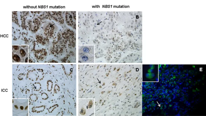
Because three of the eight missense mutations located in the binding domain of Nbs1, we examined Mre11 nuclear staining by performing IHC and IF assays (when frozen tissue was available) on all tumors with NBS1 mutations to determine whether the NBS1 mutations have functional consequences on the binding of Nbs1 to Mre11; 10 HCC and 10 ICC cases without NBS1 mutations served as controls. Disruption of the Mre11 binding domain of Nbs1 may lead to loss of Mre11 nuclear localization and increased staining for Mre11 in the cytoplasm [β1]. Strong Mre11 nuclear staining was observed in all tumors without NBS1 mutations. Down-regulation and/or loss of nuclear localization of Mre11 with cytoplasmic Mre11 staining was observed in three of the eight tumors with NBS1 mutations: case 4β5 with mutation S6γ8P, case γ75 with mutation S6γγT and case γ6β with mutation T90S (Figure γA– E). S6γ8P and S6γγT are in the Mre11 binding domain (601– 700) of Nbs1 and may impair binding of Mre11 to Nbs1, consistent with the previous report described above.
We next analyzed the impact of NBS1 mutations on Nbs1 phosphorylation by western blot and IHC analysis. Altered Nbs1 phosphorylation was not observed in any of the tumors with NBS1 mutation (Figure S1).
NBS1 mutations frequently accompanied with genetic alterations in the TP53 pathway
Genetic alterations in the TP53 pathway in the 8β analyzed cases of primary liver cancer are summarized in Table γ. TP53 mutations were identified in 1γ.4% cases (11/8β), including one frameshift mutation (616del1ins14), one stop mutation (Gβ98X), nine missense point mutations (V157P, Pγ01L, Q19βH, Rβ48G, Rβ49S, Eβ85K, Rβ7γC, Rβ86V and Yββ0C) (Figure 4A). All except the frameshift mutation are known TP53 mutations registered in the International Agency for Research on Cancer TP5γ Database (R15 release, http://www-p5γ.iarc.fr). No case had more than one TP53 mutation. MDM2 amplification, p14ARF homozygous deletion and p14ARF promoter methylation were identified in five (6.1%), seven (8.5%) and β5 (γ0.5%) cases, respectively, and 45 (54.9%) tumors contained at least one genetic alteration in the TP53 pathway (Figure 4B-D).
Of the eight tumor cases with NBS1 mutations, seven (87.5%) had at least one concomitant genetic alteration in the TP53 pathway (P =0.067β, table γ) and three carried TP53 mutations (P =0.0701, table γ). Notably, of the five cases of tumor with the five NBS1 mutations which are predicted to be damaging to Nbs1 function, three carried TP53 mutations (P =0.0159).
Discussion
Many genetic and epigenetic changes have been identified in precancerous hepatic lesions and in HCC, including
Figure 2. Distribution and type of NBS1 mutations in HCC and ICC. Mutations are located preferentially in exon 11/1β (Mre11-binding domain), but also in or close to other functional domains (FHA domain; second BRCT (BRCTβ) domain; ATM phosphorylated sites, Serβ78/Serγ4γ/Serγ97/Ser615).
chromosomal amplification, mutations, loss of heterozygosity and global DNA hypermethylation [γ,ββ]. The Wnt/ -catenin pathway is commonly disrupted in HCC, usually as a result of mutations in CTNNB1 or AXIN1, or epigenetic silencing of CDH1 [ββ]. The p5γ and Rb1 pathways are often disturbed in HCC, and somatic mutations in TP53 has been reported with the rate of 14–γ5% worldwide depending on the level of aflatoxin exposure [βγ,β4].The PIγK/Akt/mTOR pathway is also commonly disrupted, sometimes due to abnormal inactivation of tyrosine kinase receptors or as a result of constitutive activation of PI3K following loss of function of the tumor-suppressor gene PTEN [β5]. Derangements of other signal transduction pathways, such as the MAPK pathway and the TGF-β pathway, also play roles in hepatocarcinogenesis [βγ]. Therefore, HCC is characterized by remarkable molecular heterogeneity. In the present study, we provide the first evidence that mutational inactivation of NBS1, a DNA repair-associated gene, are involved in the pathogenesis of primary liver cancer.
The main functional domains of Nbs1 comprise the forkhead-associated (FHA) domain (amino acids β4–100), the breast cancer C-terminal (BRCT) domain (amino acids 105–190), the
second BRCT domain (amino acids β15–γβ4), and the Mre11-binding domain (amino acids 601–700, Mre11-binding sites 665–69γ), as well as ATM phosphorylation sites [1γ,14]. Both the FHA and BRCT domains are essential for responses to DNA damage, including the formation of ionizing radiation-induced foci, S-phase checkpoint activation and nuclear focus formation after irradiation, and play crucial roles in cell survival after radiotherapy [β6]. The Mre11-binding domain is essential for the formation of the MRN complex, which plays an important role in DNA damage-induced checkpoint control and DNA repair [7]. In the present study, two NBS1 miscoding mutations were located in the FHA domain; one NBS1 miscoding mutations was located in the second BRCT domain; three NBS1 mutations were located close to the ATM phosphorylation sites; notably, three mutations were located in the Mre11-binding domain (Figure β). Heterozygous NBS1 mutations causing loss of the Mre11-binding domain predispose carriers to common types of cancer [β7,β8]. Thus, the NBS1 mutations identified in the present study are likely to play significant roles in the pathogenesis of primary liver cancer.
Figure 3. Mre11 nuclear staining in tumor cells with or without NBS1 mutations. Upper panel: IHC of a pair of HCC cases, (A) without NBS1 mutation (case γ74) and (B) with NBS1 mutation S6γγT (case γ75). Bottom panel: IHC of a pair of ICC cases, (C) without NBS1 mutation (case γ8β) and (D) with NBS1 mutation S6γ8P (case 4β5); immunofluorescence staining of case 4β5 for which frozen tissue was available (E). The black/white arrows indicate tumor cells with nuclear or cytoplasmic staining of Mre11. Loss of Mre11 nuclear staining was observed in tumor cells from cases γ75 and 4β5 with NBS1 mutations in the Mre11-binding domain (S6γγT and S6γ8P). Original magnification: ×40.
Several studies have shown that somatic heterozygous NBS1 mutations in sporadic tumors have functional consequences for the Nbs1 protein [11,β7,β9]. Heterozygous NBS1 splicing mutation (IVS11+βinsT) detected in sporadic gastric, colorectal and lung cancers led to defects in crucial binding to Mre11, Mdc1 and BRCA1, and caused impaired ATM phosphorylation in response to low-dose irradiation in the heterozygous state [β7]. In breast cancer cells with a NBS1 missense mutation (Rβ15W), levels of radiation-induced phosphorylation of Nbs1 (Serγ4γ) were reduced to 60% of wild-type [11]. Missense NBS1 mutations (L150F and I171V) were associated with chromosomal instability in sporadic breast cancer [β9]. In the present study, three of the eight NBS1 mutations (F60γL, S6γ8P and S6γγT) are located in the Mre11-binding domain of Nbs1. Since the disruption of the Mre11 binding domain of Nbs1 may lead to loss of Mre11 nuclear localization and increased staining for Mre11 in the cytoplasm [β1], we were able to analyze the functional consequences of the three NBS1 mutations located in the Mre11-binding domain by the analysis of nuclear localization of Mre11 in tumor cells. Loss of the nuclear localization of Mre11, one of the hallmarks for Nbs1 deficiency, was observed in two tumors with the NBS1 mutation S6γ8P or S6γγT, suggesting an alteration of the function of the MRN complex, consistent with previous reports that loss of Nbs1 protein from the Mre11-binding domain disrupts the nuclear localization of Mre11 and lead to deficiency in DNA DSBs repair [β1,β7]. The role of the Table 3. Association between genetic alterations in the
TP53 pathway and NBS1 mutation in HCC and ICC.
HCC (n=64) ICC (n=18) PLC* (n=82)
NBS1 mutation YesNo P-valueaYesNo P-valueaYesNo P-valuea
TP53 mutation
Yes β 7 0.1957 1 1 0.β157 γ 8 0.0701
No 4 51 1 15 5 66
MDM2 amplification
Yes 1 γ 0.γγββ 0 1 1.0000 1 4 0.4096
No 5 55 β 15 7 70
p14 homozygous deletion
Yes 1 5 1.0000 0 1 1.0000 1 6 1.0000
No 5 5γ β 15 7 68
p14 promoter methylation
Yes β 18 1.0000 β γ 0.065γ 4 β1 0.βγ89
No 4 40 0 1γ 4 5γ
At least one of the above four alterations
Yes 5 γγ 0.γ875 β 5 0.1γ7γ 7 γ8 0.067β
No 1 β5 0 11 1 γ6
aFisher’s exact test. *PLC: primary liver cancer.
doi: 10.1γ71/journal.pone.008β4β6.t00γ
mutated Nbs1 protein on HCC progression is important to evaluate its potential functional consequence on tumorigenesis. However, NBS1 is a “caretaker” gene, which do not directly promote tumor development when inactivated. Various expression levels of Nbs1 have been detected in three HCC cell lines (HepGβ, Hep-γB and HepGβ.β.15) and one ICC cell line HCCC-9810 (Figure 5), and notably, Nbs1 null mutant cell lines is not viable due to the functional importance of MRN complex in cells. If we express exogenous mutant Nbs1 in HCC cells, we cannot rule out the possible role of endogenous Nbs1 in the cells. Thus, it is hard to conclude the role of NBS1 mutations in HCC progression by construction of the NBS1 mutation vectors and express mutated Nbs1 in HCC cell lines. It is noteworthy that, increased HCC incidence in heterozygote NBS1 mutant mice provided a strong genetic evidence for the role of NBS1 mutations in HCC [18].
Several studies have demonstrated a possible cooperation of loss of TP53 and NBS1 in the pathogenesis of neoplasms [1γ,14,16,17,γ0]. In addition to its role in DNA double-strand break repair, Nbs1 directly binds to Mdmβ, a negative regulator of p5γ, and co-localizes to sites of DNA damage caused by gamma-irradiation, while Mdmβ over-expression inhibited DNA double-strand break repair associated with Nbs1 [16]. Using a humanized p5γ knock-in allele in mice, Song et al. demonstrated that the common p5γ mutants disrupt the earliest stage of DNA double-strand break damage response by interfering with recruitment of the MRN complex to the site of DNA damage, leading to inactivation of ATM and genetic instability [γ0]. In NBS1 mutant/mutant mice expressing N-terminally truncated Nbs1, p5γ deficiency greatly facilitated the development of thymic lymphomas [17]. Moreover, mutational inactivation of the NBS1 gene in tumors is associated with TP53 mutations in sporadic medulloblastoma and primary glioblastoma [1γ,14]. On the other hand, it was recently reported that Nbs1 regulates a novel p5γ independent apoptotic pathway through BAX activation in response to DNA damage and that DNA damage-induced apoptosis is significantly reduced in Nbs1-deficient cells regardless of their p5γ status [γ1]. However, the present study notably showed the co-presence of NBS1 mutations with genetic alterations in the TP53 pathway, implying a synergistic effect of Nbs1 disruption and p5γ inactivation in liver cancer development. Although the relationship between NBS1 mutations and TP53 pathway alterations is not statistical significant, probably due to a limited number of tumor cases analyzed, our data has provided a potential possibility for such relationship, with the p-value around the threshold, and with statistic significant result (p=0.0159) when cases with the NBS1 mutations which are predicted to be damaging to Nbs1 function were analyzed.
Figure 4. Genetic alterations in the TP53 pathway in representative tumor cases. (A) Representative DNA sequencing of TP53 mutations: TP53 mutation Yββ0C in case γ8γ with NBS1 mutation Vγ48D, TP53 mutation Pγ01L in case β17 with NBS1 mutation I41M. (B) MDM2 amplification in case γ54 with NBS1 mutation S415R. The dopamine receptor (DR) sequence was used as the reference. (C) p14ARF homozygous deletion in case β17 with NBS1 mutation I41M. The GAPDH sequence was used as the reference. (D) p14ARF promoter methylation in case 4β5 with NBS1 mutation S6γ8P. U: unmethylated; M: methylated; PC: methylated DNA (Zymo Research, Irvine, CA); NC: unmethylated DNA from normal blood; M: 50 bp DNA ladder.
doi: 10.1γ71/journal.pone.008β4β6.g004
Figure 5. Endogenous expression of Nbs1 in HCC or ICC cell lines. Western blots analysis of endogenous expression of Nbs1 in three HCC cell lines (HepGβ, Hep-γB and HepGβ.β.15 ) and one ICC cell line(HCCC-9810). -actin was used as protein loading control.
Supporting Information
Figure S1. Nbs1 expression and p-Nbs1 level in tumor cells with or without NBS1 mutation. (A) Western blot analysis for detection of Nbs1 expression and p-Nbs1 level in five representative tumor cases: cases 478 (HCC) and 4β5 (ICC) with NBS1 mutation Dβ7βN and S6γ8P, Cases γβ7,γγγ and γ4γ without NBS1 mutation, -actin was used as the reference.(B) Representative IHC of Nbs1 expression and p-Nbs1 level in tumor tissue of one HCC case (case 478). Original magnification: ×40.
(TIF)
Table S1. Association between NBS1 mutations and clinical parameters in 64 cases of HCC.
(DOC)
Acknowledgements
The authors would like to thank Dr. Zheng-Yu Wei (Division of Transplantation, Department of Surgery, Thomas Jefferson University, Philadelphia, PA) for critical manuscript reading and helpful suggestions.
Author Contributions
Conceived and designed the experiments: WMT JDJ JH. Performed the experiments: YW YH ML JL YPZ JXZ. Analyzed the data: QL HY WMT JDJ JH. Contributed reagents/materials/ analysis tools: YW JL JXZ JDJ JH. Wrote the manuscript: YW WMT JDJ JH.
References
1. Theise ND, Curado MP, Franceschi S, Hytiroglou P, Kudo M et al. (β010) Hepatocellular carcinoma. In: FT BosmanF CarneiroRH HrubanND Theise. WHO Classification of Tumors of the Digestive. System: β05-β16.
β. Nakanuma Y, Curado MP, Franceschi S, Gores G, Paradis V et al. (β010) Intrahepatic cholangiocarcinoma. In: FT BosmanF CarneiroRH HrubanND Theise. WHO Classification of Tumors of the Digestive. System: β17-ββ4.
γ. Thorgeirsson SS, Grisham TW (β00β) Molecular pathogenesis of human hepatocellular carcinoma. Nat Genet γ1: γγ9-γ46. doi:10.10γ8/ ng080β-γγ9. PubMed: 1β14961β.
4. Paterlini-Bréchot P, Saigo K, Murakami Y, Chami M, Gozuacik D et al. (β00γ) Hepatitis B virus related insertional mutagenesis occurs frequently in human liver cancers and recurrently targets human telomerase gene. Oncogene ββ: γ911-γ916. doi:10.10γ8/sj.onc. 1β0649β. PubMed: 1β81γ464.
5. Zhu DQ, Huang ZY (β007) DNA damage and Iiver cancer. World Chinese Journal of Digestology.15: 1775-1780.
6. Stracker TH, Theunissen JW, Morales M, Petrini JH (β004) The MRE11 complex and the metabolism of chromosome breaks: the importance of communicating and holding things together. DNA Repair (Amst) γ: 845-854. doi:10.1016/j.dnarep.β004.0γ.014. PubMed: 15β79769. 7. Bill CA, Summers T (β004) Genomic DNA double-strand breaks are
targets for hepadnaviral DNA integration. Proc Natl Acad Sci of the USA 101: 111γ5-11140. doi:10.107γ/pnas.040γ9β5101. PubMed: 15β58β90.
8. Dandri M, Burda MR, Bürkle A, Zuckerman DM, Will H et al. (β00β) Increase in de novo HBV DNA integrations in response to oxidative DNA damage or inhibition of poly(ADPribosyl) ation. Hepatology γ5: β17-ββγ. doi:10.105γ/jhep.β00β.γ0β0γ. PubMed: 11786979.
9. Varon R, Seemanova E, Chrzanowska K, Hnateyko O, Piekutowska-Abramczuk D et al. (β000) Clinical ascertainment of Nijmegen breakage syndrome (NBS) and prevalence of the major mutation, 657del5, in three Slav populations. Eur J Hum Genet 8: 900-90β. doi: 10.10γ8/sj.ejhg.5β00554. PubMed: 1109γβ81.
10. Lee JH, Lim DS (β006) Dual role of NBS1 in the ataxia telangiectasia mutated-dependent DNA damage responseFEBS J β7γ: 16γ0-16γ6. doi:10.1111/j.174β-4658.β006.05191.x. PubMed: 166βγ700.
11. Bogdanova N, Feshchenko S, Schürmann P, Waltes R, Wieland B et al. (β008) Nijmegen breakage syndrome mutations and risk of breast cancer. Int J Cancer 1ββ: 80β-806. doi:10.100β/ijc.βγ168. PubMed: 17957789.
1β. Nowak J, Mosor M, Ziółkowska I, Wierzbicka M, Pernak-Schwarz M et al. (β008) Heterozygous carriers of the I171V mutation of the NBS1 gene have a significantly increased risk of solid malignant tumors. Eur J Cancer 44: 6β7-6γ0. doi:10.1016/j.ejca.β008.01.006. PubMed: 18β807γβ.
1γ. Huang J, Grotzer MA, Watanabe T, Hewer E, Pietsch T et al. (β008) Mutations in the Nijmegen breakage syndrome gene in medulloblastomas. Clin Cancer Res 14: 405γ-4058. doi: 10.1158/1078-04γβ.CCR-08-0098. PubMed: 1859γ981.
14. Watanabe T, Nobusawa S, Lu SQ, Huang J, Mittelbronn M et al. (β009) Mutational inactivation of the Nijmegen breakage syndrome gene (NBS1) in glioblastomas is associated with multiple TP5γ mutations. J
Neuropathol Exp Neurol 68: β10-β15. doi:10.1097/NEN. 0b01γeγ18197β4cβ. PubMed: 191516β0.
15. Chrzanowska KH, Piekutowska-Abramczuk D, Popowska E, Gładkowska-Dura M, Małdyk J et al. (β006) Carrier frequency of mutation 657del5 in the NBS1 gene in a population of Polish pediatric patients with sporadic lymphoid malignancies.Int J Cancer 118: 1β69-1β74. doi:10.100β/ijc.β14γ9. PubMed: 1615β606.
16. Alt JR, Bouska A, Fernandez MR, Cerny RL, Xiao H et al. (β005) Mdmβ binds to NBS1 at sites of DNA damage and regulates double strand break repair. J Biol Chem β80: 18771-18781. doi:10.1074/ jbc.M41γγ87β00. PubMed: 157γ474γ.
17. Kang J, Ferguson D, Song H, Bassing C, Eckersdorff M et al. (β005) Functional interaction of HβAX, NBS1, and p5γ in ATM-dependent DNA damage responses and tumor suppression. Mol Cell Biol β5: 661-670. doi:10.11β8/MCB.β5.β.661-670.β005. PubMed: 156γβ067. 18. Dumon-Jones V, Frappart PO, Tong WM, Sajithlal G, Hulla W et al.
(β00γ) Mice heterozygous for NBN (the murine homologue of NBS1) developed a wide variety of tumors affecting the liver, mammary gland, prostate, and lung, in addition to lymphomas. Cancer Res 6γ: 7β6γ-7β69. PubMed: 1461β5ββ.
19. Huang J, Zhao YP, Li Q, Zhang JX, Wang Y et al. (β01β) Association of single nucleotide polymorphisms of NBS1 gene with genetic susceptibility to primary liver cancer in a Chinese Han population. Prog Biochem Biophys γ9: 678-686. doi:10.γ7β4/SP.J.1β06.β011.005γ6. β0. Di YM, Chan E, Wei MQ, Liu JP, Zhou SF (β009) Prediction of
Deleterious Non-synonymous Single-Nucleotide Polymorphisms of Human Uridine Diphosphate Glucuronosyltransferase Genes. AAPS J 11: 469-480. doi:10.1β08/s1ββ48-009-91β6-z. PubMed: 1957ββ00. β1. Yang YG, Frappart PO, Frappart L, Wang ZQ, Tong WM (β006) A
novel function of DNA repair molecule NBS1 in terminal differentiation of the lens fibre cells and cataractogenesis. DNA Repair (Amst) 5: 885-89γ. doi:10.1016/j.dnarep.β006.05.004. PubMed: 16790γ66. ββ. Mínguez B, Tovar V, Chiang D, Villanueva A, Llovet JM (β009)
Pathogenesis of hepatocellular carcinoma and molecular therapies. Curr Opin Gastroenterol β5: 186-194. doi:10.1097/MOG. 0b01γeγβ8γβ96βa1. PubMed: 19γ87β55.
βγ. Aravalli RN, Steer CJ, Cressman EN (β008) Molecular mechanism of hepatocellular carcinoma. Hepatology 48: β047-β06γ. doi:10.100β/hep. ββ580. PubMed: 1900γ900.
β4. Ozturk M (1991) p5γ mutation in hepatocellular carcinoma after aflatoxin exposure. Lancet γγ8: 1γ56-1γ59. doi: 10.1016/0140-67γ6(91)9ββγ6-U. PubMed: 168β7γ7.
β5. Villanueva RR, Chiang DY, Newell P, Peix J, Thung S et al. (β008) Pivotal role of mTOR signaling in hepatocellular carcinoma. Gastroenterology 1γ5: 197β-198γ. doi:10.105γ/j.gastro.β008.08.008. PubMed: 189β9564.
β6. Zhao S, Renthal W, Lee EY (β00β) Functional analysis of FHA and BRCT domains of NBS1 in chromatin association and DNA damage responses. Nucleic Acids Res γ0: 4815-48ββ. doi:10.109γ/nar/gkf61β. PubMed: 1β4γγ98γ.
11158-11165. doi:10.1158/0008-547β.CAN-07-1749. PubMed: 18056440.
β8. Desai-Mehta A, Cerosaletti KM, Concannon P (β001) Distinct functional domains of nibrin mediate MRE11 binding, focus formation, and nuclear localization. Mol Cell Biol β1: β184-β191. doi:10.11β8/MCB. β1.6.β184-β191.β001. PubMed: 11βγ8951.
β9. Heikkinen K, Rapakko K, Karppinen SM, Erkko H, Knuutila S et al. (β006) RAD50 and NBS1 are breast cancer susceptibility genes
associated with genomic instability. Carcinogenesis β7: 159γ-1599. PubMed: 16474176.
γ0. Song H, Hollstein M, Xu Y (β007) p5γ gain-of-function cancer mutants induce genetic instability by inactivating ATM. Nat Cell Biol 9: 57γ-580. doi:10.10γ8/ncb1571. PubMed: 174176β7.