Arch Bone Jt Surg. 2016; 4(2): 109-115. http://abjs.mums.ac.ir
the online version of this article abjs.mums.ac.ir
Edward K. Rodriguez, MD, PhD; Jeeva Munasinghe, PhD
Research performed at Department of Orthopedic Surgery, Beth Israel Deaconess Medical Center, Boston, USA
joint morphogenesis is predetermined by genetic programming, modulated by mechanical factors, or both (13). Is motion a necessary factor throughout joint development, having a role in the modulation of gene expression that results in a normal joint phenotype? Or is the role of motion critical only at a specific time point in morphogenesis just as a means of providing a shearing force to initialize cavitation in a joint that is otherwise predetermined to form normally?. An understanding of the dependence of joint morphogenesis on embryonic
Corresponding Author: Edward K. Rodriguez, Orthopedic
Trauma Beth Israel Deaconess Medical Center, Harvard Medical School, Boston, USA
Email: [email protected]
Received: 20 June 2015 Accepted: 21 October 2015
A Chick Embryo in-Vitro Model of Knee Morphogenesis
Abstract
Background: In this feasibility study, a mechanically loaded in-vitro tissue culture model of joint morphogenesis using the isolated lower extremity of the 8 day old chick embryo was developed to assess the effects of mechanical loading on joint morphogenesis.
Methods: The developed in-vitro system allows controlled lexion and extension of the chick embryonic knee with a range of motion of 20 degrees from a resting position of 90-100 degrees of lexion. Joint morphogenesis at 2, 3, 4 and 7 days of culture was assessed by histology and micro MRI in 4 specimen types: undisturbed in-ovo control embryos, in-ovo paralyzed embryos, in-vitro unloaded limb cultures, and in-vitro loaded limb cultures. Relative glycosaminoglycan (GAG) concentration across the joint was assessed with an MRI technique referred to as dGEMRIC (delayed gadolinium enhanced MRI of cartilage) where T1 is proportional to glycosaminoglycan concentration.
Results: Average T1 over the entire tissue image for the normal control (IC) knee was 480 msec; for the 4 day loaded specimen average T1 was 354 msec; and for the 7 day loaded specimens T1 was 393 msec. The 4 day unloaded specimen had an average T1 of 279 msec while the 7 day unloaded specimen had an average T1 of 224 msec. The higher T1 values in loaded than unloaded specimens suggest that more glycosaminoglycan is produced in the loaded culture than in the unloaded preparation.
Conclusion: Isolated limb tissue cultures under lexion-extension load can be viable and exhibit more progression of joint differentiation and glycosaminoglycan production than similarly cultured but unloaded specimens. However, when compared with controls consisting of intact undisturbed embryos in-ovo , the isolated loaded limbs in culture do not demonstrate equivalent amounts of absolute growth or joint differentiation.
Keywords: chick embryo, chick embryo culture, Glycosaminoglycan, in-vitro loaded limb culture, joint morphogenesis, paralyzed chick embryo
Introduction
limb motion may provide new insight into pathologies such as arthrogryposis congenita and other conditions involving joint deformities secondary to altered embryonic motion.
To study the effects of motion on morphogenesis of the knee we developed an in-vitro chick embryo model
in which knee flexion and extension can be controlled and applied to the isolated lower extremity limb. The chick embryo is an established model for the study of joint morphogenesis and has a well described embryonic development and staging (14).. It has been
widely used for the study of joint histology, skeletal limb development, and to investigate the role of motion in joint formation (1-3, 6-13,15,16). It provides a reliable and rapidly developing animal model for the study of in-vivo limb morphogenesis and in-vitro limb tissue cultures.
The present in-vitro model was used for a feasibility
assessment of the histologic and morphometric effects of controlled flexion and extension on joint morphogenesis. Chick embryo lower limbs were obtained at incubation days 6 through 10, corresponding to Hamburger Hamilton (HH) stages 30 through 36. They were then cultured for up to 7 days under load (flexion-extension). Control groups included unloaded age-matched isolated limbs
in-vitro, intact undisturbed embryos in-ovo, and intact paralyzed embryos in-ovo. Morphometric differences in joint development were assessed by histology, and micro MRI which used to provided a measure of glycosaminoglycan concentration after 4 and 7 days of culture in unloaded vs. loaded limbs (17).
Material and Methods
Fertilized Legghorn chick embryos (SPAFAS, CT) were incubated in a desktop incubator (Lyon Electric Co., CA) in atmospheric pressure, at 99.7 degrees F of temperature, and 54-59% relative humidity with periodic rolling. Incubation times were scheduled based on estimates of fertilization time provided by the commercial supplier. Joint cavitation starting at incubation day 9-10 (HH stage 36).
Four experimental groups were defined:
Loaded isolated limbs in in-vitro culture (LIL group): Limbs harvested at days 8 through 10 of incubation and cultured under flexion-extension for 2 to 7 days .
Unloaded isolated limbs in in-vitro culture: (UIL group): Limbs harvested at days 6 through 10 of incubation, including the contralateral extremities of each limb in the LIL group. These were cultured in an unloaded state but otherwise under similar conditions as the LIL group.
Table 1a. Dimensions of normal embryos at time of harvest (IC)
Day at dissection
Stage at
Dissection n
femur length (mm+/-sdev)
tibial length (mm+/-sdev)
AP dim.At knee (mm+/-sdev)
stress free angle (degree+/-sdev)
8 34 14 4.32+/-1.16 4.35+/-1.23 1.92+/-0.49 122+/-28 9 35 11 5.76+/-0.53 6.45+/-1.13 1.57+/-0.20 108+/-12 10 36 26 6.66+/-1.56 7.83+/-1.82 1.89+/-0.36 126+/-15 11 37 4 8.27+/-0.36 9.16+/-0.40 1.58+/-0.27 131+/-23 12 38 11 9.32+/-0.93 11.59+/-1.01 2.22+/-0.34 125+/-4 14 40 12 14.29+/-1.65 19.71+/-1.03 3.61+/-0.43 87+/-11 15 41 8 15.15+/-0.94 17.89+/-1.01 4.28+/-0.71 112+/-11
Intact in-ovo controls (IC group): Age matched controls incubated in-ovo, in their shells, undisturbed to day 15.
Intact in-ovo paralyzed controls (IPC group): Age matched controls incubated in-ovo, in their shells, to day 15, after injection of a paralyzing agent at day 7 of incubation (HH stage 30-33).
Embryos were harvested at days 8 to 10 for the LIL group, days 6 to 10 for the UIL group, and at days 12 to 15 for the IC and IPC groups. Embryos in the LIL and UIL groups were staged and cultured, and those in the IC and IPC groups were staged and fixed for histology. Staging was confirmed by morphologic comparison with Hamburger-Hamilton standards.
After staging of the intact embryo assigned for culture, each specimen was euthanized by decapitation and both lower limbs were dissected free under 2.5X magnification using sterile technique. All musculature was dissected free from each specimen with preservation of an intact femur, tibia, and patella. Dissected free limb specimens were digitally photographed prior to culture or fixation. Femoral length, tibial length, and the unloaded stress-free knee flexion angle were recorded.
Each specimen in the LIL group was mounted on a hinged plastic frame with 6.0 vicryl or nylon suture [Figure 1a]. The plastic frame served to attach the specimen to the loading apparatus and supported the specimen by allowing knee flexion and extension as the
single mode of loading. A loader capable of loading 8 limbs simultaneously was designed and constructed from off-the-shelf components. The loader has four 100x15mm glass culture dishes, each modified to hold a hinged plastic frame supporting two specimens. It is powered by a tubular pull solenoid (S-15-75-28, Magnetic Sensor Systems, CA) controlled by a custom built timing circuit [Figure 1b]. Design parameters for the loader were based on studies reporting a total knee range of motion of 20 degrees in one flexion-extension cycle (LLusa-Perez 1988). From a resting angle of 90-100 degrees, each joint was flexed or extended by 20 degrees once a minute and held at the new flexion angle for 15 seconds before returning to the rest angle. Loading was done throughout the duration of the culture phase of each experiment (2 to 7 days).
Limbs in the LIL and UIL groups were cultured in F12 media with 10%FBS, 1% antibiotics (5 mg/ ml gentamycin, 100U/ml penicillin, 100 mg/ml streptomycin), and 37.5 mg/ml Ascorbate with phosphate.
Loaded limbs in the LIL group were cultured in 100x15mm glass culture dishes modified to support a hinged plastic frame. Each dish held 40-50 ml of media changed every other day. Unloaded limbs in the UIL group were cultured in independent 35x10mm dishes or in 6 well plates, each filled with 3-5ml of media, changed every day. Incubator temperature was of 37.6 degrees.
Table 1b. Dimensions of paralyzed embryos at time of harvest (IPC)
Day at dissection Stage at Dissection n femur length (mm+/-sdev)
tibial length (mm+/-sdev)
AP dim, At knee (mm+/-sdev)
stress free angle (degree+/-sdev)
14 40 6 9.24+/-0.82 12.19+/-1.52 2.24+/-0.41 99+/-25 15 41 31 11.73+/-1.59 15.63+/-1.80 2.91+/-0.42 82+/-17
Figure 2a, b. A) Normal joint development with cavitation, meniscal development, and well demarcated epiphyses in the
in-vivo controls (IC). B) Paralyzed embryos demonstrated varying degrees of fibrosis, abnormal cavitation, lack of intrarticular
The post synaptic depolarizing agent Decamethonium Bromide (DMB) was used to achieve embryonic paralysis in the IPC group. Two daily doses of 0.25 ml of DMB solution (20mg DMB in 100ml of Buffered Saline solution with 100U/ml penicillin (Mikic 2000) dropped on the chorioallantoic membrane achieved paralysis in intact embryos. Eggs were sterilized with betadine and a 2mm diameter hole was drilled under aseptic conditions. The access hole was covered with sterile tape (Tegaderm) after each application of DMB. An embryo that had a beating heart at the time of harvest, with significantly decreased muscle mass, and no observed motion for 60 seconds after removal from the shell was considered paralyzed.
All specimens were fixed in Cacodylate buffered 2% paraformaldehyde solution. Specimens for MRI were preserved fresh in media and refrigerated. Specimens for histology were dehydrated in ethanol (70-90% series) and embedded in water soluble glycomethacrylate resin (JB4 system, Polysciences). Sectioning was done with a Reichert-Jung microtome (5 mm thickness). All sections were stained with 1%
Toluidine Blue. Histology was done for the IC group and IPC group specimens at 15 days. Specimens in the LIL and UIL were studied at 2, 3 and 7 days of culture.
Intact joint morphometry and relative
glycosaminoglycan concentration in the embryonic joint tissues was assessed by MRI using a Bruker 8.45 T microimaging system (Bruker Instruments, Billerica, MA). A 4 mm solenoid radio frequency (rf) coil was used. The excised knee was removed from its preserving solution and immersed in fluorocarbon oil called Fomblin (Ashmont Inc. USA) and mounted in the rf coil. Fomblin does not produce an MRI signal, but serves to decrease edge artifacts by eliminating the tissue/air interface.
Relative glycosaminoglycan concentration across the joint was assessed with an MRI technique referred to as dGEMRIC (delayed gadolinium enhanced MRI of cartilage) (17). dGEMRIC is based on the principle that the negatively charged contrast agent Gd(DTPA)
2-(Magnevist; Berlex, NJ) will distribute in cartilage in a manner inversely proportional to the concentration of negatively charged glycosaminoglycan molecules. The Gd(DTPA)2- concentration is reflected in the MRI
parameter T1; hence T1 images were used to assess relative glycosaminoglycan (GAG) concentration across the joint.
One normal knee at 15 days of incubation, one 4 and one 7 day in-vitro loaded specimens (LIL), and the two corresponding contralateral UIL specimens were equilibrated in 400 cm3 aliquots of 0.5 mMol
Gd(DTPA)2-/Hanks solution for approx. 48 hours
and the T1 imaging was then performed. All images were acquired with an inversion recovery spin-echo imaging sequence with an in-plane resolution of 100
mm and slice thickness of 250 mm. An echo time of 6.6
ms was used while the relaxation was 3 s for the Gd equilibrated samples. These images subsequently were used to calculate T1 values for each pixel to generate T1 maps.
Results
A total of 445 eggs were used in the present study. Attrition rates were: unfertilized eggs 5-10%, dead with DMB 72% of 91 paralyzed eggs, and 24% contaminated cultures. We successfully obtained 54 in-ovo paralyzed limbs (IPC), 75 intact in-ovo control limbs (IC), 125 unloaded in-vitro limbs (UIL) and 42 loaded in-vitro limbs (LIL).
Intact in-ovo controls (IC) demonstrated the greatest
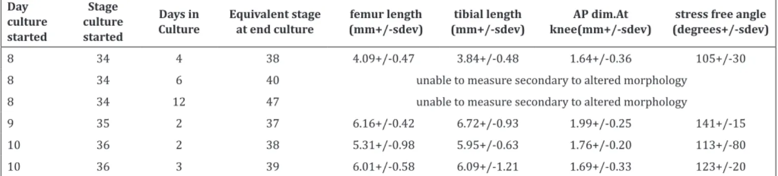
Table 2. Dimensions of unloaded cultured limbs at end culture (UIL)
Day culture
started
Stage culture
started
Days in Culture
Equivalent stage at end culture
femur length (mm+/-sdev)
tibial length (mm+/-sdev)
AP dim.At knee(mm+/-sdev)
stress free angle (degrees+/-sdev)
8 34 4 38 4.09+/-0.47 3.84+/-0.48 1.64+/-0.36 105+/-30 8 34 6 40 unable to measure secondary to altered morphology
8 34 12 47 unable to measure secondary to altered morphology
9 35 2 37 6.16+/-0.42 6.72+/-0.93 1.99+/-0.25 141+/-15 10 36 2 38 5.31+/-0.98 5.95+/-0.63 1.76+/-0.20 113+/-80 10 36 3 39 6.01+/-0.58 6.09+/-1.21 1.69+/-0.33 123+/-20
Figure 3. Histological sections of unloaded and loaded specimens after 2 and 3 days of culture. Articular surfaces in loaded specimens
at these times appear more defined with evidence of early cavitation
absolute growth with the most development of muscle mass. By day 15 of incubation femur length averaged from 8 specimens was 15.2+/- 0.9 mm, tibial length was 17.9+/-1.0 mm, and the AP dimension at the knee was 4.3+/-0.7mm. The knee flexion angle at rest was 112+/-11 degrees [Table 1a]. Intact in-ovo paralyzed controls (IPC) showed less longitudinal growth (P<0.05) [Table 1b]. This group exhibited less muscle
development, rigid joints, and an arthrogrypotic posture. In 31 specimens at 15 days of incubation, femur length was 11.7+/-1.6mm, tibia length was 15.6+/-1.8 mm and AP dimension was 2.9+/- 0.4 mm. Knee stress-free flexion angle showed a contracted joint at 82+/-17 degrees when compared with the IC specimens (P<0.05). Histological comparison showed normal joint development with cavitation, meniscal development, and well demarcated epiphyses in the in-ovo controls (IC), while the in-in-ovo paralyzed embryos demonstrated varying degrees of fibrosis, abnormal cavitation, lack of intrarticular elements, and abnormal epiphyses [Figure 2].
Isolated unloaded cultured specimens did not exhibit significant linear growth. Dimensions after 2 days of unloaded culture in 12 specimens harvested at day 9 of incubation were 6.2+/-0.4mm of femur length, 6.7+/-0.6mm of tibial length and 2.0+/-0.3mm of AP knee dimension. Stress-free knee flexion angle was 113+/-80 degrees [Table 2]. Specimens harvested at 8 days of incubation and cultured for more than 6 days had significantly altered growth geometry with loss of typical limb characteristics. Linear growth in loaded specimens was not quantified since the method to secure them to the loading hinges with sutures did not allow for free linear growth.
Minor qualitative differences in joint morphology can be appreciated when comparing unloaded and loaded specimens after 2 and 3 days of culture [Figure 3]. Articular surfaces in loaded specimens at these times appear more defined with evidence of early cavitation that is not apparent in the contralateral unloaded specimens. However, by 7 days of culture these differences did not become more apparent.
The poor resolution of joint differences using traditional histology may be in part secondary to processing artifacts resulting from dehydration, tissue fixation, embedding and sectioning. These processes may alter joint cavity size and articular geometry in unpredictable ways. MRI T1 images were used to asses glycosaminoglycan concentration in a normal specimen [Figure 4], two LIL specimens loaded for 4 and 7 days in culture, and the two contralateral UIL specimens cultured unloaded for 4 and 7 days [Figure 5]. T1 values at two different sites in each image are illustrated for each specimen in Figure 5. Average T1 over the entire tissue image for the in-ovo normal control (IC) knee was 480 msec; for the 4 day loaded specimen average T1 was 354 msec; and for the 7 day loaded specimens T1 was 393 msec. The 4 day unloaded specimen had an average T1 of 279 msec while the 7 day unloaded specimen had an average T1 of 224 msec. The higher T1 values in loaded than unloaded specimens suggest that more GAG is produced in the loaded culture than in the unloaded preparation.
Discussion
We have developed a protocol for in-vitro loading of
Figure 4. T1 imaging in a normal specimen at 15 days if incubation. Average T1 over the entire tissue image for the normal control (IC) knee was 480 msec and is proportional to GAG concentration which is fairly uniform throughout the specimen. To be contrasted with specimens in Figure 5.
the isolated limb of a chick embryo preserved in tissue culture. The present study was designed as a feasibility trial to determine if an intact embryonic joint could be viable in a mechanically stimulated tissue culture undergoing large deformations. Preliminary results using traditional histology and micro MRI suggest that limb tissue cultures under flexion-extension load are viable and that loaded knees appear to exhibit progression of joint morphogenesis that is not evident in similarly cultured but unloaded specimens. When compared with in-ovo controls, the isolated loaded limbs in culture demonstrate lesser amounts of absolute growth and joint differentiation. However, when compared with unloaded limbs cultured under similar conditions, they show further progression and preservation of joint morphology in terms of articular surface geometry, cavitation, and GAG concentration.
Paralysis of the developing limb promotes chondrification of the interarticular space and malformation of diarthrodial joints. This process has been consistently reproduced in various classical models of embryonic paralysis in the chick embryo: decamethonium bromide, botulism toxin, denervation of the lumbosacral spine, viral infection with coxasckievirus A2, and high dose reserpine (5, 7, 8) . While some studies have demonstrated that some peripheral early joint cavitation may occur in the paralyzed embryo (18,19), joints will most often fail to develop fully if motion is not allowed. Early clefting that occurs along the periphery of the joint space by apoptosis of mesenchymal and early chondral cells, was not thought to progress if joint motion is prevented but some studies suggest that joint shape morphogenesis may precede cavitation in the developing joint (12, 20). The dependence of joint cavitation on mechanical loading is still poorly understood (10,11). The natural motion of the in-vivo embryo has been well described but its causality towards joint differentiation is still unclear (19, 21). At 8-10 days of incubation knee motion has been measured at 95 degrees with +/- 8-12 degrees of flexion-extension during a motion cycle. Motion duration as described by Watson et al. can range from 134 msec to 2500 msec (21). Motion was also recorded to occur at intervals of 60 seconds or less; parameters that were used to develop our model. While it is known that embryonic limb motion starts at day 7 and peaks by day 15-17, it is not clear whether the effects of transient paralysis during gestation can be reversible (16). Studies by Mitrovic have shown that transient 3 day paralysis at an early stage when clefting is just commencing (day 8) results in a failure to develop joints similar to that observed in embryos that are paralyzed throughout incubation (20). Paralysis after joint differentiation has occurred (after day 15) results in varying degrees of fibrosis.
The present study was burdened by a high attrition rate for in-ovo paralyzed intact embryos and a high contamination rate in isolated cultures. Despite working in a sterile tissue culture facility, contamination of cultures was a problem given the extensive manipulation required to harvest specimens from
live eggs, dissect them, and mount each specimen for loading. The mortality of paralyzed embryos with DMB is high but not unusual for the method used. Titration of the DMB dose by examination of embryonic motion instead of fixed dosing may be a better approach.
The lesser overall growth exhibited by both loaded and unloaded cultures when compared to in-vivo controls may be a result of culturing the tissue in an avascular culture where diffusion is the only mechanism of nutrition. Alternative culture media, such as albumin, may improve the rate of growth. An alternative to traditional tissue culture is a vascularized culture system with a host embryo feeding isolated grafts. However, mechanical loading as implemented in the present study is not possible in vascularized cultures since the fragile capillary network would be compromised by motion.
Micro MRI, as applied in the present study, provides a morphologic assessment of the developing joint that is not affected by histological processing. By equilibrating cultured specimens with a gadolinium solution, a map of relative glycosaminoglycan distribution in terms of T1 imaging can be obtained (17). Imaging results from our trial specimens suggest that this method can differentiate morphometry non-destructively and with more reliable artifact-free resolution than traditional histology. Results suggest that loaded in vitro knees produce higher amounts of GAG than unloaded specimens. The potential also exists for nondestructive periodic assessment of a growing joint at different times in its development.
In this feasibility study, chick embryonic knees in a mechanically loaded in-vitro culture system, were used to study joint morphology after 2,3, 4 and 7 days of culture. As assessed by micro MRI and histology, the isolated loaded knee joint in tissue-culture does not appear to differentiate as extensively as in the control in-ovo embryo. However, it shows more evidence of intra-articular differentiation and relative GAG production than a similarly cultured but unloaded specimen. The present model describes a viable, mechanically stimulated, isolated, embryonic joint in tissue culture. Together with non-invasive micro MRI examination, the model provides a useful tool to examine how patterns of motion and loading may modulate gene expression and morphology.
Acknowledgments
This work was supported by an Orthopedic Research and Education Foundation resident award.
The authors report no conflict of interest concerning the materials or methods used in this study or the findings specified in this paper.
Edward K. Rodriguez MD, PhD
Orthopedic Trauma Beth Israel Deaconess Medical Center, Harvard Medical School, Boston, USA
Jeeva Munasinghe PhD
Biomech. 2011; 44(1):143-9.
12. Nowlan NC, Chandaria V, Sharpe J. Immobilized chicks as a model system for early-onset developmental dysplasia of the hip. J Orthop Res. 2014; 32(6):777-85. 13. Nowlan NC, Sharpe J. Joint shape morphogenesis
precedes cavitation of the developing hip joint. J Anat. 2014; 224(4):482-9.
14. Hamburger V, Hamilton HL. A series of normal stages in the development of the chick embryo. J Morphol. 1951; 88(1):49-92.
15. Hogg DA, Hosseini A. The effects of paralysis on skeletal development in the chick embryo. Comp Biochem Physiol Comp Physiol. 1992; 103(1):25-8. 16. Llusa-Perez M, Susu-Vergara S, Ruano-Gil D.
Recording of chick embryo movements and their correlation with joint development. Acta Anat. 1988; 132(1):55-8.
17. Allen RG, Burstein D, Gray ML. Monitoring glycosaminoglycan replenishment in cartilage explants with gadolinium-enhanced magnetic resonance imaging. J Orthop Res. 1999; 17(3):430-6. 18. Murray PD, Drachman DB. The role of movement in
the development of joints and related structures: the head and neck in the chick embryo. J Embryol Exp Morph. 1969; 22(3):349-71.
19. Rajan KT, Merker HJ. Preceedings: Joint formation in culture. Ann Rheum Dis. 1975; 34(2):200.
20. Mitrovic D. Development of the articular cavity in paralyzed chick embryos and in the chick embryo limb buds cultured on chorioallantoic membranes. Acta Anat. 1982; 113(4):313-24.
21. Watson SJ, Bekoff A. A kinematic analysis of hindlimb motility in 9- and 10-day old chick embryos. J Neurobiol. 1990; 21(4):651-60.
References
1. Fell HB. The histogenesis of cartilage and bone in the long bones of the embryonic fowl. J Morphol. 1925; 40(3):417-59.
2. Fell HB. Experiments in the differentiation in vitro of cartilage and bone. Part 1 Arch Exp Zellforsh. 1928; 18(7):390-412.
3. Fell HB, Canti RG. Experiments on the development in vitro of the avian knee-joint. Proc R Soc Lond B Biol Sci. 1934; 116(799):316-51.
4. Ruano-Gil D, Nardi-Vilardaga J, Tejedo-Mateu A. Influence of extrinsic factors on the development of the articular system. Acta Anat. 1978; 101(1):36-44. 5. Ruano-Gil D, Nardi-Vilardaga J, Teixidor-Johe A.
Embryonal hypermobility and articular development. Acta Anat. 1985; 123(2):90-2.
6. Hosseini A, Hogg DA. The effects of paralysis on skeletal development in the chick embryo. I. General effects. J Anat. 1991; 177(9):159-68.
7. Drachman DB, Sokoloff L. The role of movement in embryonic joint development. Dev Biol. 1966; 14(3):401-20.
8. Drachman DB, Weiner LP, Price DL, Chase J. Experimental arthrogryposis caused by viral myopathy. Arch Neurol. 1976; 33(5):362-7.
9. Mikic B, Johnson TL, Chhabra AB, Schalet BJ, Wong M, Hunsiker EB. Differential effects of embryonic immobilization on the development of fibrocartilagenous skeletal elements. J Rehabil Res Dev. 2000; 37(2):127-33.
10. Pitsillides AA. Early effects of embryonic movement: ‘a shot out of the dark’. J Anat. 2006; 208(4):417-31. 11. Roddy KA, Kelly GM, Van Es MH, Murphy P, Prendergast