O R I G I N A L A R T I C L E C O D E N : A A J M B G
Orthodontic treatment acceleration with corticotomy
M.B. Halkati, Smita Patil and Hemavathi Patil*
Department of Orthodontics, Al-Ameen Dental College and Hospital, Athani Road, Vijayapur-586108, Karnataka, India
Abstract: Introduction: Corticotomy-facilitated orthodontics provides a means for rapidly moving teeth purportedly with little damaging effects to the periodontium and with greatly reduced treatment time in adult patients. Our aim was to enhance the orthodontic tooth movement by reducing the cortical bone layer and to evaluate the uses and applications of the CAOT procedure, to determine rate of retraction, anchor loss and pulp vitality. Material and Method: 8 patients with angles class I and class II malocclusion with maxillary crowding requiring maxillary first premolar extraction were selected for the study. Corticotomy was performed in the maxillary anterior segment along with undermining of cortical bone distal to canine. Synthetic graft was placed and flap was repositioned with the help of sutures. Orthodontic force was applied 2 weeks later. Results:
Speedy decrowding and space closure was achieved with CAOT with mean treatment duration being almost half of what the conventional orthodontic treatment would take. There was minimal root resorption and anchor loss. Conclusion: CAOT is a promising technique that has many applications in the orthodontic treatment of adults because it helps to overcome many of the current limitations of this treatment, including lengthy duration, potential for periodontal complications, lack of growth and the limited envelope of tooth movement. Keywords: Corticotomy assisted orthodontics, PAOO, speedy orthodontics, wilckodontics
Introduction
An increasing number of adult patients are seeking orthodontic treatment. Adults have more specific objectives and concerns related to facial and dental aesthetics, the type of orthodontic appliance and the duration of treatment. As adult
patients are more prone to periodontal
complications since their teeth are confined in
non-flexible alveolar bone, this makes
orthodontic treatment of adults different and challenging as well as necessitate special concepts and procedures. The development of
corticotomy-assisted orthodontic treatment
(CAOT) opened doors and offered solutions to many limitations in the orthodontic treatment of adults. This method includes reduced treatment time, enhanced expansion, differential tooth movement, increased traction of impacted teeth and more post-orthodontic stability.
Kole in 1959 introduced a surgical procedure involving both corticotomy and osteotomy to accelerate orthodontic tooth movement, based on the concept that teeth move faster when the resistance exerted by the surrounding bone is reduced via a surgical procedure [1]. Duker performed Kole’s study on dogs and stated that
the weakening of bone by surgery and consequent orthodontic treatment reduces the dangers of injury to periodontal attachment and pulp [2]. First coined by Frost, the regional acceleratory phenomenon RAP is a collection of physiological healing events. They characterized that the initial phase of RAP as increase cortical bone porosity because of increased osteoclastic activity and speculated that bone dehiscence might occur after periodontal surgery in an area where
cortical bone is initially thin. They
summarized that RAP might be contributing factor to increased mobility of the teeth after periodontal surgery [3]. A more recent surgical orthodontic therapy was introduced by Wilckoet al. which included that the innovative strategy of combining corticotomy surgery with alveolar grafting in a technique
referred to as Accelerated Osteogenic
Orthodontics (AOO) and more recently to as
Periodontally Accelerated Osteogenic
Aims and Objectives:
1. To evaluate use of corticotomies in
orthodontic tooth movement.
2. To determine rate of retraction.
3. To determine anchor loss.
4. To assess the apical and lateral root
resorption.
5. To assess the pulp vitality of anterior teeth.
Material and Methods
8 orthodontic patients (15-27 years) who needed orthodontic treatment with extraction of upper first pre molar were included. The patients selected were undergoing orthodontic treatment
in the Department of Orthodontics and
Dentofacial Orthopedics (Fig 1 & 2).
Fig-1: Armamentarium Used During Corticotomy
Fig-2: Pre Treatment Intraoral Photographs
Clinical Procedure: 0.22 slot MBT
prescription brackets were used in the study.
Surgery was performed under local
anesthesia. Sulcular incision full thickness mucoperiosteal flaps were reflected on buccal aspect, flaps were reflected beyond the apices of the anterior teeth. Selective decortication was performed on buccal aspect with the help of no.1 or no.2 round bur of high speed hand piece, depth of the cuts was around 1.5 to 2 mm. Vertical corticotomy cuts stopping just short of alveolar crest were made between the teeth and those cuts were connected beyond the apices of the teeth with scalloped
horizontal corticotomy cut. Numerous
corticotomy perforations were made in the cortical layer around 0.5mm in depth. Also surgical undermining (2vertical groves and one oblique grove) were done in the interseptal bone distal to canine. Synthetic bone graft (Perioglas) mixed with saline was placed on buccal aspect of decorticated alveolar cortex. Mucoperiosteal flaps were then sutured. Sutures were removed after one week, and orthodontic adjustment post surgically was started.
Results
Determination of rate of retraction: Distance between the canine and second premolar was calculated to 0.1mm with the help of sliding caliper. Weekly measurements were then recorded until complete retraction was achieved. The rate of retraction was calculated by distance divided by time shown in table 1 & 2.
Table-1: Mean Rate of Retraction values
M1 M2 M3 M4 M5 M6
Left (SD)
1.90 (0.130)
1.47 (0.070)
1.32 (0.103)
1.03 (0.168)
1.06 (0.106)
0.15 (0.424)
Right (SD)
1.93 (0.168)
1.46 (0.140)
1.28 (0.099)
1.07 (0.128)
1.01 (0.064)
0.13 (0.389)
H
=
8
4
.3
0
9
P
=
2
.1
5
/1
0
1
3
Remarks : Significant
H and P value calculated by Kruskal Wallis Test
Table-2: Comparing rate of retraction between left and right side
Mean SD SEM P Value T value Results
Left 1.1550 0.5859 0.2392
Determination of the anchor loss:
• Lateral cephalograms were taken before the
first premolar extraction and after space closure.
• Cephalometric tracings were traced and
maxilla was superimposed and mesial movement of the first molar was calculated.
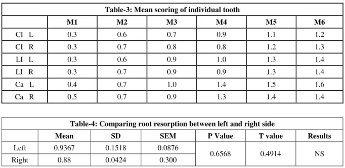
Assessment of Apical and Lateral Root Resorption: Apical and lateral root resorption of upper anterior teeth was assessed by taking OPG before treatment and after space closure. Table 3 & 4.
Table-3: Mean scoring of individual tooth
M1 M2 M3 M4 M5 M6
CI L 0.3 0.6 0.7 0.9 1.1 1.2
CI R 0.3 0.7 0.8 0.8 1.2 1.3
LI L 0.3 0.6 0.9 1.0 1.3 1.4
LI R 0.3 0.7 0.9 0.9 1.3 1.4
Ca L 0.4 0.7 1.0 1.4 1.5 1.6
Ca R 0.5 0.7 0.9 1.3 1.4 1.4
Table-4: Comparing root resorption between left and right side
Mean SD SEM P Value T value Results
Left 0.9367 0.1518 0.0876
Right 0.88 0.0424 0.300 0.6568 0.4914 NS
Determination of pulp vitality: Pulp vitality test of anterior 6 teeth was recorded with cold test. They were tested on the palatal or lingual surfaces before placing any orthodontic appliance and 1 month after the orthodontic treatment (decrowding and space closure).
Rate of Retraction: Mean treatment duration of maxillary anterior en mass retraction was recorded at 6 months. Rate of retraction was calculated by measuring the post extraction space intra orally and dividing with the amount of time taken to close the space. Space was recorded every month till six months.
Kruskal Wallis Test shows
H = 84.309 (P=2.15/1013)
Found to be statistically significant.
Rate of Root Resorption: Mean treatment duration for maxillary decrowding and space closure was 6 months. All four random examiners were independent given scores for external root resorption for individual tooth according to 4 grade ordinal scale.
Scoring For Root Resorption:
Score Examiners Remarks
0 No external root resorption
1
Slight blunting of the root apex. Slightly irregular lateral root surface; not beyond one third of the dentine width between the distal side periodontal ligament and pulp chamber
2
Moderate resorption of the root apex beyond blunting and up to one fourth of the root length. Moderate irregular lateral root surface beyond one third and up to two thirds of the dentine width between the distal side periodontal ligament and pulp chamber
3
Rate of ERR of six mean amount of ERR maxillary Ant. Teeth. = mean treatment time
= 0.92/month Left (CI+LI=Ca)
Mean 0.9367 ± 0.1518 (SD) SEM E- 0.0876
Right (CI+LI+Ca) Mean 0.88 ± 0.0424 (SD) SEM E – 0.03
Unpaired t- test between left and right side of six maxillary ant. teeth shows
T value-0.4914 (p= 0.6568) found statistically NS.
Determination of Anchor Loss: Pre-treatment
cephalograms and post space closure
cephalograms were obtained and tracings were done. Vertical line was dropped from Ptm. And distance from distal of upper first molar was calculated. Difference in values of pre and post treatment cephalograms was the amount of anchor loss.Pre and post space closure values were compared by paired t test given in table 5.
Table-5: Comparing mean of anchor loss
Mean SD SEM P Value T value Results
Pre Treatment 20.63 1.69 0.60
Post Space Closure 21.00 2.08 0.79 0.1030 1.9215 NS
The difference was not statistically significant.
Reaction to the Pulp Vitality Tester: Before placing the fixed orthodontic appliance, all the teeth were found to vital with cold testing. All of them were still under orthodontic forces when the second pulp vitality tests were performed. After completion of the space closure third tests were performed, and there were no signs of pulpal damage recorded.
Discussion
RAP was first described by Frost in 1983 the
phenomenon familiar to many
histomor-phometrists since 1966 and noted that the original injury somehow accelerated the normal regional healing processes. This acceleration is the regional acceleratory phenomenon. RAP usually occurs after a fracture, arthrodesis, osteotomy, or bone-grafting procedure, and may involve recruitment and activation of precursor cells ne-cessary for wound healing concentrated at the site of injury. Shih and Norrdin demonstrated that when intraoral cortical bone was injured by corticotomy, RAP accelerated the normal regional healing processes by transient bursts of hard- and soft-tissue remodeling [5].
Case selection: CAOT can be used to accelerate tooth movement in most of the cases requiring orthodontic treatment. It has been shown to be particularly effective in treating moderate to severe crowding, in Class I, II malocclusions
requiring expansion or extractions, and mild Class III malocclusions.
Surgical Technique
Flap design: The basic flap design is a full thickness flap reflected up to the apical portion of the tooth root. Few authors like Hessam Nowzari [6] and Derya Germec [7] have suggested to perform corticotomy only on buccal aspect of the arch to reduce the duration of surgery and patients comfort. Our flap reflection technique is similar to Hessam Nowzari’s technique (Fig 3).
Fig-3: Full thickness flap reflection
bone segments. In the CAOT procedure, decortication is performed at clinical sites without entering the cancellous bone. The corticotomies are placed on both the labial and lingual or palatal aspects of the alveolar bone, but in this study we have performed corticotomy only on buccal sidesuggested by HessanNowzariet and Germec (Fig 4).
Fig-4: Corticotomy cuts and perforations
Particulate Grafting: The materials most commonly used for grafting after decortication are synthetic grafts, deproteinized bovine bone, autogenous bone, decalcified freeze-dried bone allograft, or a combination. Grafting is done in most areas that have undergone corticotomies.We have used synthetic bone graft (PerioGlas) in our study (Fig 5)
Fig-5: Graft in place
Closure Techniques: The flap should be closed using non resorbable interrupted sutures without creating excessive tension. No packing is required. The sutures are usually left in place for 1 to 2 weeks. We have done the same procedure.
Timing of Orthodontic Treatment: The placement of orthodontic brackets is done the week before the surgical aspect of CAOT is performed. After
flap repositioning, an immediate heavy
orthodontic force can be applied to the teeth and
in all cases initiation of orthodontic force should not be delayed more than 2 weeks after surgery.This period is usually 4 to 6 months. We have followed the same protocol (Fig 6).
Fig-6: Post Space Closure
Advantages of CAOT
• Reduced treatment time. This will reduce
the treatment time up to one third the time of conventional orthodontics.
• Less root resorption due to decreased
resistance of cortical bone and shortened treatment duration.
• More bone support due to addition of
bone graft.
• It can be used to expedite the rate of
movement of individual teeth or dental segment.
Complications and Disadvantages
• Extra surgical cost.
• Mildly invasive surgical procedure. It has
• Some pain and swelling is expected, and
possibility of infection.
Retraction Mechnics: In this study the distance between the distal canine to the mesial second premolar to be recorded bilaterally with a vernier caliper directly in the patient‘s mouth right after extraction and every month until the retraction is completed and rate of retraction is calculated by distance divided by time. Canine retraction was accelerated by corticotomy in two animal studies. Both studies demonstrated faster canine retraction when compared to conventional orthodontic retraction of canines. Measurements of the rate of retraction were recorded with a vernier caliper [8].
In the present study 2 weeks after corticotomy anterior en mass retraction was started. The rate of retraction was faster in first two months followed by deceleration. The total retraction space was closed within 4 to 5 months. Hassan Noroozi in his study measured the distance between the canine cusp and the buccal cusp tip of the second premolar before and after retraction to measure the amount of retraction [9]. Similar study was done by Seher et al, they calculated the distance from lateral incisor to the first molar, from canine to second premolar and from canine to first molar before distraction and every week thereafter until the space closure [10].
Anchor Loss: AmirParviz R. Davoody measured the efficacy of anchorage control between differential moment’s mechanics and temporary anchorage devices in a clinical trial. Lateral cephalograms were taken before and after incisor retraction. The ratio of molar protraction to incisor retraction was calculated and intragroup and intergroup changes in upper lip, maxillary incisor and molar position were analyzed by paired and independent t-tests. He concluded that both anchorage modalities show statistically significant retraction of the lips during treatment. [11].
In this study, we have taken lateral cephalograms before the treatment and after the completion of
retraction. Tracings were made of that
cephalograms. And the horizontal distance from the pterygoid vertical (perpendicular to FH plane) to the distal surface of the first molar is measured. Anchor loss is calculated by subtracting pre and
post retraction values. The anchor loss in the present study was minimal i.e. 1.25mm which was statistically non-significant.
Determination of Root Resorption: Literature evidence suggests that root resorption, an adverse side effect of orthodontic therapy, may be decreased under conditions of alveolar osteopenia, a condition characterized by
diminished bone density and created
secondary to alveolar corticotomy surgery. As in CAOT duration of force applied is less, so the adverse effect like root resorption will decrease. Wilcko et al published various articles on corticotomy and concluded that there is less root resorption due to decreased resistance of cortical bone [2].
Method for evaluating root resorption: The apical rootresorption was assessed by the following score:
1. No root resorption. Smooth lateral root
surface and periodontalligament.
2. Slight blunting of the root apex. Slightly
irregular lateral root surface, not beyond one third of the dentine width between the distal side periodontal ligament and pulp chamber.
3. Moderate resorption of the root apex
beyond blunting and up to one fourth of the root length. Moderate irregular lateral root surface beyond done third and up to two thirds of the dentine width between the distal side periodontal ligament and pulp chamber
4. Excessive resorption of the root apex
beyond done fourth of the root length. Excessive irregularity of the lateral root surface beyond two thirds of the dentine width betweenthe distal side periodontal ligament and pulp chamber.
Four different randomly selected examiners evaluated the films and gave scores accordingly. All cases showed stable results efficient and speedy decrowding and space closure causing minimal root resorption.
cold test, CO2 ice and refrigerant spray were the
most accurate ones while ethyl chlorideand ice water had the least reliability. Laser Doppler flow metery and pulse oximetry showed high level of accuracy, which could be valuable specifically in children with traumatized teeth [12].
In this study we have used cold test and concluded that there was no significant pulpal damage after completion of the treatment.
Summary and Conclusion
The patients were treated with periodontally accelerated osteogenic orthodontic treatment with straight wire appliance (.022 slot MBT prescription). The rate of retraction was measured over a period of six months. The rate of retraction
for maxillary anterior left side was
1.15mm/month and right side was
1.35mm/month, (H=84.309, P=2.15/1013) being
statistically significant. The anchor loss in the present study was minimal i. e. 1.25mm which was statistically non-significant (P=.0.1030). All cases showed stable results efficient and speedy decrowding and space closure causing minimal root resorption. (P=0.6568). We have used cold test in the present study and concluded that there
was no significant pulpal damage after completion of the treatment.
The periodontally accelerated osteogenic
orthodontics procedure is gaining in
popularity with patients and doctors because of the much shorter treatment times and the increased range of treatment capabilities and
possibilities. Over time, it has been
transformedinto a successful treatment option for many orthodontic problems when used properly, including complicated cases that require a multidisciplinary in-office approach between dental specialties. It can often make the treatment of severe dental malocclusions more practical while reducing the treatment time for patients from one third to one quarter of the time typically required to treat most
dental malocclusions. Additionally, the
alveolar volume can be increased to aid in
supporting the teeth while correcting
preexisting dehiscences and fenestrations when there is a vital root surface. This technique belongs in a specialty arena where both orthodontists and periodontists work together from diagnosis through treatment and retention.
References
1. Köle H. Surgical Operations on the Alveolar Ridge to Correct Occlusal Abnormalities. Oral Surgery, Oral Medicine, Oral Pathology.1959; 12(5):515-529. 2. Düker J. Experimental Animal Research into Segmental
Alveolar Movement after Corticotomy. Journal of Maxillofacial Surgery. 1975; 3:81-84.
3. Yaffe A, Fine N and Binderman I. Regional
Accelerated Phenomenon in the Mandible Following
Mucoperiosteal Flap Surgery. Journal of
Periodontology. 1994; 65(1):79-83.
4. Wilcko WM, Wilcko T, Bouquot JE, Ferguson DJ. Rapid Orthodontics with Alveolar Reshaping: Two Case Reports of Decrowding. Int J Periodontics Restorative Dent 2001; 21:9-19.
5. Shih MS, Norrdin RW. Regional acceleration of remodeling du15. Ring healing of bone defects in beagles of various ages. Bone. 1985; 6:377-9.
6. Nowzari H, Yorita FK and Chang HC. Periodontally Accelerated Osteogenic Orthodontics Combined with
Autogenous Bone Grafting. Compendium 2008;
29(4):2-8.
7. Derya Germec¸ Bahadir Giray, Ilken Kocadereli, Ayhan Enacar. Lower Incisor Retraction with a Modified
Corticotomy. Angle Orthodontist, 2006; 76(5):2, 882-890.
8. Lotzof LP, Fine HA and Cisneros GJ. Canine Retraction: A Comparison of Two Preadjusted Bracket Systems. Am J Orthod Dentofac Orthop
1996; 110:191-6.
9. Noroozi H. A Formula to Determine the Amount of Retraction of Mandibular Canines. Angle Orthod
2000; 70:154-156.
10. Seher Sayın, A. Osman Bengi, A. Umit Gurton, Kerim Ortakoglu. Rapid Canine Distalization Using Distraction of the Periodontal Ligament: A Preliminary Clinical Validation of the Original Technique. Angle Orthod 2004; 74:304-315. 11. Davoody AR, Posada L, Utreja A, Janakiraman N,
Neace WP, Flavio Uribe et al. A prospective comparative study between differential moments and miniscrews in anchorage control. Eur J Orthod
2013; 35(5):568-576.
12. Olha Shender et al. How helpful are diagnostic tests for pulpal Conditions? www. dentistry.utoronto.ca;
2013:1-15.