Early treatment of patient with Class III skeletal and
dental patterns
Marcos Alan Vieira Bittencourt1
How to cite this article: Bittencourt MAV. Early treatment of patient with Class III skeletal and dental patterns. Dental Press J Orthod. 2015 Nov-Dec;20(6):97-109. DOI: http://dx.doi.org/10.1590/2177-6709.20.6.097-109.bbo
Submitted: October 13, 2015 - Revised and accepted: October 30, 2015
Contact address: Marcos Alan Vieira Bittencourt
Rua Eduardo José dos Santos, 147, Sala 1104, Garibaldi, Salvador, Bahia - Brazil CEP: 41940-455
Email: [email protected] » The author reports no commercial, proprietary or financial interest in the
prod-ucts or companies described in this article.
» Patient displayed in this article previously approved the use of their facial and intraoral photographs.
1Associate professor of Orthodontics, Universidade Federal da Bahia (UFBA),
Salvador, Bahia, Brazil.
INTRODUCTION
The present study reports the case of a 6 year and 3-month-old patient, in good general health, who sought treatment accompanied by her parents. Her medical history was uneventful, and she presented with good oral hygiene, without caries or restorations. She avoided smiling and the chief complaint reported by her parents was the presence of anterior crossbite and protruded chin and lower lip, which hindered facial esthetics. In general, some of these factors are deter-mined by a hereditary component; however, the etiol-ogy of the case seemed multifactorial, based on patient’s parents’ facial esthetics and absence of this type of facial features in other members of their family.
DIAGNOSIS
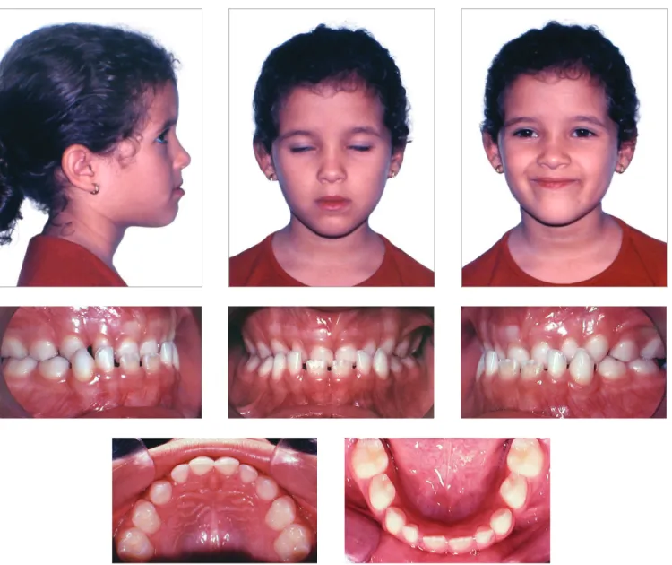
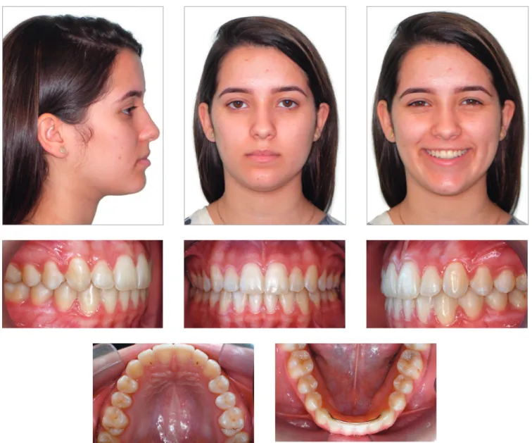
As shown in Figure 1, the patient presented with passive lip seal, slight deviation of the mandible to the right, which resulted in a slightly asymmetrical face, and deicient midface anteroposteriorly. She avoided smiling and showing her incisors teeth, which hin-dered assessment of spontaneous smile and the amount of tooth exposure at smile. The patient had a concave proile, with upper lip retrusion and lower lip protru-sion, a proportional nose and increased nasolabial and labiomental angles.
From a dental perspective (Figs 1, 2), she was in a rather incipient phase of mixed dentition, with early eruption of teeth #36, 31 and 41. She had Angle Class III malocclusion,
Class III skeletal pattern is characterized by disharmony between maxillary and mandibular basal bones anteroposteriorly, and might or might not be associated with dental changes. In general, facial esthetics is hindered significantly, which most of times is the reason why patients or patient’s guardians seek treatment. This case was presented to the Brazilian Board of Orthodontics and Dentofacial Orthopedics (BBO) in partial fulfillment of the requirements for Diplomate recertifica-tion and revalidarecertifica-tion.
Keywords:Angle Class III malocclusion. Crossbite. Interceptive Orthodontics.
DOI: http://dx.doi.org/10.1590/2177-6709.20.6.097-109.bbo
O padrão esquelético de Classe III é caracterizado pela desarmonia entre as bases ósseas maxilar e mandibular, no senti-do anteroposterior, podensenti-do ou não estar acompanhada por alterações dentárias. Em geral, o aspecto estético facial fica bastante comprometido, sendo justamente esse o fator que, na maioria das vezes, motiva o paciente ou seus responsáveis a procurar pelo tratamento. Este caso clínico foi apresentado à Diretoria do Board Brasileiro de Ortodontia e Ortopedia Facial (BBO) como parte dos requisitos para o processo de recertificação e revalidação do título de Diplomado pelo BBO.
with the terminal plane of deciduous mandibular second molars in mesial step. Also, the patient presented with
Baume type I arches,1 with primate spaces in both upper
and lower arches, 4-mm overbite and 1-mm overjet, max-illary dental midline coinciding with the median sagittal plane and 1-mm mandibular dental midline deviation to the right. In addition to anterior crossbite afecting the en-tire anterior region, she also had end-to-end bite of teeth #54 and 55 in the transverse plane, caused by mild maxil-lary atresia pronounced in the anterior region.
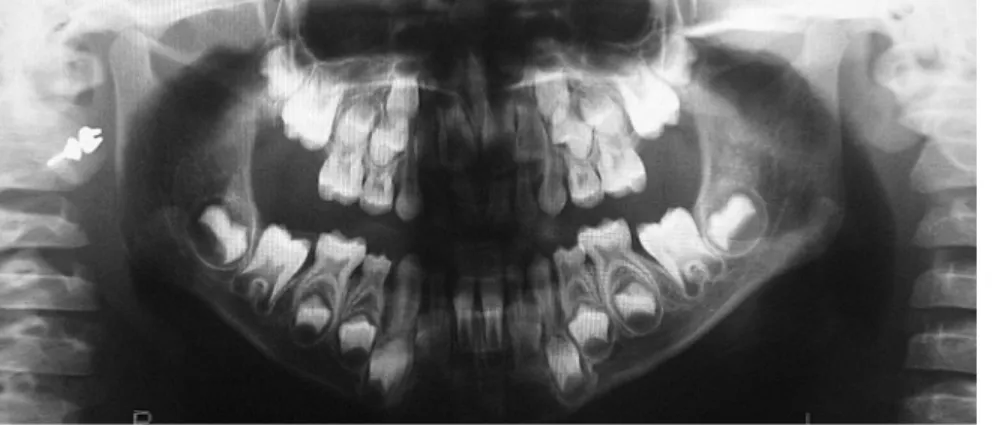
Analysis in panoramic radiograph (Fig 3) revealed normality of all deciduous and permanent teeth at diferent stages of root formation. Permanent maxil-lary and mandibular second molars were at an
ear-ly stage of development, Nolla’s stage 4,2
render-ing it impossible to identify the onset of third molars
formation. Additionally, occlusal radiograph of the maxilla (Fig 4) revealed that the region of the midpalatal suture was within normality standards.
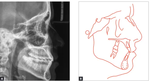
Lateral cephalograms and cephalometric tracings (Fig 5 and Table 1) revealed a Class III skeletal pattern (ANB = 0° and Wits = -7 mm), with retrusion of the maxilla in relation to the base of the skull, and protrusion of the mandible (Facial Angle = 91°). Slightly increased vertical growth pattern in the posterior region of the man-dible, with a decreased mandibular plane (FMA = 21° and Y-axis = 54°), was also identiied. As previously clinically observed, the patient had a concave proile, with upper lip retrusion (Upper Lip – S-line = -1 mm) and signii-cant lower lip protrusion (Lower Lip – S-line = 4 mm). Mandibular incisors were retruded and in slightly upright position (1-NB = 20° and 1 mm).
Figure 2 - Initial casts.
Figure 5 - Initial lateral cephalogram (A) and cephalometric tracing (B).
Figure 3 - Initial panoramic radiograph. Figure 4 - Initial occlusal radiograph of the maxilla.
As regards function, she presented with altered tongue positioning during speech and swallowing. With a view to analyzing whether anterior crossbite hindered function, patient’s closure pattern was as-sessed, revealing no shift. In other words, centric re-lation position was very close to intercuspal contact position.
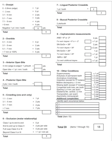
Discrepancy index (DI) was calculated and scored 18 points (Fig 6).
TREATMENT PLAN
Taking facial esthetics and disharmony between the maxilla and mandible anteroposteriorly into account, orthodontic treatment was planned to be conducted in two phases. In the irst phase, the goal was to achieve a bal-anced proile and restore lip posture as well as the skeletal relationship between the maxilla and the mandible. To this end, reverse maxillary protraction was planned to be
car-ried out by means of Petit face mask.3 In order to render
anterior repositioning of the maxilla easier, by means of weakening the sutures that hold the other facial bones to-gether, in addition to enhancing its transverse dimension,
palatal expansion4 would be performed by the modiied
Haas expansion appliance. As a result, the anteroposterior relationship between the maxilla and the mandible would also be improved and anterior crossbite corrected.
Since there was positive discrepancy in the mandible,
with potential presence of Lee Way Space5 by the end
of the mixed dentition, treatment plan included using the remaining space for retrusion of mandibular teeth. Thus, before the second phase of treatment, a lingual bar would be installed immediately before exfoliation of deciduous mandibular second molars, thereby keeping permanent irst molars in place and preserving the Lee Way Space.
In the second phase of treatment, conventional ixed orthodontic appliance would be installed in both upper and lower arches with a view to distalizing man-dibular premolars and canines, in addition to retract-ing mandibular incisors by means of takretract-ing advantage of the space preserved as a result of using the lingual bar, thus improving the relationship between the max-illa and mandible in the transverse and anteroposterior planes. At this phase, it would also be important to improve Class I molar and canine relationship, adjust lateral protrusive guidance, and align and level all max-illary and mandibular teeth.
Ater conventional treatment completion, a wrap-around removable appliance was planned to be used in the maxilla and a 0.028-in stainless steel intercanine bar to be bonded to mandibular teeth, both for retention. Special attention would also be given to the process of formation and development of third molars.
Based on the need for patient’s compliance with the face mask and the uncertainty about achieving a harmo-nious relationship between maxillary and mandibular basal bones anteroposteriorly, an alternative treatment plan by the end of growth would be to wait for facial growth to cease and dentition as well as occlusion to fully develop before conventional orthodontic-surgical treatment was conducted. This treatment modality was rejected by the patient’s parents who understood the biological limitations involved and undertook the re-sponsibility of making an efort with a view to gaining patient’ compliance.
1 – Overjet
0 - 0.9mm (edge) 1 - 3 mm 3.1 - 5 mm 5.1 - 7 mm 7.1 - 9 mm > 9 mm
= 1 pt = 0 pt = 2 pts = 3 pts = 4 pts = 5 pts = 6 pts
6 Negative 1 pt / mm / tooth
Total 2 – Overbite
0.1 - 3 mm 3.1 - 5 mm 5.1 - 7 mm > 7 mm or 100%
= 0 pt = 2 pts = 3 pts = 5 pts
2 Total Total pts pts Total = = = = =
3 – Anterior Open Bite
4 – Posterior Open Bite
0 mm (edge-to-edge)= 1 pt/tooth Open bite = 1 pt / mm / tooth
2 pts / mm / tooth
5 – Crowding (one arch only)
1.1 - 3 mm 3.1 - 5 mm 5.1 - 7 mm > 7 mm
= 1 pt = 2 pts = 4 pts = 7 pts
Total
6 – Occlusion (molar relationship)
Class I up to end to end End to end up to Class II Full cusp Class II or III Beyond Class II or III
7 – Lingual Posterior Crossbite
1 pt / tooth 2 2
2 pts/tooth
8 – Buccal Posterior Crossbite Total
Total
= 0 pt = 2 pts per side = 4 pts per side = + 1 pt / mm ad.
8 – Cephalometric measurements
ANB = 6º or -2º For each degree > 6º or < -2º SN-GoGn = 38º For each degree > 38º SN-GoGn = 26º For each degree < 26º IMPA = 99º For each additional degree
4 pts x 1 pt 2 pts x 2 pts 1 pt x 1 pt 1 pt x 1 pt
Total
10 – Other Conditions
Supernumerary Ankylosis of permanent teeth Anomalous morphology Impaction (except for 3rd molars)
Midline discrepancy ≥3 mm Missing teeth (except 3rd molars)
Congenital tooth loss, per tooth Spacing (≥ 4 mm, per arch) Diastema Mx. central ≥ 2 mm Transposition, per event Asymmetry (nonsurgical)
Additional complexities
Total 8
Identify each occurrence below:
Total(item 10)
Total DI (items 1 through 10) 18 x 1 pt x 2 pts x 2 pts x 2 pts 2 pts x 1 pt x 2 pts x 2 pts 2 pts x 2 pts 3 pts x 2 pts
1 – 2 – 3 – 4 – 5 –
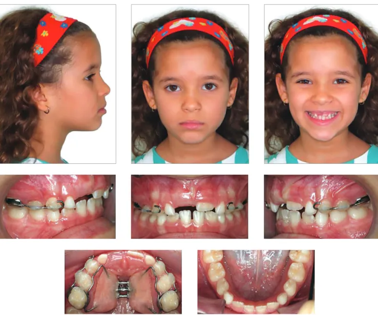
TREATMENT PROGRESS - FIRST PHASE
Orthodontic treatment began as planned, with the modified Haas appliance installed and supported by bands adapted to deciduous second molars and bonded to deciduous first molars and canines. More-over, 0.032-in stainless steel wires were adapted and welded to the orthodontic bands buccaly. They were also adapted and welded to the buccal surface of the aforementioned teeth, with hooks placed mesial to canines and used as support for maxillary protraction. The patient was advised to perform ¼ of a turn every 12 hours for 15 days. After this period, the expansion appliance was made stable and the patient began to
use the Petit face mask, with 400-g force application on each side, 18 hours a day. It is worth noting that the face mask was adapted so as to allow the elastics to form a 30° angle with the occlusal plane, thereby moving the maxilla forward without undesired
coun-terclockwise rotation.6
The patient was followed up every two months. Six months ater treatment onset, the use of the face mask decreased to eight hours a day (at night); six months ater that, the use of the face mask ceased com-pletely and the expansion appliance was removed. Im-provements resulting from the irst phase of treatment can be seen in Figures 7 to 10.
Figure 10 - Initial lateral cephalogram (A) and cephalometric tracing (B) at first treatment phase completion, immediately before removal of the Haas appliance.
Figure 8 - Panoramic radiograph at first treatment phase completion, immediately before removal of the Haas appliance.
Figure 9 - Occlusal radiograph of the maxilla at first treatment phase completion, immediately before removal of the Haas appliance.
Ater three years and six months of following up patient’s occlusion development, with appointments arranged every six months, the lingual bar was in-stalled, as planned. At this point, the patient was 11 years and one month old. Clinically, she presented with a straight proile, passive lip seal, proportional fa-cial thirds and a pleasant smile. From a dental perspec-tive, she was near the end of mixed dentition, at the second transitional period; however, with increased exfoliation in the mandible where second molars were the only deciduous teeth let. As for the maxilla, in which the exfoliation process was delayed, deciduous canines, irst and second molars were present.
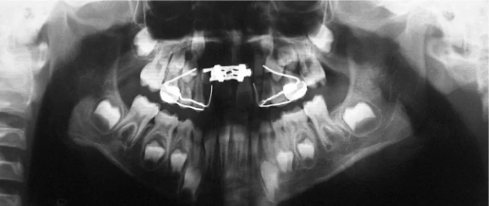
Panoramic radiograph (Fig 11) revealed teeth #35 and 45 slightly distally proclined, thereby inciting distal
root resorption of teeth #75 and 85, respectively. Mesial roots remained practically completely intact, with root resorption beginning near the cervical third.
In this scenario, it was decided to install the lin-gual bar made of 0.032-in stainless steel wire welded to orthodontic bands adapted to teeth #36 and 46. Subse-quently, the patient was referred for extraction of teeth #75 and 85 with a view to physiologically improving the eruption axis of teeth #35 and 45. As the goal was to preserve Lee Way Space in the mandible, so as to take advantage of the remaining space to move mandibular teeth posteriorly while minimizing Class III skeletal pattern expression, the patient was followed up until exfoliation of all deciduous teeth and eruption of their permanent successors.
TREATMENT PROGRESS – SECOND PHASE
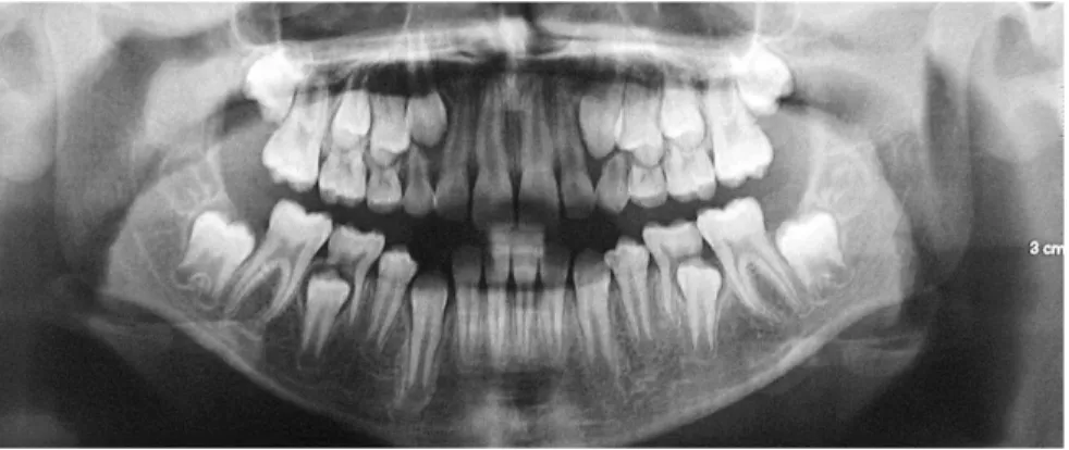
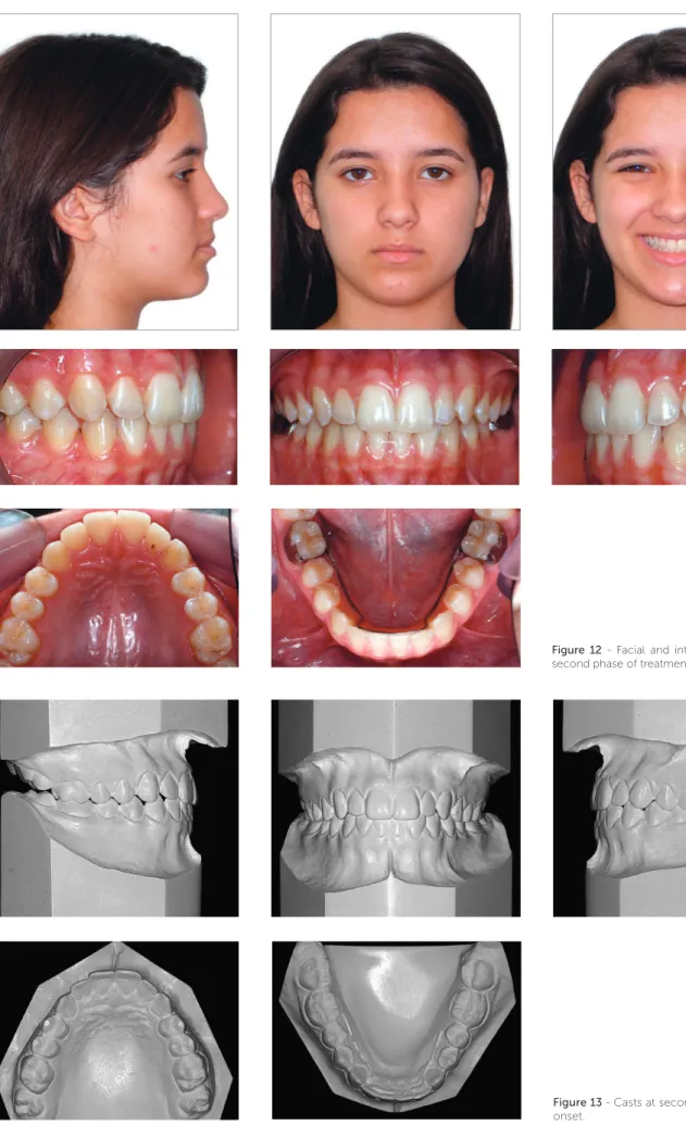
Ater three years and seven months following up patient’s occlusion development, with appointments arranged every six months, the second phase of treat-ment began. At this point, the patient was 14 years and eight months old. Clinically, as shown in Figure 12, she remained with a straight proile, satisfactory passive lip seal, proportional facial thirds and an esthetically pleas-ant smile. From a dental perspective (Figs 12 to 14), she had all permanent teeth erupted, including second mo-lars, except for third molars.
Analysis of the stages of maturation of cervical ver-tebrae C2, C3 and C4 (Fig 14) revealed that the indica-tors of skeletal maturity signaled to the end of the third
growth spurt.7
In view of the above, the second phase of treatment began with conventional orthodontic ixed appliance bonded to all teeth in both upper and lower arches, with metal brackets, Edgewise standard technique, without torques or angulations, and with 0.022 x 0.028-in slots.
Subsequently, alignment and leveling 0.014 to 0.020-in stainless steel round archwires were placed in the maxilla and mandible. Additionally, 0.018 x 0.025-in rectangular archwires were installed in the mandible. A tie together ligature was made with the 0.020-in mandibular archwire between the first and second molars on both sides. An elastomeric chain
Figure 11 - Panoramic radiograph at 11 years and one month old.
was placed to slightly move second premolars distally and correct the mild torsiversion presented by tooth #35. Subsequently, these teeth were also included as anchorage units, and elastomeric chains were indi-vidually used in order to slightly move first premo-lars and canines distally. Once the 0.018 x 0.025-in stainless steel archwire had been placed, an elastic tie back bend was crimped at the lower arch. Meanwhile, Class III intermaxillary mechanics was applied, as an-chorage reinforcement, with a view to closing the re-maining spaces mesial to canines by means of slight retraction of incisors.
At the following phase and ater all spaces had been closed, 0.019 x 0.025-in stainless steel rectangular arch-wires, with irst- and third-order bends placed as nec-essary, were placed in both upper and lower arches for treatment inishing.
Figure 12 - Facial and intraoral photographs at second phase of treatment onset.
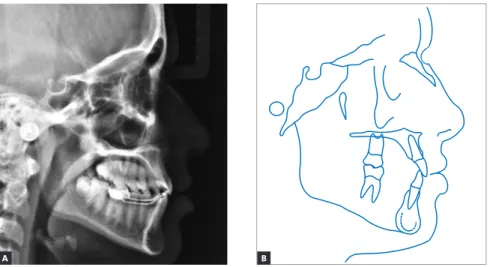
Figure 14 - Lateral cephalogram (A) and cepha-lometric tracing (B) at second phase of treat-ment onset.
RESULTS
Patient’s inal records (Figs 15 to 18) assessment revealed that all treatment objectives were achieved. As regards patient’s face, a comparison between inal and initial records taken at orthodontic treatment on-set revealed signiicant proile improvement, signiicant movement of the upper lip forward and adjustment of the lower lip, thereby resulting in an esthetic proile, with better nasolabial and labiomental angles contour. Frontal analysis revealed a balanced face, with the man-dible symmetrically positioned in relation to the median sagittal plane. The patient also presented with good pas-sive lip seal, proportional facial thirds and an estheti-cally pleasant smile, which resulted in excellent smile
arc contour and adequate buccal corridor dimensions.8
Tooth analysis revealed adequate alignment and leveling and Class I molar and canine relationship achieved on both sides. Transverse deiciency, anterior crossbite and lower midline deviation were corrected. There was slight intentional increase in maxillary in-tercanine width from 32 mm to 34 mm. Mandibular intercanine width remained at 26 mm. Functional har-mony was excellent for occlusion in protrusive excur-sion and right as well as let lateral guidance, with cen-tric relation coinciding with maximal intercuspation
position. It is worth noting that, as shown by periapi-cal radiographs taken at treatment completion (Fig 17), changes were achieved without radiographically no-ticeable apical root remodeling.
As planned, cephalometric examination revealed a number of skeletal changes. A-Point moved slightly for-ward while B-point moved backfor-wards, thereby result-ing in skeletal pattern correction, with Wits value from -7 mm to -2 mm, which is within normality standards
for female patients.9 These changes also led to facial angle
improvement from 91° to 88°, a value near normality stan-dards. In addition, there was slight opening up of the man-dibular plane angle (Y-axis from 54° to 60° and FMA from 21° to 24°), which resulted in signiicant improvement in proportionality between facial thirds in the vertical plane. These skeletal changes were also responsible for improve-ments in the relationship between upper and lower lips, with the lower lip positioned from 4 mm to 1 mm in
rela-tion to the S-line,10 which resulted in an esthetically
pleas-ant proile. These data are shown in Figure 18 and Table 1. Cephalometric superimposition revealed that, in the irst phase of treatment (Fig 19), there was signiicant maxillary advancement forward, correcting overjet; and slight clockwise rotation of the mandible, opening up the mandibular plane angle and signiicantly improving
patient’s facial proile while also enhancing upper and lower lips position. Partial cephalometric superimposi-tion revealed a decrease in maxillary length, with a re-duction between incisors and deciduous second molars width due to anchorage loss of the latter. This was po-tentially followed by displacement of the maxilla forward and slight vertical growth of the alveolar process. Slight vertical growth of the alveolar process was also seen in the mandible, followed by slight mandibular growth.
Between the first phase of treatment and treatment completion (Fig 20), there was harmonious growth of the entire facial complex forward and downward,
with profile balance preserved. In the maxilla, there was a major improvement in incisors proclination; from significant upright position to a much favor-able position. As for molars, there was slight anchor-age loss, occupying the Lee Way Space, and seek-ing improvement in Class I molar relationship with the mandibular tooth. In the mandible, there was a slight improvement in incisors proclination. As pre-dicted and desired, molars positioning remained un-changed, without anchorage loss; in addition, there was vertical growth of the alveolar process followed by mandibular growth.
Figure 16 - Final casts.
Figure 17 - Final periapical radiographs.
Figure 18 - Final lateral cephalogram (A) and cephalometric tracing (B).
Figure 19 - Total (A) and partial (B) cephalomet-ric superimpositions of initial (black) and at first treatment phase completion (blue) tracings.
Figure 20 - Total (A) and partial (B) cephalomet-ric superimpositions of at first treatment phase completion (blue) and final (red) tracings.
Table 1 - Initial (A) and final (B) cephalometric values.
Measurements Normal A A1 A2 B Dif. A/B
Skeletal pattern
SNA (Steiner) 82° 81° 84° 81° 82° 1
SNB (Steiner) 80° 81° 79° 79° 80° 1
ANB (Steiner) 2° 0° 5° 2° 2° 2
Wits (Jacobson) ♀ 0 ±2 mm
♂ 1 ±2 mm -7 mm -1 mm -2 mm -2 mm 5
Angle of convexity (Downs) 0° 1° 9° 2° 2° 1
Y-axis (Downs) 59° 54° 55° 57° 60° 6
Facial angle (Downs) 87° 91° 90° 91° 88° 3
SN-GoGn (Steiner) 32° 31° 33° 31° 31° 0
FMA (Tweed) 25° 21° 23° 22° 24° 3
Dental pattern
IMPA (Tweed) 90° 88° 87° 85° 89° 1
1.NA (degrees) (Steiner) 22° - 12° 19° 19°
-1-NA (mm) (Steiner) 4 mm - 0 mm 4 mm 5 mm
-1.NB (degrees) (Steiner) 25° 20° 19° 15° 20° 0
1-NB (mm) (Steiner) 4 mm 1 mm 4 mm 1 mm 5 mm 4
1
1- Interincisal angle (Downs) 130° - 142° 145° 140°
-1-APo (Ricketts) 1 mm 2 mm 0 mm 1 mm 5 mm 3
Proile Upper lip — S-line (Steiner) 0 mm -1 mm 1 mm -1 mm -2 mm 1
Lower lip — S-line (Steiner) 0 mm 4 mm 3 mm 2 mm 1 mm 3
A
A
B
1. Baume LJ. Physiological tooth migration and its signiicance for the development of occlusion. I. The biogenetic course of the deciduous dentition. J Dent Res. 1950 Apr;29(2):123-32.
2. Nolla CM. The development of the permanent teeth. J Dent Child.
1960;27:254-66.
3. Petit HP. The prognathic syndrome: a complete treatment plan around the facial mask. Rev Orthop Dento Faciale. 1982 Oct;16(4):381-411.
4. McNamara JA Jr. An orthopedic approach to the treatment of Class III malocclusion in young patients. J Clin Orthod. 1987 Sep;21(9):598-608. 5. Nance HN. The limitations of orthodontic treatment; mixed dentition
diagnosis and treatment. Am J Orthod. 1947 Apr;33(4):177-223.
6. Ngan PW, Hagg U, Yiu C, Wei SH. Treatment response and long-term
dentofacial adaptations to maxillary expansion and protraction. Semin Orthod. 1997 Dec;3(4):255-6.
7. Baccetti T, Franchi L, McNamara JA Jr. An improved version of the cervical vertebral maturation (CVM) method for the assessment of mandibular growth. Angle Orthod. 2002 Aug;72(4):316-23.
8. Nascimento DC, Santos ER, Machado AWL, Bittencourt MAV. Inluence
of buccal corridor dimension on smile esthetics. Dental Press J Orthod. 2012;17(5):145-50.
9. Jacobson A. The “Wits” appraisal of jaw disharmony. Am J Orthod. 1975 Feb;67(2):125-38.
10. Steiner CC. Cephalometrics for you and me. Am J Orthod. 1953;39(10):729-55.
11. Bittencourt MAV. Má oclusão Classe III de Angle com discrepância ântero-posterior acentuada. Rev Dental Press Ortod Ortop Facial. 2009;14(1):132-42.
12. Ngan P, Hägg U, Yiu C, Merwin D, Wei SH. Soft tissues and dentoskeletal proile changes associated with maxillary expansion and protraction headgear treatment. Am J Orthod Dentofacial Orthop 1996;109(1):38-48. 13. Ngan P. Early treatment of Class III malocclusion: is it worth the burden? Am
J Orthod Dentofacial Orthop. 2006 Apr;129(4 Suppl):S82-5.
14. Araújo EA, Araújo CV. Abordagem clínica não-cirúrgica no tratamento da má oclusão de Classe III. Rev Dental Press Ortod Ortop Facial. 2008;13(6):128-57.
15. Westwood PV, McNamara JA Jr, Baccetti T, Franchi L, Sarver DM. Long-term efects of Class III treatment with rapid maxillary expansion and facemask therapy followed by ixed appliances. Am J Orthod Dentofacial Orthop. 2003 Mar;123(3):306-20.
16. Baccetti T, McGill JS, Franchi L, McNamara JA Jr, Tollaro I. Skeletal efects of early treatment of Class III malocclusion with maxillary expansion and face-mask therapy. Am J Orthod Dentofacial Orthop. 1998 Mar;113(3):333-43. 17. Brunetto AR. Má oclusão de Classe I de Angle, com tendência à Classe III
esquelética, tratada com controle de crescimento. Rev Dental Press Ortod Ortop Facial. 2009 Set-Out;14(5):129-45.
18. Deguchi T, Kanomi R, Ashizawa Y, Rosenstein SW. Very early face mask therapy in Class III children. Angle Orthod. 1999 Aug;69(4):349-55. 19. Silva Filho OG, Magro AC, Capelozza Filho L. Early treatment of the Class III
malocclusion with rapid maxillary expansion and maxillary protraction. Am J Orthod Dentofacial Orthop. 1998 Feb;113(2):196-203.
20. Cericato GO, Freitas PH, Bittencourt MAV, Paranhos LR. Reliability, eicacy and reproducibility of the cervical vertebrae maturation index (CVMI). Biosc J. 2015;31(1):319-25.
21. Cericato GO, Bittencourt MA, Paranhos LR. Validity of the assessment method of skeletal maturation by cervical vertebrae: a systematic review and meta-analysis. Dentomaxillofac Radiol. 2015;44(4):20140270.
REFERENCES
FINAL CONSIDERATIONS
As previously reported,11 Angle Class III
malocclu-sion, characterized by anteroposterior dental discrep-ancy, is rendered more severe when in association with skeletal disharmony which might result from maxillary deiciency, mandibular excess or a combination of both. These changes hinder facial proile, oten leading to psy-chosocial consequences.
The therapeutic possibilities available to correct the anomaly imply in a number a factors. For patients in the pre-pubertal growth spurt phase, an early approach is rec-ommended; for instance, the use of face mask for reverse maxillary protraction usually associated with palatal
ex-pansion.12 With this approach, as performed in the case
reported herein, a more forward positioning of the max-illa is expected, so as to enhance its relationship with the mandible, in addition to providing satisfactory oc-clusion and pleasant facial esthetics. Nevertheless, pa-tient’s guardians should be informed about a number of uncertainties involved in the potential outcomes to be achieved, especially regarding the direction of patient’s
facial growth13 and the degree of patient’s compliance.
Thus, in spite of agreeing with the role heredity plays in the etiology of Class III skeletal malocclusion, several authors believe it is possible not only to change growth pattern and direction by means of a non-surgical ap-proach, but also to minimize or even treat the
maloc-clusion successfully.14,15 Younger individuals tend to
yield more favorable results, in which case the ideal age ranges between 4 and 10 years old, although patients aged between 10 and 14 years old also yield positive
out-comes.16,17 Deguchi et al18 assessed, during six months,
variations in Wits value for children treated with the same treatment modality described herein. The authors found an increase of 2 mm. For the clinical case report-ed by the present study, there was a greater improve-ment: 5 mm of change in Wits value.
In the irst phase of treatment, maxillary
protrac-tion by means of Petit face mask3 was conducted in
combination with rapid maxillary expansion, with a view to enhancing its transverse dimension while weakening the sutures that hold the other facial bones together, thereby rendering maxillary displacement
forward efective.19 Importantly, this was the main
procedure carried out for the present clinical case, which allowed patient’s mid- and lower face to grow and develop accordingly, thus signiicantly minimizing
the complexity of the following phase. Furthermore, the stability of procedures carried out during the irst phase of treatment reveals that the biological limits
were respected,20,21 which resulted in favorable