Periodontal impact of surgically induced dental lesions in mandibular
osteodistraction: An animal study
*Pedro E. G. SOARES CORREIA1, Heiner WEHRBEIN2, Maurice Y. MOMMAERTS3
1
Unit of Oro-Maxillo-Facial Surgery, Hospital CUF Descobertas, Lisbon, Portugal; 2
Department of Orthodontics, University of Mainz, Germany;3Division of Maxillo-Facial Surgery, General Hospital St. Jan, Bruges, Belgium
SUMMARY. Aim: The objective of the study was to evaluate the impact of dental lesions on the periodontium, in
a canine model of mandibular osteodistraction. Material and methods: In six adult male Beagle dogs, an osteot-omy was made between the right second lateral incisor and canine, and a distraction device placed. The roots adjacent to the osteotomy were deliberately damaged by the reciprocating saw and chisel, with preservation of the attached gingiva. The osteodistraction protocol used was: latency of 7 days, rate of distraction 1 mm per day, and rhythm once a day for 5 days. Vital staining was carried out with tetracycline, Xylenol Orange and Calcein Green. The dogs were sacrificed after 12 weeks of consolidation and the specimens were evaluated with light microscopy (native, polarized light, fluorescence, and after toluidin blue staining). Results: The periodontal ligament (PDL) regeneration was observed in the 2500 slices examined. Cementum and dentine le-sions were repaired by cellular cementum. Loose dentine and cementumedentine fragments were embedded in regenerated PDL and their surface repaired by cementum. By means of light microscopic examination and within the limited observation time, no degenerative pulpal changes were found, when the pulp canal was not entered. Extensive pulp exposure and destruction resulted in ingrowth of the PDL and bone-like tissue. In that case, cellular cementum also lined the dentine surface of the pulp canal. Conclusion: Although there was an extensive reparative response to the para-pulpal lesions, none of the changes observed showed evidence of a loss of functional integrity of the periodontium at the distraction site. The fate of the tooth with exposed pulp canal remains uncertain. Ó2008 European Association for Cranio-Maxillofacial Surgery
Keywords:osteogenesis, distraction, mandible, periodontium, animal experimentation
INTRODUCTION
Symphyseal widening by means of distraction osteogen-esis is a less popular treatment option for severe irregu-larity and crowding of anterior mandibular teeth due to a narrow anterior apical base. Indeed, an inter-dental os-teotomy is required and besides its morbidity, permanent damage to the periodontium and teeth may occur. In an-imal experiments (Bell et al., 1997, 1999; Del Santo et al., 2000) and clinical investigations (Guerreroet al., 1997; Weil et al., 1997; Kewittand Van Sickels, 1999; Copeet al., 2002) inter-dental callus formation and tooth movements were studied, and occasionally accidental damage to teeth and periodontal structures was reported.
Mommaertset al. (2005)described the periodontal status
and tooth vitality changes during the distraction and con-solidation phase more in depth in a clinical study. Also in this study, however, the surgeon tried to avoid damage to the dental structures.
The aim of the present study was to assess any repair mechanisms after deliberately induced lesions to teeth and periodontal tissues adjacent to the regenerate bone during transversal mandibular osteodistraction.
MATERIALS AND METHODS
The study project was approved by the Board of Health Defense and Animal Welfare of the Ministry of Agricul-ture and Fisheries of Portugal (#14.MAI 01 04603). Six purebred male adult Beagle dogs with a difference in age of less than 15 days, and weighing between 11 and 15 kg, were acquired from the Animal Experimentation and Production Service of the University of Granada, Spain, in accordance with the EC Rules. All were cleared for facial malformation and oral pathology. The surgeries took place at the ‘‘Estac¸~ao Zoote´cnica Nacional’’, San-tare´m, Portugal, in the Institute of Biomedical Technol-ogy. The dogs were microchip tagged but for convenience of the reader, numbered 1e6 according to
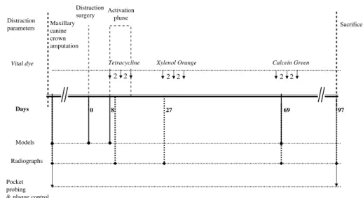
the chronology of the operations. The chronology of all procedures, including surgical procedures, dye injec-tions, impressions, radiographs, periodontal probings, and the distraction parameters, is depicted inFig. 1.
*
This paper was presented at the 80th European Orthodontic Meet-ing in Aarhus, Denmark, 2004, and at the 17th MeetMeet-ing of the European Association for Cranio-Maxillo-Facial Surgery Tours, France, 2004.
174
Ó2008 European Association for Cranio-Maxillofacial Surgery
Anaesthesia
Preliminary maxillary canine crown amputation, osteo-distraction surgery itself, and sacrifice of the animals were done with the following anaesthesia protocol. Thirty minutes before the surgical intervention, the ani-mals were tranquilized with an intramuscular injection of 0.25 mg/kg acepromazine (CalmiveteÒ, Ve´toquinol, Lure, France). They were placed on the operating table in a lateral position. Anaesthesia induction was obtained with sodium thiopental (PentothalÒ, Abbot Laboratories, Abbott Park, Illinois, USA) with a dosage of 15 mg/kg by intravenous bolus. After oro-tracheal intubation, the anesthetic state was maintained with 100% oxygen (2 l/m) and 1.5%halothane (FluothaneÒ, Abbott Labora-tories, Abbott Park, Illinois, USA).
Periodontal pocket probing, together with the taking of impressions and radiographs, was done using 5 mg/kg of ketamine (Imalge`neÒ, Merial, Toulouse, France) and 0.1 mg/kg of diazepam (ValiumÒ, Roche, F. Hoffmann-La Roche Ltd., Basel, Switzerland) intramuscularly.
Surgery
Because the canines were in cross-bite, being a character-istic of the breed, the crowns of the upper canines were sectioned at the cemento-enamel junction followed by a conventional gutta-percha endodontic treatment. This was done as a preliminary procedure to avoid traumatic occlusion during mandibular expansion, since a maloc-clusion in the sense of a cross-bite tendency will occur during symphyseal widening.
After disinfection, a horizontal buccal sulcus incision was made, approximately 3 cm in length, 5 mm caudally to the mucogingival margin, from canine to canine, in the lower jaw. Cranially and caudally based mucoperiosteal flaps exposed the osteotomy site on the right side. Care was taken not to disrupt the gingival fibre system. A transmandibular distractor (Mommaerts, 2001; TMDÒ, Surgi-Tec, Bruges, Belgium) was adapted to the bony surface, fixed with six bicortical screws, and temporarily
removed. The TMD device is made entirely of pure tita-nium grade 2. It consists of two vertical footplates for fix-ation, each provided with an offset extension that pierces the vestibular incision. Two parallel distraction rods con-nect the intra-oral extensions, with an opposite thread running along each side. The device is activated with a screwdriver than can be inserted at either end of the rods. Every full turn equals 1 mm expansion.
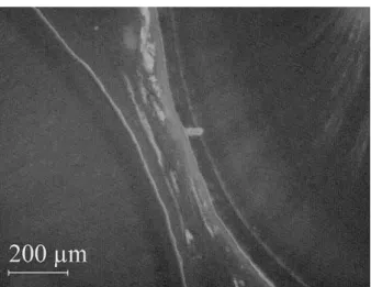
With a reciprocating saw (Nouvag, Basle, Switzer-land), the blade of which is 0.5 mm thick, a unilateral paramedian osteotomy of the mandible was carried out between the second lateral incisor and the canine tooth, in between the holes made for the osteosynthesis screws. A chisel was used to split the crestal bone between the roots, with preservation of the gingival cuff. Digital con-trol maintained the integrity of the lingual mucoperios-teum. The space between the roots at the crestal level was so limited (Fig. 2) that root damage was inevitable. The distractor was finally fixed again and the wound su-tured in a watertight fashion with 3-0 polyglactin 910 (VicrylÒ, Ethicon, Neuilly, France). The antibiotic amox-icillin (7 mg/kg) (ClavamoxÒ, Pfizer Animal Health, NY, USA) was administered intramuscularly every 24 h for three days. Dipirone and hioscine (DiplabiolÒ, Sabiol, Lisbon, Portugal) were administered intramuscularly for analgesia, at a dosage of 2 ml/day, during three days.
The dogs were sacrificed after 12 weeks of callus con-solidation. After intravenous injection of thiopental, the common carotid arteries were bilaterally clamped with vessel loops, and injected with 120 ml formaldehyde in a 10% concentration. Hence, fixation of the tissues oc-curred in vivo and cardiac arrest within a few minutes. After the sacrifice, the anterior segment of the mandible was removed with an oscillating autopsy saw (Stryker Corporation, Kalamazoo, USA).
Distraction policy
The animals were kept on soft granulated diet with waterad libitum(Selection 7eEvolution
Ò
, Royal Canin, Emargues, France) until the 8th week of the consolidation phase. Oral
Calcein Green Xylenol Orange
Tetracycline
0 8 27 69 97
2 2
Days Vital dye Distraction
parameters Maxillarycanine
crown amputation
Activation phase
Sacrifice
Models
Radiographs
Pocket probing & plaque control
2
2 2 2
Distraction surgery
hygiene was ensured with a daily cleansing with a 20/100 brush (Pierre Fabre, Castres, France), and fluoride tooth-paste (Elgidium, Pierre Fabre, Castres, France). Active dis-traction started on day 8, with a rate of 1 mm once a day, corresponding to a 360rotation of both distractor rods.
An-aesthesia was not necessary. The activation period lasted for 5 days, the consolidation phase for 84 days.
Data acquisition
Periodontal pocket probing and bacterial plaque control were performed at the time of canine crown amputation, and at the time of animal sacrifice. Radiological evalua-tion was done at the time of maxillary crown amputaevalua-tion (day 8), at the time of the first activation, and after 2 weeks (day 27), 8 weeks (day 69) and 12 weeks (day 97) of consolidation. Impressions were made at the time of the preliminary procedure (for individual tray fabrication), at the time of the distraction surgery and
the first activation, and after 8 weeks (day 69) of consol-idation (Fig. 1).
The periodontal pockets were measured with a cali-brated probe (HU-FriedyÒ, Chicago, Illinois, USA). For all eight front teeth, pockets were measured at six surfaces; buccal, buccomesial (BM), buccodistal (BD), lingual, linguomesial, and linguodistal. Bacterial plaque was controlled with erythrosine (DentoplaqueÒ, Pierre Fabre, France). Plaque was assessed according to the Pla-que Index (Pl.I;Lo¨e andSillness, 1963) and also scored on 6 surfaces of all eight front teeth. Occlusal films (Ko-dak EktaspeedÒ, Eastman Kodak, Rochester, USA) were fitted into a PVC frame for standardization purposes. This device comprised a custom-made cylinder adapted to the X-ray tube (Oralix 65, PhilipsÒ, Eindhoven, the Netherlands). Impressions were made with polyvinyl si-loxane (ExpressÒ, 3M, Minneapolis, USA). Models were made of acrylic (Orthosin-Uni, Schu¨tz Dental, Ros-bach, Germany). The intercanine distance, the distance between the second lateral incisors at their distal sur-faces, and the distance between the adjacent surfaces of the canine and second lateral incisor related to the osteot-omy line were measured with a digital caliper (Mitutoyo CD-15DC, Kanagawa, Japan) (Fig. 3).
Histology protocol
A vital staining protocol was used as recommended by
Rahn (1999). Oxytetracyclin (Pfizer AG, Zurich, Swit-zerland) in a dosage of 25 mg/kg, diluted in 10 ml of a 0.9% saline solution, was injected subcutaneously three times, with a two-day interval, covering the activa-tion period (days 8, 10, 12) (Fig. 1). Xylenol Orange (Fluka AG, Buchs, Switzerland), at a dose of 90 mg/ kg, diluted in a 9%distilled water solution, was injected subcutaneously in the first part of the consolidation phase (days 27, 29, 31). Calcein Green (Fluka AG, Buchs, Switzerland) at a dosage of 5 mg/kg of a 0.5%distilled water solution was injected in the second part of the con-solidation period (days 69, 71, 73).
After sacrifice, the marked mandibular specimens were placed in appropriate containers with a 4%formalin so-lution and sent to the University of Aachen, Germany, for histological preparation and processing. After gradual dehydration by ethanol wash, the specimens were infil-trated with methacrylate. The histological preparation method was the cuttingegrinding EXAKT/Donath
tech-nique (DonathandBreuner, 1982). The slices had inter-vals of 800e900m and were 70e90m thick. The specimens were evaluated by light microscopy (native, polarized light, fluorescence, and after toluidin blue staining) with magnifications 0.8, 1.6, 1.8, 2.5,
5, 10and 20.
Quantification and statistical analysis were not per-formed because of the small number of animals, which was in turn because of moral restrictions. Hence, the analysis is descriptive.
RESULTS
Specific tooth movements during the activation and early consolidation phase are listed inTable 1. The ‘‘walking Fig. 2eSection of the canine root (left) and lateral incisor (right) with
crestal bone in between, at the control side. The distance between the root surfaces in all dogs was too narrow to allow the 0.5 mm thick saw blade to split the crestal bone. Root surface damage was automatic and inevitable.
Fig. 3eGraphical representation of the distances measured on the acrylic models made from impressions taken at the time of the first day of activation and at 8 weeks of consolidation. 1, Intercanine cusp distance; 2, distance between the distal surfaces of both second lateral incisors; 3, distance between the mesial surface of the right canine and the distal surface of the right second lateral incisor.
tooth phenomenon’’ can be deducted from the increase in distance 2, being the distance between the distal sur-faces of the second lateral incisors (Fig. 3). On average, the distance over the six incisors increased by 2.2 mm (standard deviation [SD] 1.1). The changes in the inter-canine distance (distance 1) did not correlate with the width of the distraction gap measured at the dental level (distance 3).
The differences in pocket depths of the teeth adjacent to the distraction gap and the contralateral control teeth are listed in Table 2. The probings of the other areas did not add valuable information. Dog 5 was excluded from the calculations because it had accidentally trauma-tized its oral vestibule with pathological pocket forma-tion (10 mm) and delayed consolidaforma-tion as a result. The pocket depths at the experimental side were slightly in-creased. Because of the size of the sample, differences between the two sides were not statistically analyzed.
The Pl.I at time of crown amputation was on average 1, to increase to 1.83 at time of sacrifice (Table 3). The differ-ence was significant (P.0.001epairedt-test) but there
was no correlation (Pearson Product Moment Correlation). Occlusal radiographs (Fig. 4) were useful to correlate the type of dental lesion with the findings of the histology slices. Callus formation was evident in all occlusal radio-graphs already after the second week of consolidation (day 27), except in those of Dog 5, in which there was no expansion.
The axial histology slices were grouped according to the apical, middle and coronal levels of the second lateral incisor, and viewed with different magnifications, light-ing and stainlight-ing (a total of 2500 pictures). In all the out-fractured root fragments embedded in the distraction regenerate, signs of resorption and of deposition of min-eralized material with an important cellular component could be observed. Fibrous tissue resembling periodontal ligament (PDL) was surrounding the fragments, with fi-bres running parallel to the fragment’s surface. There were no signs of inflammation or of foreign body reac-tion (Fig. 5). A pattern of regeneration was observed in the PDL adjacent to the sectioned root surface. Although there were some inconsistent aspects, e.g. localized signs of ankylosis alternating with others of scar tissue for-mation, the dominant appearance was that of bundles of collagen fibres with an altered orientation almost coin-cidental with the direction of the distraction forces. These aspects were more evident in the polarized light pictures (Fig. 6). In none of the sectioned areas could inflamma-tion be observed, except in Dog 5. However, magnifica-tions of up to 10 are not detailed enough to make
a definitive statement.
A fluorescent green line (compatible with Calcein Green fixation) could be consistently seen covering tooth zones with saw cut lesions (Fig. 7). The intensity of the fixation had considerable variation, being almost nonex-istent (but still present) in Dog 5 with the laceration. Cor-relation of the native histology with the fluorescence revealed that this green line most probably represented a layer of regenerated cementum at different levels of in-tensity. The presence of bone-like tissue is observed in the pulpal canal after an oblique lower third root section in Dog 2 (Fig. 8). This tissue is formed by several small grossly kidney shaped structures that are present not only in the open zone of the pulp canal but also in the contig-uous upper slices (inside the pulpal chamber). The fluo-rescence reveals a high degree of fixation. Those could Table 1eThe difference in the distances (mm) depicted inFig. 3,
between the first day of activation (day 8) and 8 weeks of consolidation (day 69), indicative for the ‘‘walking tooth phenomenon’’ occurring in the activation and first half of the consolidation phase
Intercanine distance (1)
Distance between the lateral surfaces of the 2nd lateral incisors (2)
Distance between the proximal surfaces of the canine and 2nd lateral incisor adjacent to the distraction gap (3)
Dog 1 1.7 1.3 2.6
Dog 2 4 4.1 3.8
Dog 3 3.6 1.6 3.4
Dog 4 4.4 1.2 5
Dog 5 0.2 2.3 1.6
Dog 6 1.5 2.5 0.8
Table 2eDifferences in BM and BD pocket depth, measured at the teeth adjacent to the distraction gap at the experimental side, and at the contralateral teeth at the control side, between the preoperative situation and the situation at sacrifice
Pocket depth
Experimental side Control side
Canine 2nd Lateral
incisor
2nd Lateral incisor
Canine
BD BM BD BM BM BD BM BD
Mean 1.2 0.8 0.8 0.2 0.2 0.4 0 0.2
SD 0.8 1.3 1.1 0.4 0.4 0.9 0 0.4
Depth in millimeter.
Table 3ePlaque index of each dog for each tooth surface, averaging all 8 mandibular front teeth. The index was calculated at the time of canine crown amputation (‘‘initial’’) and at the moment of sacrifice (‘‘final’’)
Buccal Lingual BM BD Linguomesial Linguodistal
Initial Final Initial Final Initial Final Initial Final Initial Final Initial Final
DOG 1 1 2 1 2 1 2 1 2 1 2 1 2
DOG 2 1 2 1 2 1 2 1 2 1 2 1 2
DOG 3 1 1 1 2 1 2 1 2 1 2 1 2
DOG 4 0 1 1 2 1 1 1 1 1 1 1 1
DOG 5 1 2 1 2 1 2 1 2 1 2 1 2
DOG 6 1 2 2 2 1 2 1 2 1 2 1 2
be bone fragments dislocated by the saw at the moment of the osteotomy although its presence in the upper slices is not congruous with this vision.
The fact that Dog 5 had a traumatic gingival disruption induced by a convulsion (apparently caused by the sub-cutaneous injection of Xylenol Orange) turned out to be a key issue in this study. The presence of bacterial pla-que on the surface of the coronal thirds of the roots adja-cent to the osteotomy had induced inflammation in the surrounding soft tissues. Regeneration of periodontal structures was almost nonexistent (Fig. 9). The fluores-cence observed on the root surfaces may represent sites of resorption or perhaps simply be a site of mechanical matrix injury.
Whereas all alveolar crests in the uninjured animals had maintained their original height after consolidation, there was significant resorption down to the level of the second lateral incisor apex in Dog 5.
When osteosynthesis screws extended into the root canal, the pulp remained viable (Fig. 10a). The screw thread may transplant bone fragments into the root canal. This bone may stay viable and undergo extensive remod-elling (Fig. 10a). The periodontium reacted similarly to tangential trauma from a screw thread as to a clean cut from a saw or chisel; periodontal-like tissue encom-passed the root surface (Fig. 10b) which showed cemen-tum regeneration under polarized light.
DISCUSSION
The changes in the intercanine distance (distance 1) do not reflect the width of the distraction gap measured at the dental level (distance 3), because the distraction had an important antero-posterior vector and the un-known degree of mandibular segment tilting will have
had an influence as well. The tendency for anterior teeth to close the gap mesially is a known phenomenon in an-terior mandibular osteodistraction with midline osteot-omy (‘‘walking tooth phenomenon’’; Weilet al., 1997). In the present experiment, the histological section showed that only the lateral incisors tended to close the gap by distal movement, probably because the dog ca-nine has such a long, firmly anchored root.
Crestal bone loss has been observed with immediate expansion (DorfmanandTurvey, 1979), and with imme-diate expansion of 1 mm followed by a latency phase of 5 days to 2 weeks prior to further expansion. The authors suggested that it was caused by radicular surface expo-sure, resulting from inter-dental osteotomies in narrow inter-dental spaces (Bell et al., 1997, 1999). When Fig. 4e(a) Occlusal radiographs at the moment of maxillary canine crown amputation. Note the midline synarthrosis, (b) at day 1 of activation, (c) at day 27 (2 weeks of consolidation), (d) at the time of sacrifice (12 weeks of consolidation). The cone was positioned under the symphysis. The left side of the radiograph shows the right side of the mandible.
Fig. 5eChipped fragment showing signs of localized resorption and cementum regeneration. The fragment is embedded in a fibrous tissue collar of the thickness and appearance of PDL. (A) Native histology (0.8), arrow pointing at the fragment, (B) fluorescent histology (5) (C) polarized light histology, magnification (5).
Fig. 6eSectioned surface of the canine tooth with regenerated PDL-like tissue regeneration () demonstrating fibres directed according to the distraction vector. Polarized light histology (1.6).
a true latency period of 5e7 days was respected, crestal
bone loss was not observed and the attached gingiva maintained its preoperative level (Guerrero et al., 1997; Kewitt and Van Sickels, 1999; Niederhagen et al., 1999; Mommaerts et al., 2005). These findings, and the ones from the present experiment, suggest that keeping the junctional epithelium attached, or allowing its reattachment, may be more important than keeping bone on the roots. The periodontal response can though be greatly influenced by the preoperative periodontal sta-tus and maintenance of good oral hygiene habits in the postoperative period (Kewitt and Van Sickels, 1999;
Mommaertset al., 2005).
Andreasenet al. (1990)defined periodontal healing by ‘‘a newly formed PDL space surrounding the entire root periphery.’’ In this investigation, not yet-well organized connective tissue between the distraction callus and the damaged root surface was observed. All exposed tooth surfaces, being cementum, dentine (at the external and pulpal surface), or dental particles, were lined by a tissue indistinctive from normal PDL.
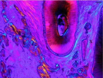
Fig. 7eSlice taken at the middle part of the second lateral incisor root within healthy distraction callus (native histology, 1.6). The inset shows the sectioned surface of the incisor covered by a regenerated layer, which may represent cementum repair (fluorescent histology, 10). In the pulp canal a fragment of bone ingrown bone is recognisable (Fig. 8).
Fig. 8eSlice taken at the apical part of the specimen ofFig. 7. PDL-like tissue is covering the inner surface of the root canal and is contiguous with the regenerated PDL-like tissue covering the sectioned root surface. This tissue is again contiguous and similar in texture with the ligament covering the normal root surface within the newly formed distracted bony callus. (A) Native histology (1.6), (B) native histology (5), and (C) fluorescent histology (5).
Bavitzet al. (2000)discovered a clinically small but sta-tistically significant difference in cementum regeneration using distraction osteogenesis to regenerate tissues in supracrestal defects. This cementum regeneration oc-curred on root surfaces that had been exposed to plaque for 3 months. Whether cementoblasts, pluripotential cells, or other cells within the PDL resulted in cementum deposition could not be determined. In Dog 5 with the accidental gingival laceration, cementum repair was not observed on the plaque covered root surfaces. This is not in contradiction with the findings of Bavitz et al. (2000), since in this dog the root surfaces concerned re-mained exposed until sacrifice. Cementum repair was manifest on all surfaces temporarily exposed by the os-teotomy. Small focal regions of root resorption were seen on each of the teeth that moved. However, some de-gree of root resorption occurs with any type of orthodon-tic tooth movement and approximately 75%is repaired by cellular cementum (Copeet al., 1999;Roberts-Harry andSandy, 2004). Some basal level of root surface turn-over may be normal in these dogs (Verna and Melsen, 2003).
The PDL of the teeth contiguous with the distraction gap showed a widening and an altered orientation of the collagen fibres that coincided with the direction of the distraction forces. This phenomenon has also been observed by Bell et al. (1997) and is identical to what is observed during orthodontic tooth movement in non-distracted bone (Verna andMelsen, 2003).
Bone tissue with osteocytes and osteoblasts has been observed in replanted and autotransplanted immature teeth whether the original pulp tissue was removed
(Claus et al., 2004), or not (Skoglund and Tronstad,
1981). The amount of atubular hard tissue these authors found was very small and did not occupy large parts of the cavity or walls. Interestingly, this bone-like tissue, formed centrally in the pulp was in some teeth connected to the alveolar bone through the open apical foramen (Skoglundand Tronstad, 1981).
An interesting finding was that tooth fragments, whether small after being cut off tangentially, or large following extensive pulp exposure, were not expelled by an inflammatory reaction. On the contrary, until the end of the experiment, the reparation tissues surrounding or extending into these fragments resembled a normal
periodontal apparatus, with oriented bundles of collagen, cellular cementum and supportive cell rich woven bone. Tangential root damage from screws has been exam-ined by Asscherickxet al. (2005), also in Beagle dogs, with fluorescence and toluidin staining techniques. Their results corroborate with the present findings; tooth pres-ervation is not jeopardized by tangential titanium screw lacerations as can occur during maxillo-facial trauma or orthognathic surgery, absolute anchorage for orthodon-tics or osteodistraction procedures.
CONCLUSIONS
Deliberate surgical injury to the teeth and surrounding periodontium was examined histologically and radio-graphically in Beagle dogs receiving transverse mandib-ular osteodistraction surgery. Cementum and dentine lesions were repaired by cellular cementum, and the damaged root surfaces surrounded by PDL-like collagen bundles. Loose dentine and cementumedentine
frag-ments were consistently embedded in regenerated perio-dontium and their surface repaired by cementum. The pulp remained viable when damaged by osteosynthesis screws. Extensive pulpal evisceration resulted in in-growth of the PDL and cell rich woven bone. The dentine surface of the pulp canal was also lined by cellular ce-mentum. These severely damaged teeth survived as giant fragments. Loss of attached gingiva in an inadver-tently traumatized dog resulted in 10 mm pathological pocket formation with histological signs of inflammation and no bone formation. In none of the dogs was the dis-traction callus jeopardized by the root lesions, except in the traumatized dog.
Future research should focus on ultramicroscopic ex-amination of the regenerative tissue in the periodontium, and on the phenomena occurring in the pulpal structures. The increased use of bone borne devices in modern or-thodontics emphasizes its importance.
ACKNOWLEDGEMENTS
The authors thank Prof. G. Pessanha Alcoforado, Univer-sity of Lisbon; Prof. R. Mascarenhas, Biomedical Tech-nology Institute of Santare´m, Portugal; Prof. B. Rahn Fig. 10eDamage by titanium screws. (a) Screw entering the pulp tissues. Bony fragments are pushed inside the pulp along with the thread. These fragments show extensive remodelling. The pulp shows signs of vitality. Fluorescent histology (1.6). (b) Screw cutting a root surface. The root surface is covered by collagen bundles, as a regenerative response. Polarized light histology (2.5).
and PD S. Milz, AO Research Institute, Davos, Switzer-land; Prof. P. Diedrich and Mrs. U. Ernst, Bone Labora-tory e Department of Orthodontics, University of
Aachen; Prof. J. Galv~ao Leit~ao, University of Lisbon; and the Surgi-Tec company, Bruges, Belgium for their support of this study.
References
Andreasen JO, Paulsen HU, Yu Z, Schwartz O: A long-term study of 370 autotransplanted premolars. Part III. Periodontal healing subsequent to transplantation. Eur J Orthod 12: 25e37, 1990
Asscherickx K, Vande Vannet B, Wehrbein H, Sabzevar MM: Root repair after injury from mini-screw. Clin Oral Implants Res 16: 575e578, 2005
Bavitz JB, Payne JB, Dunning D, Glenn A, Koka R: The use of distraction osteogenesis to induce new suprabony periodontal attachment in the beagle dog. Int J Periodontics Restorative Dent 20: 596e603, 2000
Bell WH, Harper RP, Gonzalez M, Cherkasin AM, Samchukov ML: Distraction osteogenesis to widen the mandible. Br J Oral Maxillofac Surg 35: 11e19, 1997
Bell WH, Gonzalez M, Samchukov ML, Guerrero CA: Intraoral widening and lengthening of the mandible in baboons by distraction osteogenesis. J Oral Maxillofac Surg 57: 548e562 and discussion 563, 1999
Claus I, Laureys W, Dermaut L: Histologic analysis of pulpal revascularisation of autotransplanted immature teeth after removal of the original pulp tissue. Am J Orthod Dentofacial Orthop 125: 93e99, 2004
Cope J, Samchukov ML, Muirhead DE: Distraction osteogenesis and histogenesis in Beagle dogs: the effect of gradual mandibular osteodistraction on bone and gingival. J Periodontol 73: 271e282, 2002
Cope JB, Harper RP, Samchukov ML: Experimental tooth movement through regenerate alveolar bone. A pilot study. Am J Orthod Dentofacial Orthop 116: 501e505, 1999
Del Santo Jr M, Guerrero CA, Buschang PH, English JD,
Samchukov ML, Bell WH: Long-term skeletal and dental effects of mandibular symphyseal distraction osteogenesis. Am J Orthod Dentofacial Orthop 118: 485e493, 2000
Donath K, Breuner G: A method for the study of undecalcified bones and teeth with the attached soft tissues: the Sage Schliff (sawing and grinding) technique. J Oral Pathol 11: 318e326, 1982
Dorfman HS, Turvey TA: Alterations in osseous crestal height following interdental osteotomies. Oral Surg Oral Med Oral Pathol 48: 120e125, 1979
Guerrero CA, Bell WH, Contasti GI, Rodriguez AM: Mandibular widening by intraoral distraction osteogenesis. Br J Oral Maxillofac Surg 35: 383e392, 1997
Kewitt GF, Van Sickels JE: Long-term effect of mandibular midline distraction osteogenesis on the status of the temporomandibular joint, teeth, periodontal structures, and neurosensory function. J Oral Maxillofac Surg 57: 1419e1425 and discussion 1426, 1999 Lo¨e M, Sillness J: Periodontal diseases in pregnancy. I. Prevalence and
severity. Acta Odontol Scand 21: 533e551, 1963
Mommaerts MY: Bone anchored intraoral device for transmandibular distraction. Br J Oral Maxillofac Surg 39: 8e12, 2001
Mommaerts MY, Polsbroek R, Santler G, Correia PEGS, Abeloos JVS: Anterior transmandibular osteodistraction: clinical and model observations. J Craniomaxillofac Surg 33: 318e325, 2005 Niederhagen B, Braumann B, Schmolke C, Appel T, von Lindern JJ,
Berge S: Tooth-borne distraction of the mandible. An experimental study. Int J Oral Maxillofac Surg 28: 475e479, 1999
Rahn BA: Intra vitam staining techniques. In: von Recum AF (ed.), Handbook of biomaterials evaluation. scientific, technical and clinical testing of implant materials, Philadelphia: Taylor and Francis, 727e738, 1999
Roberts-Harry D, Sandy J: Orthodontics. Part 11: orthodontic tooth movement. Br Dent J 196: 391e394, 2004
Skoglund A, Tronstad L: Pulpal changes in replanted and
autotransplanted immature teeth of dogs. J Endod 7: 309e316, 1981 Verna C, Melsen B: Tissue reaction to orthodontic tooth movement
in different bone turnover conditions. Orthod Craniofac Res 6: 155e163, 2003
Weil TS, Van Sickels JE, Payne CJ: Distraction osteogenesis for correction of transverse mandibular deficiency: a preliminary report. J Oral Maxillofac Surg 55: 953e960, 1997
Maurice MOMMAERTS, MD, DMD, PhD, FEBOMFS Division of Maxillo-Facial Surgery
GH St. Jan av Ruddershove 10 B-8000 Bruges, Belgium
Tel.: +32 50 45 22 60 Fax: +32 50 45 22 79 E-mail:maxfac@azbrugge.be