Phenotypic and Immunomodulatory
Properties of Equine Cord Blood-Derived
Mesenchymal Stromal Cells
Laurence Tessier1, Dorothee Bienzle2, Lynn B. Williams3, Thomas G. Koch1,4*
1Department of Biomedical Sciences, Ontario Veterinary College, University of Guelph, Guelph, Canada,
2Department of Pathobiology, Ontario Veterinary College, University of Guelph, Guelph, Canada,
3Department of Clinical Studies, Ontario Veterinary College, University of Guelph, Guelph, Canada,4The Orthopaedic Research Lab, Aarhus University, Aarhus, Denmark
*[email protected](TGK)
Abstract
Multipotent mesenchymal stromal cells (MSC) have attracted interest for their cytotherapeu-tic potential, partly due to their immunomodulatory abilities. The aim of this study was to test the robustness of our equine cord blood (CB) MSC isolation protocol, to characterize the CB-MSC before and after cryopreservation, and to evaluate their immunosuppressive phe-notype. We hypothesized that MSC can be consistently isolated from equine CB, have unique and reproducible marker expression andin vitrosuppress lymphoproliferation. Pre-liminary investigation of constitutive cytoplasmic Toll-like receptor (TLR) 3 and 4 expression was also preformed due to their possible association with anti- or pro-inflammatory MSC phenotypes, respectively. Surface markers were assessed for antigen and mRNA expres-sion by flow cytometry and quantitative polymerase chain reaction (qPCR). Immunomodula-tory properties were evaluated in mixed lymphocyte reaction assays, and TLR3 and TLR4 expression were measured by qPCR and immunocytochemistry (ICC). CB-MSC were iso-lated from each off nine cord blood samples. CB-MSC highly expressed CD29, CD44, CD90, and lacked or had low expression of major histocompatibility complex (MHC) class I, MHC-II, CD4, CD8, CD11a/18 and CD73 before and after cryopreservation. CB-MSC sup-pressedin vitrolymphoproliferation and constitutively expressed TLR4. Our findings con-firmed CB as a reliable MSC source, provides an association of surface marker phenotype and mRNA expression and suggest anti-inflammatory properties of CB-MSC. The relation-ship between TLRs and lymphocyte function warrants further investigation.
Introduction
Cell-based therapies are increasingly in demand for treatment of a variety of conditions, in-cluding equine osteoarthritis [1]. However, our understanding of stem and stromal cell proper-ties is evolving slower than clinical applications are being pursued. The use of variably
characterized stromal cell preparations has led to discrepancies between predicted and OPEN ACCESS
Citation:Tessier L, Bienzle D, Williams LB, Koch TG
(2015) Phenotypic and Immunomodulatory Properties of Equine Cord Blood-Derived Mesenchymal Stromal Cells. PLoS ONE 10(4): e0122954. doi:10.1371/ journal.pone.0122954
Academic Editor:Zoran Ivanovic, French Blood
Institute, FRANCE
Received:October 22, 2014
Accepted:February 16, 2015
Published:April 22, 2015
Copyright:© 2015 Tessier et al. This is an open
access article distributed under the terms of the Creative Commons Attribution License, which permits unrestricted use, distribution, and reproduction in any medium, provided the original author and source are credited.
Data Availability Statement:All relevant data are
within the paper.
Funding:This project was supported by the Danish
Agency for Technology, Production and Innovation, Equine Guelph and the Department of Biomedical Sciences at the University of Guelph. The funders had no role in study design, data collection and analysis, decision to publish, or preparation of the manuscript.
Competing Interests:T. G. Koch acts in a volunteer
observed efficacy, and between different studies [1–3]. Development of safe and efficacious cell-based treatments is crucial for clinical application and to define potential value as novel therapy.
Multipotent mesenchymal stromal cells (MSC) are potential candidates for cell-based thera-py [2,4–6]. These cells are most commonly derived or isolated from bone marrow (BM), adi-pose tissue (AT) or umbilical cord blood (CB). In human research, the term MSC is often associated with mesenchymal stem cells rather than mesenchymal stromal cells. Here, we ex-clusively refer to mesenchymal stromal cells. Stem cells are characterized by long-term self-re-newal and differentiation abilities [7]. In horses, MSC differentiation abilities are still poorly understood, and no evidence ofin vivolong-term self-renewal ability have been published. Therefore, mesenchymal stem cells remain uncharacterized in this species, precluding their ref-erence in the present paper. In humans, MSC are evaluated based on minimal classification cri-teria that were established by the International Society for Cellular Therapy (ISCT). Cricri-teria include plastic adherence, osteo-, chondro- and adipogenic differentiation, and cell surface ex-pression of CD73, CD90, and CD105 concurrent with absent exex-pression of CD11b or CD14, CD45, CD34, CD79a or CD19, and human leukocyte antigen (HLA)-DR [8]. However, MSC remain incompletely characterized, and the above marker panel is largely exclusive rather than inclusive. MSC derived from animals are less well defined, and may also differ from human MSC. Therefore, improved and consistent culture methods for MSC and more comprehensive phenotypic characterization are required.
Equine MSC are less well characterized than human MSC, and inconsistent surface marker profiles have been observed. Surface expression of CD29, CD44 and CD90 was reported in sev-eral studies. However, unlike for human MSC, variable identification of CD73 and CD105 on MSC has precluded establishment of a consensus panel for horse MSC [9–17]. In addition, sev-eral investigators reported expression of MHC-II, CD31, CD34, CD45 and CD79a to be low or absent [9–28]. Hence, surface antigens characteristic of equine MSC have neither been clearly established nor confirmed with quantitative gene expression assays, although clinical applica-tion of such cells has been widely implemented [6]. Furthermore, MSC are often cryopreserved for potential future clinical use. Phenotypic and functional stability during such storage is unknown.
MSC progenitor function is defined by ability to differentiate into multiple cell types includ-ing osteo-, chondro- or adipogenic tissue [8]. MSC were also suggested to influence proximal cellsin vivoby secreting trophic and immunomodulatory factors [29]; a non-progenitor cell function that attracted much attention due to potential for also treating conditions associated with aberrant immune responses.In vivoimmunosuppressive function was reported for human MSC that were used for successful treatment of steroid-resistant stage IV graft-versus-host disease (GVHD) [30]. Promising results for treatment of other inflammatory diseases, such as osteoarthritis, with MSC has also been reported for several species [6,31]. Analysis of short-term outcomes indicated reduced inflammatory cytokine production and enhanced car-tilage regeneration.In vitroanti-proliferative effect of equine MSC on lymphocyte proliferation was reported [23]. However, results fromin vitroandin vivostudies are inconsistent, and evi-dence of long-term therapeutic efficacy is lacking [6,32].
The phenotype of MSC with immunomodulatory capability is unknown, which precludes derivation of cell populations for such purpose. It has been suggested that MSC, similarly to macrophages, might include MSC-1 and -2 subpopulations with pro-inflammatory and anti-inflammatory properties, respectively. MSC-1 and -2 subpopulations have been hypothesized to be associated with expression of TLR4 or TLR3, respectively [33]. MSC from different spe-cies or tissues expressed distinct TLR transcripts and antigens [34–36], suggesting functional Canada, a company for which T. G. Koch’s research
properties might be linked to tissue origin. Equine CB-MSC suppressed lymphoproliferationin vitro[23], but correlation with TLR3 or TLR4 expression was not determined.
The aims of this study were to determine the efficacy of deriving MSC from equine CB, to determine phenotype before and after cryopreservation, to evaluate whether CB-MSC suppress lymphocyte proliferation, and to assess expression of TLR3 and TLR4. We report highly effi-cient derivation of MSC with consistent phenotype from equine CB. Phenotypic and functional characteristics were preserved through cryostorage and associated with expression of TLR4.
Materials and Methods
Ethics statement
This study was specifically approved by the University of Guelph Animal Care Committee with regard to the procedures of collection of equine peripheral blood lymphocytes and equine umbilical cord blood (animal use protocols 1756 and 1570). Additional research conducted using specimens of this kind does not require review by the Animal Care Committee (falls under CCAC Category of Invasiveness A) and therefore the mixed lymphocyte reactions can be considered to have been conducted in accordance with the institutional ethics guidelines. Collection of peripheral blood and cord blood was add-on procedures to the routine care of the horses. No animals were sacrificed during the study. Equine umbilical cord blood was collected on two privately owned commercial farms in Southern Ontario. Four of five samples were col-lected on one farm from Thoroughbred foals. One sample was colcol-lected on another farm from a Warmblood foal. Informed consent was obtained in writing from the horse owners/agents prior to sampling. All the data for this study was subsequently anonymized. The broodmares on the foaling farms are housed in large foaling boxes. Both farms are staffed 24/7 and mares are under constant video surveillance and carrying foaling alarms to allow for observed foaling and assisted delivery if needed. Umbilical cord blood was collected by the farm staff after re-ceiving instruction by Dr. Koch. Instruction included video-review of cord blood collection. Cord blood was collected from an isolated segment of the umbilical cord after the umbilical cord had been clamped and detached from the foal. Peripheral venous blood was obtained from the Equine Research Herd owned by the Ontario Ministry of Agriculture and Food (OMAF). Once investigators have an approved animal care protocol from the University of Guelph Animal Care Committee access to these research horses are granted. In this study pe-ripheral blood was collected from 5 adult mixed-bred horses. The adult horses on the research farm are housed in smaller groups with run-in sheds throughout the year. The horses are on pasture during the summer and during the winter they have access to large paddocks with gravel surface. Peripheral blood was collected by Drs. Williams or Koch. Peripheral blood was collected under mild sedation (Xylazine HCl, 0.35–0.40 mg/kg bwt IV; Bayer, Toronto, ON) from the jugular vein following which manual pressure was applied for several minutes to aid hemostasis. All the data for this study was subsequently anonymized. This study was also spe-cifically approved by the University of Guelph Research Ethics Board with regard to the proce-dures of collection of Human peripheral blood. Human peripheral blood was collected in agreement with Institutional Research Ethics Board guidelines (protocol 12FE008). Informed written consent was obtained from the participants.
Experimental design
CB collection and shipping
CB was collected from nine foals immediately after foaling, as previously described [37]. CB collection was approved by the Institutional Animal Care Committee (protocol 1570). Veni-puncture of the umbilical vein was performed with a 16G hypodermic needle attached to a 450 mL blood transfusion collection bag (Fenwal, Baxter, Deerfield, IL) containing citrate phos-phate dextrose adenine as the anticoagulant solution. The blood was stored at 4–8°C for up to 24 hours before and up to 12 hours after being transported overnight by courier. A
Greenbox thermocontrol device (Greenbox System, ThermoSafe Brands; Arlington Heights, VA) was used for shipping and temperature control for up to 48 hours to account for possible delay during shipping. No samples encountered shipping delay.
CB-MSC isolation and culture
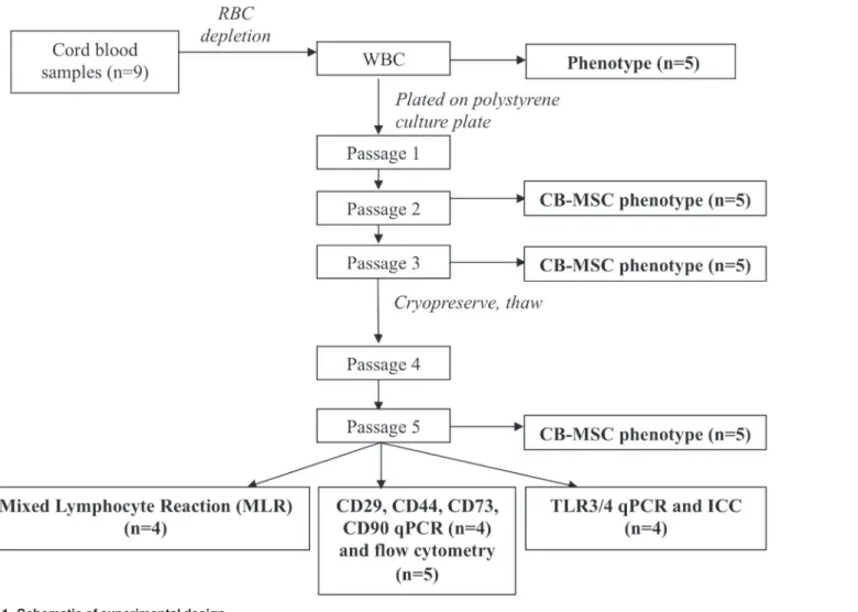
CB-MSC isolation and culture were adapted from a previously described protocols [38]. In brief, the white blood cell (WBC) fraction was isolated using PrepaCyte-WBC (PC, CytoMedi-cal Design Group; St. Paul, MN). Whole CB was mixed 1:1 with PC medium in 50 mL BD Fal-con Fal-conical tubes (BD Biosciences, Mississauga, ON) for 5 minutes, and then incubated for 25 minutes at room temperature (RT). Supernatant was collected, pooled and spun at 400gfor 10 Fig 1. Schematic of experimental design.
minutes at RT. Pellets were suspended in isolation medium (IM) consisting of Dulbecco’s mod-ified eagle medium (DMEM)-low glucose (1 g/L; Lonza, Wakersville, MD), 30% fetal bovine serum (FBS; Invitrogen, Burlington, ON), penicillin (100 IU/mL; Invitrogen), streptomycin (0.1 mg/mL; Invitrogen), L-glutamine (2 mM; Sigma-Aldrich, St. Louis, MO), and dexametha-sone (10-7M; Sigma-Aldrich). Cells were then incubated at 5% CO
2at 38°C in humidified
at-mosphere. Live and dead cells were counted with an automated cell counter (NucleoCounter NC-100, Mandel Scientific, Guelph, ON), and plated at 1x10⁶/cm2live cells in 75 cm²
polysty-rene cell culture flasks (Corning, Sigma-Aldrich). Primary colonies were detached using tryp-sin/ethylene diamine tetraacetic acid (EDTA; 0.04%/0.03%; Sigma-Aldrich) and hereafter expanded in the same culture medium without dexamethasone (expansion medium, EM). During expansion, cells were seeded at 5,000/cm2. Cells were cryopreserved in EM containing
10% dimethyl sulfoxide (DMSO; Sigma-Aldrich) at -1°C/min controlled freezing rate to -80°C for 24 hours, prior to long-term storage in liquid nitrogen. Cell concentration in the freezing medium was 1x10⁶/mL.
Cell morphology, colony counting, CB-MSC progenitor frequency, and
population doubling time
After eight days of incubation, cultures were inspected daily for presence of colonies. Confluent colonies were detached using trypsin/EDTA and re-seeded in T175 culture flasks. To docu-ment cell morphology, digital images were obtained prior to detachdocu-ment at passage 1, 3, 4 and post-thawing passage 5 using phase-contrast microscopy and Q-Capture software (Q-Imaging, Surrey, BC). CB-MSC progenitor frequency (PF) and doubling times were calculated as:
PF=Colony number/Number of WBC x100 Doubling times were calculated according to:
1. cell–doubling number(CD) = In(Nh/Ns)/In2
In=natural Logarithm
Nh=harvest cell number
Ns=seed cell number
2. Cell doubling time (DT)=CT/CD
CT=cell culture time
Flow cytometry
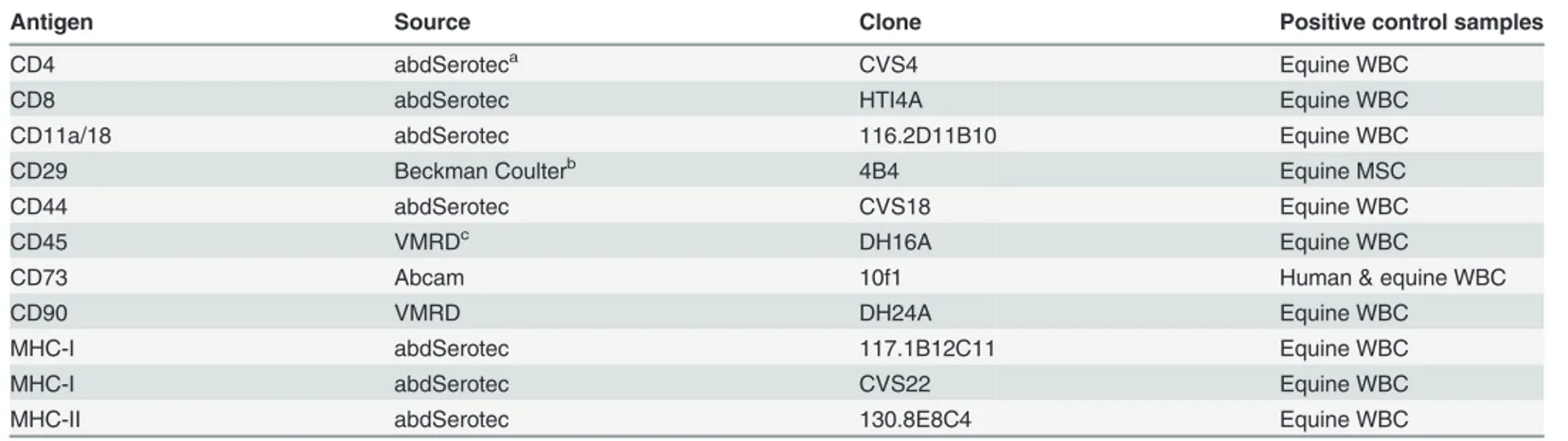
and blood samples from horses according to an approved animal care protocol (#11R034). In addition, CD73 clone 10f1 was reported to cross-react with equine cells using flow cytometry and confirmed with confocal microscopy [16]. All other antibodies used in this study were test-ed on equine WBC as part of the feasibility studies (data not shown). Incubations were at 4°C in the dark for 15 minutes, followed by a wash, and secondary antibody incubation at 4°C for 15 minutes in the dark. Rat anti-mouse IgM-FITC, goat anti-mouse IgG1-FITC (both Abcam, Toronto, ON) or donkey anti-mouse IgG (H+L)-FITC (Jackson ImmunoResearch Laborato-ries Inc., West Grove, PA) were used as secondary antibodies. Prior to staining, ammonium chloride (Sigma-Aldrich) hypertonic red blood cell (RBC) lysis was performed on peripheral and CB for leukocyte isolation, followed by a wash with flow buffer ((phosphate buffer saline (PBS); Sigma Aldrich), 5mM EDTA, 1% horse serum (HS), and 0.1% sodium azide). Cultured CB-MSC were chemically detached with Accumax (Stemcell Technologies Inc., Vancouver, BC) and washed with flow buffer prior to antibody incubation. WBC and CB-MSC at passage 2, 3 and 5, were assessed for CD4, CD8, CD11a/18, CD90, MHC-I and MHC-II surface expres-sion, and CB-MSC at passage 5 for CD29, CD44 and CD73 expression. Negative control sam-ples were cells incubated with only secondary antibody, and with isotype-matched non-binding primary antibody plus fluorescent secondary antibody. A minimum of 10,000 events were acquired for each antibody with CellQuest software (BD) and analyzed with FlowJo soft-ware (Tree Star Inc., Ashland, OR). Gates to identify WBC or CB-MSC populations were main-tained consistent throughout all experiments.
Quantitative PCR
Total RNA was extracted from WBC and CB-MSC at passage 3, 4, and 5 using themirVana miRNA Isolation Kit (Ambion, Life Technologies, Burlington, ON) following the manufactur-er’s instructions. RNA was quantified using a spectrophotometer (NanoDrop ND-1000, Thermo Fisher Scientific, Waltham, MA), aliquoted, and stored at -80°C for reverse transcrip-tion. For complementary DNA (cDNA) synthesis, RNA was thawed, and any potential residual genomic DNA was digested using DNase I amplification grade treatment (Invitrogen), directly followed by reverse transcription using the SuperScript II Reverse Transcriptase kit (Invitro-gen) with random priming of RNA. RNA quality was assessed by capillary electrophoresis Table 1. Antibody panel for flow cytometric analysis.
Antigen Source Clone Positive control samples
CD4 abdSeroteca CVS4 Equine WBC
CD8 abdSerotec HTI4A Equine WBC
CD11a/18 abdSerotec 116.2D11B10 Equine WBC
CD29 Beckman Coulterb 4B4 Equine MSC
CD44 abdSerotec CVS18 Equine WBC
CD45 VMRDc DH16A Equine WBC
CD73 Abcam 10f1 Human & equine WBC
CD90 VMRD DH24A Equine WBC
MHC-I abdSerotec 117.1B12C11 Equine WBC
MHC-I abdSerotec CVS22 Equine WBC
MHC-II abdSerotec 130.8E8C4 Equine WBC
aRaleigh, NC bMississauga, ON cPullman, WA
(Bioanalyzer 2100,AgilentTechnologies, Mississauga, ON) and only samples with RNA integ-rity numbers (RIN) above 9 were used.
Relative quantification was performed in a CFX Real-Time PCR Detection System (Bio-Rad, Kitchener, ON) by two-step real-time qPCR. Primers (Table 2) were designed using Invi-trogen OligoPerfect Designer or theNational Center for Biotechnology Information(NCBI) primer-BLAST software, or adapted from previously published sequences [39]. Beta-2-micro-globulin (B2M) and class II MHC transactivator (CIITA) sequences were used to detect MHC-I and-II, respectively. Amplicons were separated by 2% agarose gel electrophoresis, ex-tracted and purified using the QIAquick Gel Extraction kit (Qiagen, Hilden, Germany). DNA was sequenced and sequences were analyzed using the NCBI basic local alignment search tool (BLAST) against the equine database. Minimal identity cut-off was set at 96%. All primers ex-cept those for CD4 and CD73 incorporated at least one intron-exon spanning junction. To en-sure specificity of all primers, sample without reverse transcription (RT-control) and samples lacking cDNA template (NT-control) were included in each run. PCR assays were performed in triplicate in a 10μL total volume consisting of 5μL of PCR SsoFast EvaGreen Supermix (Bio-Rad), 0.2μM of primer mix and cDNA template. After an initial incubation at 95°C for Table 2. Nucleotide sequence of primers.
Gene Primers
CD4 50CCAGACTGACCAGACTGCAA 30
50TTGGATTCCAGCAGGACTTT 30
CD8 50AGTGGCTGGACTTCGACTGT 30
50CAAACACGTCTTCGGTTCCT 30
CD11a/18 50TTCAGCCAGCAACAAGAAGA 30
50GACAGCTGTGTTCCCACTGA 30
CD29 50CCCTTGCACAAGTGAACAGA 30
50ATTCCTCCAGCCAATCAATG 30
CD44 50ATCCTCACGTCCAACACCTC 30
50CTCGCCTTTCTTGGTGTAGC 30
CD73 50TGATCTTTCCCGAAAACCTG 30
50GGAATCCATCTCCACCATTG 30
CD90 50TGCCTGAGCACACATACCGCTC 30
50GCTTATGCCCTCGCACTTGACC 30
B2Ma 50TCGGGCTACTCTCCCTGACT 30
50ATTTCAATCTCAGGCGGATG 30
CIITAb 50GGTGCTACTTCGAGCTTTCG 30
50CCAACGTAGAGTCCGGTGAG 30
TLR4 50CCCACATCAACCAAGGAACT 30
50ATGGTTGAGGCCCTGATATG 30
TLR3c 50CAAACCCTGGTGGTCCTGTT 30
50GAAGGCCTCTGCTGGGATCT 30
β-actin 50TGGGCCAGAAGGACTCATAC 30
50GGGGTGTTGAAGGTCTCAAA 30
S18 50ACTGAGGATGAGGTGGAACG 30
50GCCCGTATCTTCTTCAGTCG 30
aB2M sequence was used for gene expression detection of MHC-I bCIITA sequence was used for gene expression detection of MHC-II cPreviously published sequence [39].
3 min, reactions were cycled 40 times with denaturation at 95°C for 5s, annealing for 5s at tem-peratures specific for each primer pair, and extension at 72°C for 15s. Amplification specificity was determined with melting-curve analysis whereby each amplicon was heated from 65 to 95°C. Efficiency for each primer pair was calculated from a standard curve generated from cDNA of leukocytes or CB-MSC. qPCR data were analyzed by the delta-delta Ct method (∆∆Ct) [40] using two endogenous reference genes chosen following the minimum informa-tion for publicainforma-tion of quantitative real-time PCR experiment (MIQE) guidelines [41]. Expres-sion stability analysis of the reference genes was performed with geNorm software. Initial selection of housekeeping genes was based on review of current literature [14,42], and com-prisedβ-actin, S18, B2M, and hypoxanthine phosphoribosyltransferase 1 (HPRT1). Based on geNorm [43] analysis of minimal variability in expression across samples, two housekeeping genes were selected. Assessment of variability in expression of different genes was based on the M-value calculated as the average pairwise variation of a gene compared to the other house-keeping genes [44]. The initial cut-off M-value was<1.5, and S18 andβ-actin were selected to
be used together as reference genes for subsequent relative quantification.
Lymphocyte proliferation assay
Blood was obtained from the jugular vein of five adult horses of variable breed and sex with an 18G hypodermic needle attached to a 450 mL blood transfusion collection bag (Fenwal, Baxter, Deerfield, IL). Mononuclear cells (MNC) were isolated a using Ficoll-density gradient. At RT, 15 mL of Ficoll-Paque Plus (density 1.078 g/mL, Stemcell Technologies) was loaded in a 50 mL tube with 35 mL of whole blood. Gradients were centrifuged at 300gfor 30 min at RT with no brake. Interphases containing the MNC fraction were carefully removed, pooled and washed with PBS. Supernatant was then removed and pellets were washed again with 10 mL of PBS. Pellets were then resuspended in 10 mL of Roswell Park Memorial Institute (RMPI) 1640 me-dium supplemented with penicillin (100 IU/mL; Invitrogen), streptomycin (0.1 mg/mL; Invi-trogen) and 10% FBS (InviInvi-trogen), and a live cell count was performed with an automatic cell counter (NucleoCounter NC-100). Cells were resuspended in freshly prepared cryomedium consisting of RPMI-1640 medium supplemented with penicillin (100 IU/mL; Invitrogen), streptomycin (0.1 mg/mL; Invitrogen), 10% FBS (Invitrogen) and 10% DMSO (Sigma-Aldrich) at a concentration of 6x10⁶cells/mL. One mL of cell suspension was placed in a 2 mL cryovial (Corning, Sigma-Aldrich) on ice for 30 minutes before gradual freezing over 12–18 hours to -80°C, and storage in liquid nitrogen.
acquired per triplicate sample. Controls consisted of unstained cells and cells incubated with only FITC-conjugated antibody.
TLR immunocytochemistry
MSC (5,000) were cultured in 250μL of EM in 8-well Permanox chamber slides (Thermo Fish-er Scientific) until 60–80% confluency. Cells were exposed to 1μg/mL of polyinosinic:polycy-tidylic acid (poly I:C; Aldrich) and 10 ng/mL of lipopolysaccharide (LPS; Sigma-Aldrich) for one hour to induce TLR3 and TLR4 expression. For ICC, cells were washed thrice with PBS, fixedin situfor 5 minutes with 4% paraformaldehyde (PFA; Invitrogen), washed thrice again with PBS and permeabilized with 0.1% Triton for 15 minutes. Cells were then washed again and treated with 3% hydrogen peroxide for 15 minutes. Non-specific antibody binding was blocked by incubation with 5% FBS for 10 minutes followed by one hour incuba-tion with polyclonal primary antibody to TLR3 or TLR4 (both Imgenex, San Diego, CA). Cells were then washed thrice with PBS and incubated for one hour with goat anti-rabbit secondary antibody (Abcam). Bound antibody linked to horseradish peroxidase (HRP) was detected using 3,3'-diaminobenzidine chromogen (Dako, Burlington, ON). Cells were counterstained with Harris hematoxylin to visualize nuclei, and images were acquired on a phase
contrast microscope.
Statistical analysis
All data were analysed with statistical analysis software (SAS Institute, Cary, NC). Effects were considered significant atP<0.05. Mixed lymphocyte reaction data and comparison between
passages for flow cytometry data were analyzed using a three-factor ANOVA without replica-tion with covariate (negative control). Comparison between markers for flow cytometry data were analyzed as single assays using a two-factor factorial ANOVA and Pmax as a covariate. Data were plotted as percent positive staining with 95% confidence intervals (CI). qPCR results was analyzed using a two-factor factorial in a randomized block with sub-sampling, accommo-dating unequal variances among targets to meet ANOVA assumption. Multiple comparisons were subsequently adjusted using Tukey’s honest significant difference (HSD) method. Data were plotted as relative expression with 95% CI. For CD8 and CIITA, transcripts were detected only in WBC, precluding comparisons. Analysis of residuals was performed to assess the ANOVA assumption. Residuals were plotted against the predicted values and explanatory vari-ables used in the model. The residuals were tested for normality using the four tests offered by SAS: Shapiro-Wilk, Kolmogorov-Smirnov, Cramer-von Mises, and Anderson-Darling.
Results
MSC progenitor frequency, population doubling time, and cell
morphology
CB-MSC from all nine CB samples were expandable to allow cryopreservation of 1.42x10⁶to 7.82x10⁶cells after passage 3 (average of 3.8 x10⁶cells).
Loss of WBC markers and retention of CD90 in CB-MSC
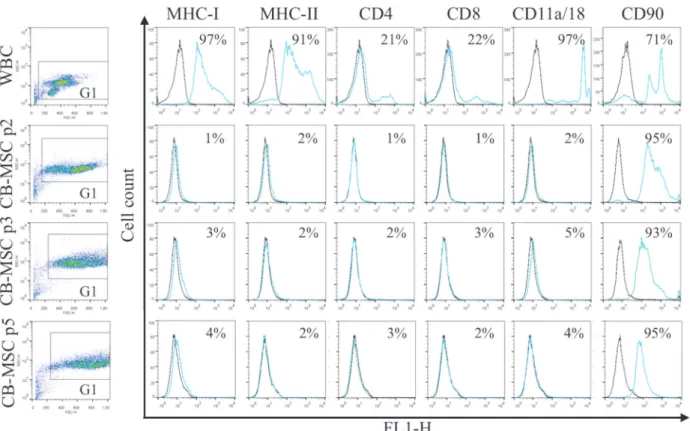
All antibodies yielded expected reactions except antibody to CD45 which did not react with equine cells. MHC-II was detected on WBC but not CB-MSC. Flow cytometric analysis (Fig 2) showed that the WBC fraction in CB contained cells that uniformly expressed MHC-I, MHC-II and CD11a/18, and variably frequent proportions of CD4+, CD8+, and CD90+cells (Fig 2). With derivation of CB-MSC, expression of MHC-I, MHC-II, CD4, CD8 and CD11a/18 disappeared. A large proportion of WBC expressed CD90, which persisted throughout differ-entiation into CB-MSC. Statistical analysis revealed significant decreases in the proportion of cells expressing each marker except CD90. Changes in surface marker expression between the WBC fraction and the CB-MSC population at passage two was significant for all markers (p<0.0001). No significant changes in marker expression between CB-MSC at passage 2, 3
and 5 were noted for CD90 (p = 0.50 (passage 2 to 3), p = 0.60 (passage 3 to 5)). Low/negative surface expression of other markers on CB-MSC precluded relevant statistical analysis. In WBC, two populations of cells with distinct CD90 expression were detected, possibly reflecting lymphocytes and neutrophils, while in CB-MSC CD90 expression was more homogeneous (Fig 2). To confirm the lack of surface expression of MHC-I on CB-MSC at passage 5, MHC-I clone CVS22 was additionally assessed and showed similar lack of expression (data not shown). Fig 2. Antigen expression on WBC in CB, and CB-MSC at passage (p) 2, 3 and 5.Consistent gates were applied for all cell analyses. Control sample fluorescence is indicated by black lines, fluorescence of samples incubated with specific antibody by light blue/gray lines. Results were similar for samples from 5 animals. Numbers in each plot represent the mean proportion of cells expressing each antigen.
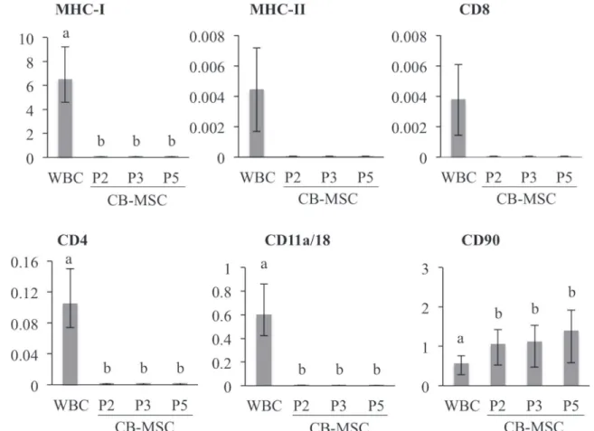
Relative quantification of gene transcripts correlated with flow cytometric detection of anti-gen expression (Fig 3). Transcripts of antigens expressed on WBC were either not detected or extremely low in CB-MSC at each passage. However, relative CD90 transcript abundance was significantly higher in CB-MSC at passage 2 than in WBC (p = 0.04), and remained high throughout CB-MSC culture. Findings were similar for all samples.
CD29 and CD44 are highly expressed on CB-MSC
Expression of CD29, CD44 and CD73 was assessed on passage 5 CB-MSC. Flow cytometric analysis indicated CD29 and CD44 were expressed on99% of CB-MSC, which was a signifi-cantly greater proportion of CB-MSC than those expressing CD90 (p<0.0001) (Fig 4A).
CD73 was either very low or undetectable on CB-MSC. Analysis of mRNA expression of each of these genes also indicated high abundance of CD29, CD44 and CD90 mRNA, and relatively low expression of CD73 mRNA (Fig 4B).
CB-MSC decrease lymphocyte proliferation in mixed lymphocyte
reactions
Culture of allogeneic mononuclear cells yielded lymphoproliferation typical of mixed lympho-cyte reactions (MLR) while culture of autologous mononuclear cells, as expected, yielded a low proportion of proliferating cells (Fig 5). Addition of CB-MSC reduced allogeneic lymphoproli-feration to levels below those of both allogeneic and autologous cells. In all four CB-MSC
Fig 3. Relative mRNA expression in WBC and CB-MSC.Gene expression were assessed with qPCR and normalized to S18 andβ-actin, and is shown relative to the lowest average CT. Bars represent CI; different letters indicate significant differences (p<0.05) between groups. Transcripts for MHC-II and
cultures suppression was significant compared to allogeneic cells (p<0.0001 for all CB-MSC
cultures). In three of four CB-MSC experiments there was significant suppression relative to autologous cells (p = 0.0032 (CB-1213), p = 0.0028 (CB-1216), p = 0.0007 (CB-1216)).
CB-MSC constitutively express TLR4
In order to determine possible contributions of TLR3 and TLR4 to functional properties of CB-MSC, expression of each receptor by mRNA quantification and ICC was determined. Nei-ther TLR3 mRNA nor protein was detected by qPCR and ICC assays (data not shown). Howev-er, TLR4 mRNA was highly expressed in CB-MSC, and protein was detected in untreated as well as LPS-treated cells (Fig 6). CB-MSC constitutively highly expressed TLR4 mRNA with minimal increase in expression after LPS stimulation.
Fig 5. CB-MSC decrease proliferation in mixed lymphocyte reactions.Autologous lymphocytes had minimal proliferation, while culture of allogeneic lymphocytes resulted in proliferation. Addition of CB-MSC to allogeneic lymphocyte cultures reduced proliferation as detected by BrdU incorporation and flow cytometry. Different letters indicate significant differences (p<0.05) between groups, bars indicate CI.
Fig 4. CD29, CD44 and CD90 are highly and consistently expressed on CB-MSC.A. Fluorescence of control samples (black line) and specific antibody (light blue/gray) with the mean proportion of MSC expressing each antigen. Results are representative of 5 experiments. B. Relative mRNA expression. Bars represent CI, and different letters indicate significant differences (p<0.05) between groups. CB-MSC had consistently high expression of CD29, CD44 and CD90 relative to CD73.
Discussion
Equine CB-MSC were derived from each of nine CB samples, suggesting this culture protocol was highly efficient. Although collection of CB is relatively non-invasive and yields highly pro-liferative MSC with chondrogenic potency [37,45,46], relatively low isolation success has limit-ed their utility to date [47,48]. As a result, CB-MSC are less well characterized relative to BM-and AT-derived MSC. In initial studies of equine CB-MSC, cell lines were derived from 4 of 7 [37], 41 of 51 [49] and 13 of 17 samples [50], e.g. 57–80% isolation frequency. Subsequently, improved culture methods generated MSC from 5 of 5 [38] and 6 of 6 CB samples [12]. In hu-mans, successful derivation of MSC from CB ranges from 29 to 48% irrespective of methodo-logical differences [47,48]. Generation of CB-MSC differs between humans and horses due to the available volume of CB and collection methods. The method described here consistently yielded CB-MSC cultures, even from samples up to 48 hours old. Therefore, in light of defini-tion of robust culture methods, focus may now be placed on timely and consistent collecdefini-tion of CB from mares.
Leukocytes in CB consisted of cells with light scatter properties and antigen expression typi-cal of lymphocytes, neutrophils and monocytes. Derivation of CB-MSC was associated with loss of WBC and appearance of a uniform population of large fibroblast-like cells with very high forward light scatter. CB-MSC were characterized by lack of expression of typical WBC antigens, and by expression of CD29, CD44 and CD90. High expression of CD29, CD44 and CD90 and low expression of CD73 was previously reported for equine MSC [14,16,51]. CD29 and CD44 are molecules that function in cell adhesion, and high expression is associated with stem cell phenotype [52]. CD90 is glycosylated immunoglobulin-family member protein wide-ly expressed on many types of stem cells and WBC [53,54]. CD73 is an ectonuclease and the CD73 (clone 10f1) used in this study was expressed on human and horse WBC, with nucleotide identity of 88% between horses and humans (data no shown). Hence, the antibody utilized was expected to detect equine CD73, which implies that our CB-MSC are indeed low/negative for CD73. In rodents, CD73 expression has been associated with small AT-derived MSC, while large AT-derived MSC lacked CD73 [55]. Expression of CD73 correlated with differentiating properties of MSC. Whether similar functional properties apply to CB-MSC remains to be determined.
Consistent low or absent expression of MHC-I on equine CB-MSC is in contrast to findings by Carrade et al. (2012) and De Schauwer et al. (2014). In those studies, CB-, umbilical cord matrix (UCM)-, peripheral blood (PB)-, BM- and AT-derived MSC expressed MHC-I [12,23]. Lack of MHC-I expression on CB-MSC at passage 5 was also noted with antibody CVS22 as used by Carrade et al. (2012) (data not shown). Lack of detection of MHC-I with different anti-bodies by flow cytometry, and lack of mRNA detection, indicate that CB-MSC derived under conditions described here do not express MHC-I. Lack of MHC-I and-II expression may con-tribute to or account for lymphosuppressive properties of MSC during co-culture with mono-nuclear cells from a range of unrelated donors [56]. Nevertheless, since expression of MHC-I and-II on MSC has been suggested to be unstable [23,32,57], transient downregulation during the first 5 passages cannot be ruled out. Additional investigations on stability and mechanisms of MHC regulation in MSC are needed. Resolving presence or absence of highly polymorphic Fig 6. CB-MSC constitutively expresses TLR4 with minimal up-regulation after exposure to LPS.A. TLR4 mRNA was normalized to S18 andβ-actin. Bars indicate CI. Different letters indicate significant differences (p<0.05) between groups. B. Untreated (top) and LPS-treated (bottom) CB-MSC have similarly intense TLR4 immunoreactivity. Inset: Omission of primary antibody.
molecules such as MHC may aid in their potential therapeutic application in allogeneic patients.
Equine CB-MSC suppressed proliferation of allogeneically stimulated lymphocytes in co-culture experiments. Carrade et al. (2012) reported similar findings for CB and other tissue-de-rived MSC. However, results fromin vivostudies are controversial. MHC compatibility and MSC concentration may influence the suppressive effect of MSC [58]. Heterogeneous ability of MSC to suppress immune response was also suggested to account for lack of immunosuppres-sive effect in recent clinical trials [59]. Immunomodulatory mechanisms of MSC are poorly un-derstood and antigens predictive of lymphosuppressive ability have not been identified, therefore, such functions cannot be ascribed to specific cells within a heterogeneous culture of MSC. Human pro-inflammatory and anti-inflammatory macrophages have discrete antigen expression and cytokine production [60]. Among these, expression of TLR4 was suggested to correspond to an anti-inflammatory or immunosuppressive phenotype [33]. Based on reports of plasticity of TLRs [33,61], up-regulation of TLR3 and TLR4 following stimulation with poly (I:C) and LPS, respectively, was expected. However, TLR4 mRNA and protein were constitu-tively expressed in untreated CB-MSC, and treatment with LPS upregulated expression only slightly. Constitutive expression of TLR3 mRNA or protein was undetectable or minimal in CB-MSC (data not shown). These findings suggest that CB-MSC derived under conditions em-ployed here have distinct expression of TLR4 but not TLR3, which may correspond to their ability to suppress allogeneic lymphoproliferation. Future studies need to be directed at evalu-ating different culture conditions and multiple time points following exposure to TLR ligands, and at investigating other molecules that may convey immunomodulatory function [33,61]. Furthermore, TLR3 and TLR4 may not respond to identical stimuli in different species, and more extensive optimization of protocols for equine cells may be required [62,63].
Conclusion
A protocol for consistent generation of MSC from equine CB is described. Resulting CB-MSC have a cell antigen phenotype that is cryotolerant and consists of high expression of CD29, CD44 and CD90, and low or absent expression of MHC-I, MHC-II, CD4, CD8, CD11a/18 and CD73. CB-MSC constitutively expressed TLR4 but not TLR3, and suppressed lymphocyte pro-liferation in allogeneic co-cultures. These findings comprise an important advance in deriva-tion of CB-MSC for clinical use.
Acknowledgments
MHC-I (Serotec clone CVS22) was kindly provided by D.L. Borjesson, UC Davis. CD11a/ CD18 (clone #116.2D11B10), MHC-I (clone #117.1B12C11) and MHC-II (clone #130.8E8D9) antibodies were kindly provided by D.F. Antczak, Cornell University. We would like to thank C.H. Hackett, Cornell University, for sharing protocols for several antibodies, to Monica Antenos and Tasma Revay for their guidance with qPCR experiments, and to William Sears for help with the statistical analysis. We are most grateful to Norse Ridge Farms, King City, On-tario, and Manning Equine Veterinary Services, Orton, Ontario for umbilical cord
blood collection.
Author Contributions
References
1. Cyranoski D. Stem cells boom in vet clinics. Nature. 2013; 496: 148–149. doi:10.1038/496148aPMID: 23579655
2. Frisbie DD, Kisiday JD, Kawcak CE, Werpy NM, McIlwraith CW. Evaluation of adipose-derived stromal vascular fraction or bone marrow-derived mesenchymal stem cells for treatment of osteoarthritis. J Orthop Res. 2009; 27: 1675–1680. doi:10.1002/jor.20933PMID:19544397
3. Frisbie DD. Future directions in treatment of joint disease in horses. Vet Clin North Am Equine Pract. 2005; 21: 713–24, viii. doi:10.1016/j.cveq.2005.07.001PMID:16297729
4. Koch TG, Berg LC, Betts DH. Current and future regenerative medicine—principles, concepts, and therapeutic use of stem cell therapy and tissue engineering in equine medicine. Can Vet J. 2009; 50: 155–165. PMID:19412395
5. Koch TG, Berg LC, Betts DH. Concepts for the clinical use of stem cells in equine medicine. Can Vet J. 2008; 49: 1009–1017. PMID:19119371
6. Fortier LA, Travis AJ. Stem cells in veterinary medicine. Stem Cell Res Ther. 2011; 2: 9. doi:10.1186/ scrt50PMID:21371354
7. Chagastelles PC, Nardi NB. Biology of stem cells: an overview. Kidney Int Suppl. 2011; 1: 63–67. doi: 10.1038/kisup.2011.15
8. Dominici M, Le Blanc K, Mueller I, Slaper-Cortenbach I, Marini F, Krause D, et al. Minimal criteria for de-fining multipotent mesenchymal stromal cells. The International Society for Cellular Therapy position statement. Cytotherapy. 2006; 8: 315–317. doi:10.1080/14653240600855905PMID:16923606
9. Paebst F, Piehler D, Brehm W, Heller S, Schroeck C, Tárnok A, et al. Comparative immunophenotyping of equine multipotent mesenchymal stromal cells: An approach toward a standardized definition. Cy-tometry A. 2014; 85: 678–687. doi:10.1002/cyto.a.22491PMID:24894974
10. Kang JG, Park SB, Seo MS, Kim HS, Chae JS, Kang KS. Characterization and clinical application of mesenchymal stem cells from equine umbilical cord blood. J Vet Sci. 2013; 14: 367–371. PMID: 23820166
11. Mohanty N, Gulati BR, Kumar R, Gera S, Kumar P, Somasundaram RK, et al. Immunophenotypic char-acterization and tenogenic differentiation of mesenchymal stromal cells isolated from equine umbilical cord blood. In Vitro Cell Dev Biol Anim. 2014; 50: 538–548. doi:10.1007/s11626-013-9729-7PMID: 24414976
12. De Schauwer C, Goossens K, Piepers S, Hoogewijs MK, Govaere JL, Smits K, et al. Characterization and profiling of immunomodulatory genes of equine mesenchymal stromal cells from non-invasive sources. Stem Cell Res Ther. 2014; 5: 6. doi:10.1186/scrt395PMID:24418262
13. Radtke CL, Nino-Fong R, Esparza Gonzalez BP, Stryhn H, McDuffee LA. Characterization and osteo-genic potential of equine muscle tissue- and periosteal tissue-derived mesenchymal stem cells in com-parison with bone marrow- and adipose tissue-derived mesenchymal stem cells. Am J Vet Res. 2013; 74: 790–800. doi:10.2460/ajvr.74.5.790PMID:23627394
14. Ranera B, Ordovás L, Lyahyai J, Bernal ML, Fernandes F, Remacha AR, et al. Comparative study of equine bone marrow and adipose tissue-derived mesenchymal stromal cells. Equine Vet J. 2012; 44: 33–42. doi:10.1111/j.2042-3306.2010.00353.xPMID:21668489
15. Ranera B, Lyahyai J, Romero A, Vázquez FJ, Remacha AR, Bernal ML, et al. Immunophenotype and gene expression profiles of cell surface markers of mesenchymal stem cells derived from equine bone marrow and adipose tissue. Vet Immunol Immunopathol. 2011; 144: 147–154. doi:10.1016/j.vetimm. 2011.06.033PMID:21782255
16. De Schauwer C, Piepers S, Van de Walle GR, Demeyere K, Hoogewijs MK, Govaere JL, et al. In search for cross-reactivity to immunophenotype equine mesenchymal stromal cells by multicolor flow cytometry. Cytometry A. 2012; 81: 312–323. doi:10.1002/cyto.a.22026PMID:22411893
17. Braun J, Hack A, Weis-Klemm M, Conrad S, Treml S, Kohler K, et al. Evaluation of the osteogenic and chondrogenic differentiation capacities of equine adipose tissue-derived mesenchymal stem cells. Am J Vet Res. 2010; 71: 1228–1236. doi:10.2460/ajvr.71.10.1228PMID:20919912
18. Lovati AB, Corradetti B, Lange Consiglio A, Recordati C, Bonacina E, Bizzaro D, et al. Comparison of equine bone marrow-, umbilical cord matrix and amniotic fluid-derived progenitor cells. Vet Res Com-mun. 2011; 35: 103–121. doi:10.1007/s11259-010-9457-3PMID:21193959
20. Iacono E, Brunori L, Pirrone A, Pagliaro PP, Ricci F, Tazzari PL, et al. Isolation, characterization and dif-ferentiation of mesenchymal stem cells from amniotic fluid, umbilical cord blood and Wharton's jelly in the horse. Reproduction. 2012; 143: 455–468. doi:10.1530/REP-10-0408PMID:22274885
21. Lange-Consiglio A, Corradetti B, Meucci A, Perego R, Bizzaro D, Cremonesi F. Characteristics of equine mesenchymal stem cells derived from amnion and bone marrow: in vitro proliferative and multili-neage potential assessment. Equine Vet J. 2013; 45: 737–744. doi:10.1111/evj.12052PMID: 23527626
22. Guest DJ, Smith MR, Allen WR. Monitoring the fate of autologous and allogeneic mesenchymal pro-genitor cells injected into the superficial digital flexor tendon of horses: preliminary study. Equine Vet J. 2008; 40: 178–181. doi:10.2746/042516408X276942PMID:18267891
23. Carrade DD, Lame MW, Kent MS, Clark KC, Walker NJ, Borjesson DL. Comparative Analysis of the Immunomodulatory Properties of Equine Adult-Derived Mesenchymal Stem Cells. Cell Med. 2012; 4: 1–11. doi:10.3727/215517912X647217PMID:23152950
24. de Mattos Carvalho A, Alves AL, Golim MA, Moroz A, Hussni CA, de Oliveira PG, et al. Isolation and immunophenotypic characterization of mesenchymal stem cells derived from equine species adipose tissue. Vet Immunol Immunopathol. 2009; 132: 303–306. doi:10.1016/j.vetimm.2009.06.014PMID: 19647331
25. Arnhold SJ, Goletz I, Klein H, Stumpf G, Beluche LA, Rohde C, et al. Isolation and characterization of bone marrow-derived equine mesenchymal stem cells. Am J Vet Res. 2007; 68: 1095–1105. doi:10. 2460/ajvr.68.10.1095PMID:17916017
26. Violini S, Ramelli P, Pisani LF, Gorni C, Mariani P. Horse bone marrow mesenchymal stem cells ex-press embryo stem cell markers and show the ability for tenogenic differentiation by in vitro exposure to BMP-12. BMC Cell Biol. 2009; 10: 29. doi:10.1186/1471-2121-10-29PMID:19383177
27. Raabe O, Shell K, Würtz A, Reich CM, Wenisch S, Arnhold S. Further insights into the characterization of equine adipose tissue-derived mesenchymal stem cells. Vet Res Commun. 2011; 35: 355–365. doi: 10.1007/s11259-011-9480-zPMID:21614641
28. Pascucci L, Curina G, Mercati F, Marini C, Dall'Aglio C, Paternesi B, et al. Flow cytometric characteriza-tion of culture expanded multipotent mesenchymal stromal cells (MSCs) from horse adipose tissue: to-wards the definition of minimal stemness criteria. Vet Immunol Immunopathol. 2011; 144: 499–506. doi: 10.1016/j.vetimm.2011.07.017PMID:21839521
29. Caplan AI, Dennis JE. Mesenchymal stem cells as trophic mediators. J Cell Biochem. 2006; 98: 1076– 1084. doi:10.1002/jcb.20886PMID:16619257
30. Le Blanc K, Rasmusson I, Sundberg B, Götherström C, Hassan M, Uzunel M, et al. Treatment of severe acute graft-versus-host disease with third party haploidentical mesenchymal stem cells. Lancet. 2004; 363: 1439–1441. PMID:15121408
31. Singh A, Goel SC, Gupta KK, Kumar M, Arun GR, Patil H, et al. The role of stem cells in osteoarthritis: An experimental study in rabbits. Bone Joint Res. 2014; 3: 32–37. doi:10.1302/2046-3758.32.2000187 PMID:24526748
32. Schnabel LV, Pezzanite LM, Antczak DF, Felippe MJ, Fortier LA. Equine bone marrow-derived mesen-chymal stromal cells are heterogeneous in MHC class II expression and capable of inciting an immune response in vitro. Stem Cell Res Ther. 2014; 5: 13. doi:10.1186/scrt402PMID:24461709
33. Waterman RS, Tomchuck SL, Henkle SL, Betancourt AM. A new mesenchymal stem cell (MSC) para-digm: polarization into a pro-inflammatory MSC1 or an Immunosuppressive MSC2 phenotype. PLoS One. 2010; 5: e10088. doi:10.1371/journal.pone.0010088PMID:20436665
34. Raicevic G, Najar M, Stamatopoulos B, De Bruyn C, Meuleman N, Bron D, et al. The source of human mesenchymal stromal cells influences their TLR profile as well as their functional properties. Cell Immu-nol. 2011; 270: 207–216. doi:10.1016/j.cellimm.2011.05.010PMID:21700275
35. Liotta F, Angeli R, Cosmi L, Filì L, Manuelli C, Frosali F, et al. Toll-like receptors 3 and 4 are expressed by human bone marrow-derived mesenchymal stem cells and can inhibit their T-cell modulatory activity by impairing Notch signaling. Stem Cells. 2008; 26: 279–289. doi:10.1634/stemcells.2007-0454PMID: 17962701
36. Pevsner-Fischer M, Morad V, Cohen-Sfady M, Rousso-Noori L, Zanin-Zhorov A, Cohen S, et al. Toll-like receptors and their ligands control mesenchymal stem cell functions. Blood. 2007; 109: 1422– 1432. doi:10.1182/blood-2006-06-028704PMID:17038530
37. Koch TG, Heerkens T, Thomsen PD, Betts DH. Isolation of mesenchymal stem cells from equine umbil-ical cord blood. BMC Biotechnol. 2007; 7: 26. doi:10.1186/1472-6750-7-26PMID:17537254
39. Figueiredo MD, Salter CE, Andrietti AL, Vandenplas ML, Hurley DJ, Moore JN. Validation of a reliable set of primer pairs for measuring gene expression by real-time quantitative RT-PCR in equine leuko-cytes. Vet Immunol Immunopathol. 2009; 131: 65–72. doi:10.1016/j.vetimm.2009.03.013PMID: 19376596
40. Livak KJ, Schmittgen TD. Analysis of relative gene expression data using real-time quantitative PCR and the 2(-Delta Delta C(T)) Method. Methods. 2001; 25: 402–408. doi:10.1006/meth.2001.1262 PMID:11846609
41. Bustin SA, Benes V, Garson JA, Hellemans J, Huggett J, Kubista M, et al. The MIQE guidelines: mini-mum information for publication of quantitative real-time PCR experiments. Clin Chem. 2009; 55: 611– 622. doi:10.1373/clinchem.2008.112797PMID:19246619
42. Radcliffe CH, Flaminio MJ, Fortier LA. Temporal analysis of equine bone marrow aspirate during estab-lishment of putative mesenchymal progenitor cell populations. Stem Cells Dev. 2010; 19: 269–282. doi: 10.1089/scd.2009.0091PMID:19604071
43. Vandesompele J, De Preter K, Pattyn F, Poppe B, Van Roy N, De Paepe A, et al. Accurate normaliza-tion of real-time quantitative RT-PCR data by geometric averaging of multiple internal control genes. Genome Biol. 2002; 3: research0034–research0034.11. Available:http://medgen.ugent.be/~ jvdesomp/genorm/.
44. Nailis H, Coenye T, Van Nieuwerburgh F, Deforce D, Nelis HJ. Development and evaluation of different normalization strategies for gene expression studies in Candida albicans biofilms by real-time PCR. BMC Mol Biol. 2006; 7: 25. doi:10.1186/1471-2199-7-25PMID:16889665
45. Kögler G, Sensken S, Wernet P. Comparative generation and characterization of pluripotent unrestrict-ed somatic stem cells with mesenchymal stem cells from human cord blood. Exp Hematol. 2006; 34: 1589–1595. doi:10.1016/j.exphem.2006.07.011PMID:17046580
46. Kern S, Eichler H, Stoeve J, Klüter H, Bieback K. Comparative analysis of mesenchymal stem cells from bone marrow, umbilical cord blood, or adipose tissue. Stem Cells. 2006; 24: 1294–1301. doi:10. 1634/stemcells.2005-0342PMID:16410387
47. Zhang X, Hirai M, Cantero S, Ciubotariu R, Dobrila L, Hirsh A, et al. Isolation and characterization of mesenchymal stem cells from human umbilical cord blood: reevaluation of critical factors for successful isolation and high ability to proliferate and differentiate to chondrocytes as compared to mesenchymal stem cells from bone marrow and adipose tissue. J Cell Biochem. 2011. 112: 1206–1218. doi:10. 1002/jcb.23042PMID:21312238
48. Bieback K, Kern S, Klüter H, Eichler H. Critical parameters for the isolation of mesenchymal stem cells from umbilical cord blood. Stem Cells. 2004. 22: 625–634. doi:10.1634/stemcells.22-4-625PMID: 15277708
49. Schuh EM, Friedman MS, Carrade DD, Li J, Heeke D, Oyserman SM, et al. Identification of variables that optimize isolation and culture of multipotent mesenchymal stem cells from equine umbilical-cord blood. Am J Vet Res. 2009; 70: 1526–1535. doi:10.2460/ajvr.70.12.1526PMID:19951125
50. De Schauwer C, Meyer E, Cornillie P, De Vliegher S, van de Walle GR, Hoogewijs M, et al. Optimization of the isolation, culture, and characterization of equine umbilical cord blood mesenchymal stromal cells. Tissue Eng Part C Methods. 2011; 17: 1061–1070. doi:10.1089/ten.tec.2011.0052PMID:21870941
51. De Schauwer C, van de Walle GR, Piepers S, Hoogewijs MK, Govaere JL, Meyer E, et al. Successful isolation of equine mesenchymal stromal cells from cryopreserved umbilical cord blood-derived mono-nuclear cell fractions. Equine Vet J. 2013; 45: 518–522. doi:10.1111/evj.12003PMID:23206252
52. Geng S, Guo Y, Wang Q, Li L, Wang J. Cancer stem-like cells enriched with CD29 and CD44 markers exhibit molecular characteristics with epithelial-mesenchymal transition in squamous cell carcinoma. Arch Dermatol Res. 2013; 305: 35–47. doi:10.1007/s00403-012-1260-2PMID:22740085
53. Haeryfar SM, Hoskin DW. Thy-1: more than a mouse pan-T cell marker. J Immunol. 2004; 173: 3581– 3588. PMID:15356100
54. Calloni R, Cordero EA, Henriques JA, Bonatto D. Reviewing and updating the major molecular markers for stem cells. Stem Cells Dev. 2013; 22: 1455–1476. doi:10.1089/scd.2012.0637PMID:23336433
55. Li Q, Qi LJ, Guo ZK, Li H, Zuo HB, Li NN. CD73+ adipose-derived mesenchymal stem cells possess higher potential to differentiate into cardiomyocytes in vitro. J Mol Histol. 2013; 44: 411–422. doi:10. 1007/s10735-013-9492-9PMID:23456425
56. Hass R, Kasper C, Böhm S, Jacobs R. Different populations and sources of human mesenchymal stem cells (MSC): A comparison of adult and neonatal tissue-derived MSC. Cell Commun Signal. 2011; 9: 12. doi:10.1186/1478-811X-9-12PMID:21569606
58. Pigott JH, Ishihara A, Wellman ML, Russell DS, Bertone AL. Investigation of the immune response to autologous, allogeneic, and xenogeneic mesenchymal stem cells after intra-articular injection in hors-es. Vet Immunol Immunopathol. 2013; 156: 99–106. doi:10.1016/j.vetimm.2013.09.003PMID: 24094688
59. Galipeau J. The mesenchymal stromal cells dilemma—does a negative phase III trial of random donor mesenchymal stromal cells in steroid-resistant graft-versus-host disease represent a death knell or a bump in the road? Cytotherapy. 2013; 15: 2–8. doi:10.1016/j.jcyt.2012.10.002PMID:23260081
60. Verreck FA, de Boer T, Langenberg DM, van der Zanden L, Ottenhoff TH. Phenotypic and functional profiling of human proinflammatory type-1 and anti-inflammatory type-2 macrophages in response to microbial antigens and IFN-gamma- and CD40L-mediated costimulation. J Leukoc Biol. 2006; 79: 285– 293. doi:10.1189/jlb.0105015PMID:16330536
61. Tomchuck SL, Zwezdaryk KJ, Coffelt SB, Waterman RS, Danka ES, Scandurro AB. Toll-like receptors on human mesenchymal stem cells drive their migration and immunomodulating responses. Stem Cells. 2008; 26: 99–107. doi:10.1634/stemcells.2007-0563PMID:17916800
62. Ferwerda B, McCall MB, Verheijen K, Kullberg BJ, van der Ven AJ, Van der Meer JW, et al. Functional consequences of toll-like receptor 4 polymorphisms. Mol Med. 2008; 14: 346–352. doi: 10.2119/2007-00135.FerwerdaPMID:18231573