Human
a
4
b
2 Nicotinic Acetylcholine Receptor as a Novel
Target of Oligomeric
a
-Synuclein
Qiang Liu1, Sharareh Emadi2, Jian-Xin Shen3, Michael R. Sierks2, Jie Wu1,3,4*
1Divisions of Neurology, Barrow Neurological Institute, St. Joseph’s Hospital and Medical Center, Phoenix, Arizona, United States of America,2Department of Chemical Engineering, Arizona State University, Tempe, Arizona, United States of America,3Department of Physiology, Shantou University of Medical College, Shantou, People’s Republic of China,4Department of Basic Medical Sciences, University of Arizona College of Medicine Phoenix, Arizona, United States of America
Abstract
Cigarette smoking is associated with a decreased incidence of Parkinson disease (PD) through unknown mechanisms. Interestingly, a decrease in the numbers ofa4b2 nicotinic acetylcholine receptors (a4b2-nAChRs) in PD patients suggests an a4b2-nAChR-mediated cholinergic deficit in PD. Although oligomeric forms ofa-synuclein have been recognized to be toxic and involved in the pathogenesis of PD, their direct effects on nAChR-mediated cholinergic signaling remains undefined. Here, we report for the first time that oligomerica-synuclein selectively inhibits humana4b2-nAChR-mediated currents in a dose-dependent, non-competitive and use-independent manner. We show that pre-loading cells with guanyl-59-yl thiophosphate fails to prevent this inhibition, suggesting that thea-synuclein-induced inhibition ofa4b2-nAChR function is not mediated by nAChR internalization. By using a pharmacological approach and cultures expressing transfected human nAChRs, we have shown a clear effect of oligomeric a-synuclein on a4b2-nAChRs, but not on a4b4- or a7-nAChRs, suggesting nAChR subunit selectivity of oligomeric a-synuclein-induced inhibition. In addition, by combining the size exclusion chromatography and atomic force microscopy (AFM) analyses, we find that only large (.4 nm) oligomerica -synuclein aggregates (but not monomeric, small oligomeric or fibrillara-synuclein aggregates) exhibit the inhibitory effect on humana4b2-nAChRs. Collectively, we have provided direct evidence thata4b2-nAChR is a sensitive target to mediate oligomerica-synuclein-induced modulation of cholinergic signaling, and our data imply that therapeutic strategies targeted towarda4b2-nAChRs may have potential for developing new treatments for PD.
Citation:Liu Q, Emadi S, Shen J-X, Sierks MR, Wu J (2013) Humana4b2 Nicotinic Acetylcholine Receptor as a Novel Target of Oligomerica-Synuclein. PLoS ONE 8(2): e55886. doi:10.1371/journal.pone.0055886
Editor:Ashley I. Bush, University of Melbourne, Australia
ReceivedMay 17, 2012;AcceptedJanuary 7, 2013;PublishedFebruary 20, 2013
Copyright:ß2013 Liu et al. This is an open-access article distributed under the terms of the Creative Commons Attribution License, which permits unrestricted use, distribution, and reproduction in any medium, provided the original author and source are credited.
Funding:This work was supported by grants from the Michael J. Fox Foundation and the Arizona Biomedical Research Commission. The funders had no role in study design, data collection and analysis, decision to publish, or preparation of the manuscript.
Competing Interests:The authors have declared that no competing interests exist. * E-mail: [email protected]
Introduction
Parkinson disease (PD) is one of the most common neurode-generative disorders affecting more than half a million people in the United States, with annual costs estimated at 10 billion dollars [1]. The neuropathological hallmarks of PD are progressive loss of dopaminergic neurons in the substantia nigra pars compacta (SNc) and microscopic proteinaceous inclusions, composed mainly of aggregated fibrillar a-synuclein in neurons and glia [2,3]. a -Synuclein, an abundant presynaptic protein in the central nervous system (CNS), consists of a 140 amino-acid sequence that is highly homologous across human, rat and mouse [3]. Although the precise mechanisms of PD pathogenesis are only partially un-derstood, it is now widely accepted that the accumulation and aggregation ofa-synuclein plays a crucial role in the pathogenesis of PD.a-Synuclein has been tightly linked to PD [4,5] and other related neurodegenerative disorders such as multiple systems atrophy (MSA), Hallervorden-Spatz disease, neurodegeneration with brain iron accumulation type-1, and Niemann-Pick Type C Disease [6,7]. Additionally, over expression of a-synuclein in transgenic models has been shown to induce the formation of PD-like pathological phenotypes and behavior, despite absence of neuronal loss in the CNS [8,9]. a-Synuclein is considered a cytosolic protein, and consequently its pathogenic effect was
assumed limited to the cytoplasm of single cells [10]. However, recent studies have suggested thata-synuclein also has extracel-lular pathogenic effects [11,12,13,14]. a-Synuclein has been detected in blood plasma and cerebrospinal fluid in both monomeric and oligomeric forms [11,12,13,14], and the presence of significantly elevated levels of oligomeric species ofa-synuclein has been reported in plasma and cerebrospinal fluid samples from patients with PD [12]. Furthermore, various studies have shown that the extracellular addition of aggregateda-synuclein to culture medium is cytotoxic [15,16,17,18,19,20,21].
nAChRs and a-synuclein, the major pathogen in PD, remains obscure and undefined, and there is little evidence indicating whether a-synuclein, particularly different forms of a-synuclein, can directly affect nAChRs function.
Considering the significant loss of nAChRs in PD brain witha -synuclein over expression, the neurotoxicity ofa-synuclein to SNc dopaminergic neurons, the extensive distribution of cholinergic innervations and their receptors in SNc dopaminergic neurons, and the neuroprotective effects provided by nAChR activation [27,28,29], it is reasonable to hypothesize thata-synuclein might perturb cholinergic signaling by impairing nAChRs function. To test this hypothesis, in the present study we employed patch-clamp techniques combined with size exclusion chromatography and atomic force microcopy (AFM) analyses to examine and elucidate the acute effects of specific forms ofa-synuclein on the function of humana4b2-nAChRs heterologously expressed in the human SH-EP1 cell line.
Methods
Heterologously Expressed Humana4b2-,a7- anda4b4 nAChRs in SH-EP1 Cells
Human a4, a7, b2, and b4 subunits were subcloned into pcDNA3.1-zeocin and pcDNA3.1-hygromycin vectors, and trans-fected using established techniques [34,35,36] into native nAChR-null SH-EP1 cells [37] to create the SH-EP1-ha4b2 cell line. For this and all other methods, manipulations were conducted at room temperature (2361uC) unless otherwise noted. Briefly, 3 million SH-EP1 cells in 0.5 ml of 20 mm HEPES, 87 mm NaCl, 5 mm KCl, 0.7 mm NaHPO4, 6 mm dextrose, pH 7.05, in an
electro-poration cuvette were mixed witha7,a4+b2, ora4+b4 subunit cDNA constructs. Samples were subjected to electroporation (Bio-Rad Gene Pulsar model 1652076) at 960 microfarads and 200 volts. After electroporation, cells were suspended to 5 ml in complete medium [38], and 1-ml aliquots were added to 12-ml aliquots of medium in each of five 100-mm dishes before returning Figure 1. Characterization ofa-synuclein oligomeric species by size exclusion chromatography. A. AFM image ofa-synuclein pre-incubated at 37˚C for 7 days.B. Fibrillar aggregates ofa-synuclein.CandD.The size of particles was measured on the AFM height images by using Scanning Probe Image Processor 4.5.5 (Image Metrology A/S, Hoesholm, Denmark). The typical height ofa-synuclein oligomers (C) is at 2–3 nm, comparatively the height of fibrils (D) is at 7–8 nm.
doi:10.1371/journal.pone.0055886.g001
the cells to an incubator at 37uC. 48 h later, positive selection of incubated cells was initiated by supplementing the medium with 0.25 mg/ml zeocin (Invitrogen, NY) and 0.4 mg/ml hygromycin (Calbiochem, CA). Colonies of surviving cells were selected by ring cloning and expanded before being screened for radioligand binding and functional evidence for nAChR expression, which led to selection of the clone designated as the SH-EP1-human nAChR cell line. Cells were maintained at low passage numbers in medium with 0.25 mg/ml zeocin and 0.4 mg/ml hygromycin to ensure stable expression of phenotype and passaged once weekly by splitting just-confluent cultures 1/10 to maintain cells in pro-liferative growth.
Patch-clamp Whole Cell Recordings
Conventional whole cell current recording, coupled with techniques for fast application and removal of drugs (two-barrel tubes, Warner Instrument), was applied in this study as previously described [39,40,41]. Briefly, cells plated on polylysine-coated 35-mm culture dishes were placed on the stage of an inverted microscope (Olympus iX7, Lake Success, NY) and continuously superfused with standard external solution (2 ml/min). Glass microelectrodes (3–5 MV resistance between pipette and extra-cellular solutions) were used to form tight seals (.2 GV) on the cell
surface until suction was applied to convert to conventional whole cell recording. Cells were then voltage-clamped at a holding potential of260 mV, and ion currents in response to application of ligands were measured (Axon Instruments 200 B amplifier, Molecular Devices, Sunnyvale, CA), typically using data filtered at 2 kHz, acquired at 10 kHz, displayed and digitized on-line (Axon Instruments Digidata 1322 series A/D board), and stored on hard media for subsequent off-line analysis. Both pipette and whole cell current capacitance were minimized, and the series resistance was routinely compensated to 80%. Before series resistance compen-sation, whole cell access resistance less than 20 MVwas accepted. Data acquisition and analyses were done using Pclamp9.2 (Axon Instruments, Molecular Devices, Sunnyvale, CA), and results were plotted using Origin 5.0 (Microcal, North Hampton, MA). The drugs used in this study are nicotine bitartrate, acetylcholine, choline and GDP-b-S, which were purchased from Sigma Aldrich (St. Louis, MO). Amyloid peptide 1–42 was purchased from r-Peptide (Bogart, GA), anda-synuclein was provided by Dr. Sierks.
Production and Purification ofa-synuclein
a-Synuclein was prepared and purified as previously described [19,42]. Briefly,a-synuclein plasmid was transformed into BL-21 competent cells, plated onto LB-agar plates (supplemented with Figure 2.a-Synuclein acutely modulates ha4b2-nAChR-mediated currents.A. Effects of monomeric, oligomeric and fibrillar forms ofa -synuclein on ha4b2-nAChR- mediated whole-cell currents induced by nicotine. B. Bar graph summarizes results (peak and steady-state currents) from replicate studies. The horizontal dashed line indicates the control value (normalized as 1.0) for the specified parameters for the nicotinic response (induced by 3mM nicotine) beforea-synuclein treatment. Double asterisk meansp,0.01.C. Comparison of effects of 10 nma-synuclein (oligomer)
on nicotine-(with or without pretreatment of 10 nma-synuclein) and ACh-induced currents. Whole-cell current response traces induced by nicotine recorded from the same cell without (Black) or witha-synuclein (Red), and currents induced by ACh from another cell without (Black) or witha -synuclein (Red), respectively.D. Bar graph summarizes results (peak and steady-state currents) from replicate studies. In all recordings, the cells were held at a holding potential (VH) of260 mV.
doi:10.1371/journal.pone.0055886.g002
100mg/ml ampicillin), and grown overnight at 37uC. Single colonies of BL21 (DE3) were grown and purified essentially as described [42].a-Synuclein was lyophilized and stored at –80uC.
Production and Determination of Oligomeric and Fibrillar
a-synuclein
The lyophilized a-synuclein stock was dissolved in buffer (25 mM Tris-HCl and 150 mM NaCl, pH 7.4) to a concentration of 70mM. Oligomeric aggregates ofa-synuclein were obtained by incubating at 37 uC for 7–10 days without shaking. Fibrils were obtained upon longer incubation up to 35 days. Atomic force microscopy (AFM) was used to determine the morphologies of the synthetically prepareda-synuclein aggregates. Topographic AFM images were obtained in air at room temperature using a Tapping Mode AFM with a Nanoscope IIIa controller (Veeco, Santa Barbara, CA). Images were acquired using oxide sharpened Si3N4
AFM tips (k = 40 N/m, fo,300 kHz) (Model: OTESPA, Veeco, Santa Barbara, CA) at scan rates of 2–3 Hz and at scan resolution of 512 samples per line. Images were subjected to 2nd order polynomial flattening as needed to reduce the effects of image bowing and tilt. AFM images were analyzed with the Scanning Probe Imaging Processor (SPIP) software (Image Metrology, www. imagemet.com) to generate height distribution histograms for each sample.
Data Analysis and Statistics
nAChR acute desensitization (the decline in inward current amplitude over the course of agonist application) was analyzed for decay half-time (t; t= 0.693/k for decay rate constant k), peak current (Ip), and steady-state current (Is), by fitting to the mono- (or
double-) exponential expressionI= [(Ip2Is)e2
kt
]+Is(orI= [(Ip2Ii)
e2k1t]+[(Ii2Is)e2
k2t
]+Is, whereIiis the intermediate level of current
and k1 and k2 are rate constants from the two separate decay
processes). The statistical significance of the comparison between two groups of matched data sets was assessed asp,0.05 using two-tailed Student’st-test. All values are expressed as mean6SEM. For statistical analysis of data from multiple groups of data, one-way or multivariate ANOVA followed by appropriate test were applied. All experiments were performed at room temperature (2361uC). Dose-response profiles were fit to the Hill equation and analyzed using Prizm 3.0.
Results
Oligomerica-synuclein Inhibits ha4b2-nAChR-mediated Whole-cell Currents
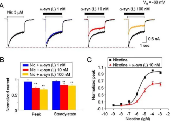
Morphologically distinct oligomeric and fibrillar forms of a -synuclein were generated by incubating monomeric a-synuclein for different lengths of time and aggregate morphologies were analyzed by AFM (Fig. 1A and B). Initial experiments were designed to examine the acute effects of different forms of a -synuclein (10 nM monomeric equivalent) on ha4b 2-nAChR-mediated currents. Oligomeric but not monomeric or fibrillar forms ofa-synuclein inhibited nicotine-induced whole-cell currents (Fig. 2A). Within 2 min of pretreatment, oligomeric a-synuclein inhibited the nicotine-induced peak (reduced to 77.964.1% of control values for nicotine,n= 8,p,0.01, t-test; Fig. 2A and B) and steady-state currents (reduced to 82.862.3% of control values, n= 8, p,0.01, t-test; Fig. 2B). Similarly, oligomeric a-synuclein inhibited the ACh-induced peak (reduced to 81.564.2% of control Figure 3. Effects of different morphological sizes ofa-synuclein oligomers on ha4b2-nAChR function. A. Typical traces illustrating the effects of 10 nM large oligomerica-synuclein [a-syn (L), .4 nm] on 3mM nicotine-induced whole-cell currents recorded from ha4b2-nAChRs expressed in SHEP1 cells.B. Representative typical traces of nicotine-induced whole-cell currents in the absence and presence of monomeric or small oligomerica-synuclein (2–4 nm).C.Summary of experimental results fromAandB. Each symbol was averaged from 6 cells tested. The vertical bars indicate Mean6SE. The single asterisk meansp,0.05, the double asterisk meansp,0.01.
doi:10.1371/journal.pone.0055886.g003
values, n= 8,p,0.01, t-test) and steady-state whole cell currents (reduced to 78.361.9% of control values, n = 8, p,0.01, t-test; Fig. 2C and D). However, without pretreatment, 10 nM oligomeric a-synuclein (co-application with nicotine) did not exhibit significant inhibition of peak current responses to nicotine (Fig. 2C and D; 93.7%68.2, n = 8,p.0.05, one-way ANOVA). Taken together, these data suggest that oligomeric a-synuclein aggregates acutely inhibit ha4b2-nAChR function.
a-Synuclein Differentially Inhibits
ha4b2-nAChR-mediated Whole-cell Currents Depending on Aggregate Morphology
To further characterize the specific aggregate morphologies of a-synuclein involved in altering ha4b2-nAChR function, we used size-exclusion chromatography to separate a 7-day pre-aggregated a-synuclein sample into different aggregated species. The 7-day aggregated sample contains high concentrations of different oligomeric a-synuclein species. We obtained three distinct aggregated forms of a-synuclein particles from 7-day oligomeric aggregates and determined the height distribution of these particles by AFM as previously described [10], where the largest aggregate have heights greater than 4 nm, smaller aggregates have heights between 1 and 4 nm and monomeric particles have heights less than 1 nm [10]. Therefore, we can obtain samples predominantly containing large oligomeric (.4 nm), small oligo-meric (1–4-nm), or monooligo-merica-synuclein.
We compared the effects of the morphologically distinct oligomeric a-synuclein aggregates on ha4b2-nAChR-mediated whole-cell currents. While the large oligomeric a-synuclein aggregates significantly inhibited ha4b2-nAChR-mediated whole-cell currents (Fig. 3A), the small aggregates or monomers did not (Fig. 3B). Statistical analysis showed that after 10 min exposure of large oligomeric, small oligomeric or monomerica -synuclein, the nicotinic responses (normalized peak amplitude) were reduced to 63.563.9% (n = 6, p,0.05 or p,0.01, multi-variate ANOVA), 93.265.6% (n = 6, p.0.05, multi-variate ANOVA) or 92.566.3% (n = 6,p.0.05, multi-variate ANOVA), respectively. These findings suggest that the inhibition of ha4b 2-nAChR-mediated whole-cell currents by oligomerica-synuclein is mediated by large size oligomerica-synuclein.
a-Synuclein Inhibits Humana4b2-nAChR-mediated Currents in Non-competitive
To characterize the inhibitory effects on ha4b2-nAChR function induced by large oligomerica-synuclein, we performed a series of studies using different concentrations of aggregateda -synuclein. Nicotine (3mM,,EC50 concentration) was used as an agonist to activate ha4b2-nAChR expressed in SH-EP1 cells. a -Synuclein inhibited nicotine-induced whole-cell currents in a concentration-dependent manner (Fig. 4A and B; n = 6). Dose-dependent profiles of nicotine-induced whole-cell peak currents in the presence or absence of 10 nM aggregated a-synuclein Figure 4.a-Synuclein inhibits ha4b2-nAChR-mediated currents in a dose-dependent and non-competitive manner. A. Representative typical traces (recorded from the same cell) of nicotine-induced whole-cell currents in the presence of different concentrations of large oligomerica -synuclein.B. Summary of pool results for the effects of different concentrations ofa-synuclein.C. Effects ofa-synuclein (10 nM, large oligomeric) on the concentration-response curves of ha4b2-nAChR-mediated whole-cell currents. Functional fit to the logistic equation indicates that in the presence ofa-synuclein, the maximal current response in the agonist dose-response profile was significantly reduced without change of apparent EC50for agonist (nicotine), suggesting a non-competitive inhibition. All symbols were normalized to the peak current induced by 100mM nicotine, (averaged from 6 cells for nicotine, and 6 cells for nicotine plus 10 nMa-synuclein). In all recordings (A,BandC), the cells were held at a holding potential (VH) of260 mV. InB, the double asterisk meansp,0.01. Vertical bars indicate SEM.
doi:10.1371/journal.pone.0055886.g004
(monomeric equivalent) showed a significant reduction in the maximal concentration of the nicotine-induced peak current but no change in nicotine EC50values and Hill coefficients (2.760.6
and 1.060.08, for nicotine alone,n= 8; 3.660.8 and 1.160.07 for nicotine plus 10 nMa-synuclein,n= 8, p.0.05; t-test, Fig. 4C). These results suggest a non-competitive mechanism of a -synuclein–mediated inhibition.
Effects of Large Oligomerica-synuclein on Different nAChR Subtypes
Since large oligomeric a-synuclein inhibited ha4b2-nAChR currents, we studied if there was a differential effect of this a -synuclein aggregate species on different nAChR subtypes. We compared effects of the large oligomeric a-synuclein on ha4b2-, ha7- and ha4b4-nAChRs heterologously expressed in an SH-EP1 cell line. The ha4b2- and ha4b4-nAChRs were activated using 3mM nicotine, while ha7-nAChR was activated using 3 mM choline. The results indicate that large oligomerica-synuclein (at a pathophysiologically relevant concentration of 10 nM mono-meric equivalent) [43] inhibited nicotinic peak current responses mediated by ha4b2-nAChRs, but not the current responses
mediated by either ha7- or ha4b4-nAChRs (Fig. 5A). Statistical analysis showed that large oligomerica-synuclein species reduced the peak amplitude of ha4b2-nAChR-mediated current to 62.963.7% (n = 6, p,0.01, multi-variate ANOVA); of ha 7-nAChR-mediated current to 93.167.9% (n = 6, p.0.05, multi-variate ANOVA); and of ha4b4-nAChR-mediated current to 96.563.8% (n = 6, p.0.05, multi-variate ANOVA), respectively. These results indicate that the ha4b2-nAChRs are more sensitive toa-synuclein than ha7- and ha4b4-nAChRs.
Mechanisms Involved ina-synuclein-induced Inhibition of ha4b2-nAChR Function
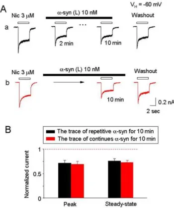
Since oligomerica-synuclein selectively inhibits ha4b2-nAChRs function, we next determined whethera-synuclein was functioning through either of two different potential mechanisms: 1) via a mechanism of open channel block, or 2) via induction of ha4b 2-nAChR internalization. To test whether a-synuclein-induced inhibition operated through the open channel block mechanism, we repeatedly applied 3mM nicotine in the continuous presence of 10 nM large oligomeric a-synuclein. Repeated application of nicotine (2 min interval) in the presence of 10 nMa-synuclein for 10 min led to a reduction of nAChR response (Fig. 6Aa), while continuous application of a-synuclein for 10 min without re-Figure 5. a-Synuclein inhibits ha4b2- but not a7- or a4b
4-nAChR function. A. Representative whole-cell current traces com-paring effects of 10 nMa-synuclein ona4b2-,a7-, anda4b4-nAChRs function.B. Summary of the effects of 10 nMa-synuclein on the peak currents for responses mediated by humana4b2-nAChRs (triangle),a 7-nAChRs (diamond), or a4b4-nAChRs (square). The asterisk indicates
p,0.05 and the double asterisk indicatesp,0.01. Vertical bars indicate
SEM.
doi:10.1371/journal.pone.0055886.g005
Figure 6.a-Synuclein is not an open-channel blocker to ha4b 2-nAChR. A. Representative ha4b2-nAChR mediated whole-cell currents induced by repetitive applications nicotine (4 sec exposure at an interval of 2 min). The first response was recorded as controls. InAa, nicotine exposures were repeated at 2-min intervals in the presence of 10 nMa-synuclein for 10 min, and subsequent response to nicotine was recorded after 6 min of washout ofa-synuclein. InAb, nicotine was applied at the end of the a-synuclein treatment (10 min), and a subsequent response to nicotine was recorded after 6 min of washout ofa-synuclein.B. Bar graph summarizes replicated recordings of effects of 10 nMa-synuclein on nicotinic responses with and without repeated nicotine exposure. Data were collected from 8 cells in each group tested.
doi:10.1371/journal.pone.0055886.g006
petitive exposure to nicotine led to similar inhibition (Fig. 6Ab). The peak component of repetitive nicotine-induced current was reduced to 70.865.8% (Fig. 6Aa, n = 8, p,0.01, t-test) and by non-repetitive challenges of nicotine, the peak current was reduced to 67.566.9% (Fig. 6Ab, n = 8, p,0.01, t-test). No statistical significance in the nicotine-induced currents was observed between the two protocols described above (Fig.6B, n = 8, p.0.05, t-test). Thus, 10 nM large oligomeric a-synuclein did not show clear signs of use-dependent inhibition of ha4b 2-nAChRs (Fig. 6A and B). The absence of a use-dependence feature suggests that non-competitive inhibition of ha4b2-nAChR function bya-synuclein is not mediated through an open channel block. To investigate whether or nota-synuclein-induced ha4b 2-nAChR internalization is involved, patched cells were preloaded with GDP-b-S (600mM) for 20 min, which has previously been reported to preventa -amino-3-hydroxy-5-methyl-4-isoxazolepro-pionate receptor endocytosis [44]. GDP-b-S treatment neither preventeda-synuclein-mediated inhibition nor improved ha4b 2-nAChR functional recovery from washout ofa-synuclein (Fig. 7, A and B). These data suggest that inhibition of ha4b2-nAChR function by a-synuclein is also not mediated by ha4b2-nAChR internalization through a process affectinga -amino-3-hydroxy-5-methyl-4-isoxazolepropionate receptors.
Discussion
In the present study, we show that pathologically-relevant levels (10 nM monomeric equivalent) [43] of aggregated a-synuclein inhibit human a4b2-nAChR function. We find that larger oligomeric a-synuclein aggregate species (.4 nm) but not monomeric, fibrillar or smaller a-synuclein aggregates (2–4 nm) are responsible for this partial inhibitory effect ona4b2-nAChRs. The partial inhibitory effect of a-synuclein on ha4b2-nAChRs exhibits a mechanism that is dose-dependent, non-competitive, non use-dependent and non-internalization based. Interestingly, the a-synuclein-induced inhibition occurs more profoundly in a4b2-nAChRs than in other nAChR subtypes such as a7- or a4b4-nAChRs, indicating subtype selectivity.
Distinguish Inhibitory Effects on nAChRs by Different Forms ofa-synuclein
Misfolding and aggregation ofa-synuclein has been implicated in the pathogenesis of numerous neurodegenerative diseases, particularly in PD [4,45]. Different oligomerica-synuclein species can be generated as intermediate species during the transition from monomeric to fibrillar aggregates [46,47]. Accumulating evidence indicates that oligomeric a-synuclein is the most toxic species responsible for neurodegeneration and neuronal loss in PD [48,49,50]. nAChRs have been linked to pathogenesis of PD and recent evidence suggests possible roles for nAChRs as potential Figure 7. Inhibition of ha4b2-nAChRs bya-synuclein is not mediated through receptor turnover or internalization. A. Representative whole-cell current traces using typical tris-filled pipettes (Aa) and the tris-filled pipettes supplemented with 600mM GDP-b-S (Ab). Initial responses to nicotine were measured after 20 min of the formation of conventional whole-cell recording (infusion of intracellular GDP-b-S into recorded cell). Sequence of drug applications are indicated as horizontal bars.B. Temporal patterns show the similar effects of 10 nMa-synuclein on nicotinic responses with (n = 8) and without (n = 8) GDP-b-S in the pipette solution. The double asterisks indicatep,0.01 compared before and aftera -synuclein exposure, while#meansp.0.05 compared between the pipette solution with (red) and without (black) GDP-b-S.
doi:10.1371/journal.pone.0055886.g007
targets fora-synuclein-induced neurotoxicity manifest as cholin-ergic hypofunction in PD [26,27,33]. However, whether or nota -synuclein directly modulates nAChR function, especially whether specific aggregated morphologies of a-synuclein interact with different subtypes of nAChRs has not been examined previously. Several studies have shown that different aggregateda-synuclein forms added extracellularly to the culture medium can have different cytotoxic effects [15,16,17,18,20,21]. Here we show that oligomeric but not monomeric or fibrillar a-synuclein directly inhibits the function of ha4b2-nAChRs. To further study the effects of oligomeric a-synuclein on ha4b2-nAChRs, we de-termined whether there are any differences in the inhibitory effects of different aggregate forms of a-synuclein toward ha4b 2-nAChRs. Size exclusion chromatography was used to separate several distinct aggregate species of a-synuclein. We show that a large oligomerica-synuclein aggregate species (predominantly,
.4 nm, 99.6%), but not small aggregate species (2–4 nm, 87.4%) significantly inhibited ha4b2-nAChRs function, indicating that morphologically distinct forms of a-synuclein result in different nAChRs inhibition potency. These studies support the hypothesis that aggregated a-synuclein, particularly oligomeric species, may target ha4b2-nAChRs expressing dopaminergic neurons during the pathogenesis of PD and may account for the loss of cholinergic input to dopaminergic neurons [30,51].
Possible Mechanisms ofa-synuclein-mediated Inhibition of ha4b2-nAChR Function
These results clearly demonstrate that oligomeric a-synuclein selectively and partially inhibits ha4b2-nAChR function. The finding of non-competitive antagonism of ha4b2-nAChRs by oligomeric a-synuclein suggests that the large oligomeric a -synuclein species acts as a non-competitive antagonist of ha4b 2-nAChRs under our experimental conditions. Our results also demonstrate that oligomeric a-synuclein, at concentrations from 1 nM to 1mM, failed to directly induce whole-cell current responses from cells expressing ha4b2- or ha7-nAChRs or from untransfected cells (data not shown). These results indicate thata -synuclein in our hands does not have properties of a nAChR agonist.
Additionally, our data show no use-dependence of large oligomeric a-synuclein-induced inhibition on ha4b2-nAChR function, suggesting that the inhibitory effects are not mediated by open channel block, although the persistence in functional block after washout of fluid phasea-synuclein suggests a ‘‘linger-ing’’ effect of the aggregates. We found that pretreatment with large oligomeric a-synuclein is necessary to induce convincing inhibition of ha4b2-nAChR-mediated whole-cell currents. How-ever, the nature of this inhibition is still unknown. One potential explanation is that there is an aggregateda-synuclein-driven long-lasting closed conformation of nAChRs. This idea is supported by the present observation that long exposure times (10 min) to large oligomerica-synuclein aggregates leads to persistent loss of ha4b 2-nAChR function, and any loss of 2-nAChR function does not appear to be mediated via a-synuclein-induced ha4b2-nAChR internal-ization. This conclusion is based on the observation that GDP-b-S (600mM) fails to prevent the loss of ha4b2-nAChR function induced by a-synuclein pre-incubation. On the other hand, the inhibition of ha4b2-nAChR function by a-synuclein is partial, non-competitive, and reversible, which is similar to amyloid-induced inhibition on ha4b2-nAChR [5]. In fact, pre-treatment with oligomeric amyloid 1–42 (1 nM) for 10 min prevented a -synuclein-induced inhibition in ha4b2-nAChR-mediated currents (Figure S1), suggesting a mechanism reminiscent of a negative allosteric inhibitor. The relatively large size of the inhibitory
species may be indicative of a physical interaction partially inhibiting ha4b2-nAChR function but not totally blocking it.
In addition, it is also interesting that a-synuclein selectively inhibits ha4b2-nAChR subtype rather than ha7- or ha4b 4-nAChR subtypes. Although it has been reported thata7 nAChRs, shown to be unaffected in the present report bya-synuclein, are upregulated in PD [31], there are experimental differences between our studies and that of Guan et al. that make direct comparison difficult. For example, we examined direct effects of acute exposure of a-synuclein on nAChR function using transfected nAChRs in cell lines, while Guan et al. reported the nAChR subunit expression and binding using PD brain tissue [31]. The potential effects of a-synuclein-induced inhibition remain to be examined on other nAChR subtypes, such as ha6b2-, ha6b2b3- and ha3b4-nAChR, which may serve as a potential target for PD therapeutics as well. Thesea6-containing nAChR subtypes may be important since they show significant declines in PD animal models. However, due to difficulties in stably expressing these heterologous a6-containing receptors in SH-EP-1 cell lines, we were not able to test the effects of a -synuclein on these subtypes of nAChRs here.
Pathological Relevance ofa4b2-nAChR Dysfunction and PD
Neuronal nicotinic receptors that bind radiolabeled nicotine with the highest affinity containa4 subunits (a4*-nAChR) [52,53]. Immunoassays have shown that the predominant, naturally expressed form of thea4*-nAChR in the vertebrate brain contains a4 and b2 subunits (a4b2-nAChR) [54,55]. Evidence indicates that a consistent, significant loss of a4*-nAChRs has been observed at autopsy in PD brain [31,56,57,58]. The major pathological features of PD are a-synuclein protein deposition, lewy body formation, and a severe dopaminergic deficit [32]. It has been shown that thea-synuclein protein is a major constituent of lewy bodies, a neuropathologic hallmark of PD [32]. However, links between soluble a-synuclein accumulation and cholinergic dysfunction remain unclear. The present study characterized the aggregated morphologies of a-synuclein by size exclusion chro-matography and AFM. This enabled us to distinguish different aggregated a-synuclein species. Oligomeric a-synuclein, particu-larly the larger oligomeric a-synuclein aggregates studied here, selectively inhibits ha4b2-nAChR function in a dose-dependent and non-competitive manner, providing the basis for a new hypothesis thata-synuclein can directly modulate ha4b2-nAChR function, which in turn may contribute to cholinergic signaling deficits in PD.
Although a partial inhibitory effect of a-synuclein on ha4b 2-nAChR function was observed at pathophysiology relevant concentrations, further investigation is needed to determine whether such an effect will be large enough to be clinically relevant. It is also noteworthy thata7-nAChR binding sites are increased in PD brain tissues, suggesting thata7-nAChR might be affected bya-synuclein during PD pathogenesis. However, under our conditions, direct acute exposure ofa-synuclein fails to affect ha7-nAChR function. One possible interpretation is that there may be other confounders during chronica-synuclein accumula-tion to affect a7-nAChR expression in vivo, which cannot be mimicked by acute, in vitro experiments. Our perspective on a primary role for ha4b2-nAChRs in low concentration effects of a-synuclein complements other findings that the modulation of nAChR function bya-synuclein could be pathologically relevant [27,32,33].
Conclusion
Collectively, our findings demonstrate for the first time that pathologically-relevant concentrations of aggregated oligomerica -synuclein directly inhibit neuronal humana4b2-nAChR function. We find that large oligomerica-synuclein aggregates (.4 nm), but not monomeric or fibrillara-synuclein, selectively inhibit ha4b 2-nAChR function starting at 10 nM (monomeric equivalent). Specifically, we show that predominantly larger oligomeric a -synuclein aggregates (.4 nm) but not smaller species (,4 nm) potently inhibit ha4b2-nAChRs mediated whole-cell currents. Furthermore, we elucidate pharmacological mechanisms of a -synuclein–induced inhibition, which includes dose-dependent, non-competitive, non-use-dependent manners, and this inhibition is not mediated through nAChR internalization. Finally, we demonstrate that the functional inhibition bya-synuclein exhibits nAChR subunit selectivity, occurring more profoundly ina4b 2-nAChRs than in other nAChR subtypes such as a7- or a4b 4-nAChRs. Our findings, along with previous reports on the roles of a6b2*-nAChRs in PD pathogenesis [33], suggests that nAChRs are sensitive targets for a-synuclein toxicity. Thea4b2-nAChRs are sensitive to morphologically specific and pathologically relevant concentrations of a-synuclein, suggesting that novel strategies for PD therapy could involve amelioration of specific aggregated a-synuclein-induceda4b2-nAChR functional deficits and/or perhaps preservation ofa4b2-nAChR function.
Supporting Information
Figure S1 Effects of pretreatment of oligomeric amyloid (Ab 1-42) on a-synuclein-induced inhibition of human a4b2-nAChRs
heterologously expressed in SH-EP1 cell line. We found that after 10 min pre-treatment with 1 nM oligomeric Ab1-42, 3mM nicotine (around EC50 concentration)-induced inward current was reduced (Figure S1A, blue trace). Thereafter, we immediately added 10 nMa-synuclein (in the continuous presence of 1 nM Ab1-42) for 10 min, and then tested nicotinic response. However, we did not observe further reduction of nicotine-induced inward current (Figure S1A, red trace). Statistic analysis showed that Ab 1-42 pre-treatment significantly reduced both peak and steady-state components of nicotine-induced-whole-cell current (Figure S1B, n = 6,p,0.01), while in the presence of Ab1-42,a-synuclein failed to further reduce this current response (p.0.05 between Ab1-42 anda-synuclein treated group), indicated as no significance (NS) in the figure. These results suggest that both oligomeric molecules of Ab1-42 and a-synuclein likely bind to a common negative allosteric site to reduce humana4b2-nAChR function.
(DOC)
Acknowledgments
We thank Dr. Ronald J. Lukas for helpful discussion, Dr. Min Wang for AFM assistance, J. Brek Eaton for maintenance of SHEP1-ha4b2 cells, and Dr. Andrew George for his assistance for manuscript edition.
Author Contributions
Conceived and designed the experiments: QL JW. Performed the experiments: QL SE MS. Analyzed the data: QL SE JS MS. Contributed reagents/materials/analysis tools: QL SE MS JW. Wrote the paper: QL JS MS JW.
References
1. Brown RE, McKenna JT, Winston S, Basheer R, Yanagawa Y, et al. (2008) Characterization of GABAergic neurons in rapid-eye-movement sleep control-ling regions of the brainstem reticular formation in GAD67-green fluorescent protein knock-in mice. Eur J Neurosci 27: 352–363.
2. Son JH, Winzer-Serhan UH (2008) Expression of neuronal nicotinic acetylcholine receptor subunit mRNAs in rat hippocampal GABAergic interneurons. J Comp Neurol 511: 286–299.
3. Tamamaki N, Yanagawa Y, Tomioka R, Miyazaki J, Obata K, et al. (2003) Green fluorescent protein expression and colocalization with calretinin, parvalbumin, and somatostatin in the GAD67-GFP knock-in mouse. J Comp Neurol 467: 60–79.
4. Baba M, Nakajo S, Tu PH, Tomita T, Nakaya K, et al. (1998) Aggregation of alpha-synuclein in Lewy bodies of sporadic Parkinson’s disease and dementia with Lewy bodies. Am J Pathol 152: 879–884.
5. Liu Q, Wu J (2006) Neuronal nicotinic acetylcholine receptors serve as sensitive targets that mediate beta-amyloid neurotoxicity. Acta Pharmacol Sin 27: 1277– 1286.
6. Allison DW, Wilcox RS, Ellefsen KL, Askew CE, Hansen DM, et al. (2011) Mefloquine effects on ventral tegmental area dopamine and GABA neuron inhibition: a physiologic role for connexin-36 GAP junctions. Synapse 65: 804– 813.
7. Texido L, Ros E, Martin-Satue M, Lopez S, Aleu J, et al. (2005) Effect of galantamine on the human alpha7 neuronal nicotinic acetylcholine receptor, the Torpedo nicotinic acetylcholine receptor and spontaneous cholinergic synaptic activity. Br J Pharmacol 145: 672–678.
8. Aracava Y, Pereira EF, Maelicke A, Albuquerque EX (2005) Memantine blocks alpha7* nicotinic acetylcholine receptors more potently than n-methyl-D-aspartate receptors in rat hippocampal neurons. J Pharmacol Exp Ther 312: 1195–1205.
9. Fayuk D, Yakel JL (2004) Regulation of nicotinic acetylcholine receptor channel function by acetylcholinesterase inhibitors in rat hippocampal CA1 interneur-ons. Mol Pharmacol 66: 658–666.
10. Emadi S, Kasturirangan S, Wang MS, Schulz P, Sierks MR (2009) Detecting morphologically distinct oligomeric forms of alpha-synuclein. J Biol Chem 284: 11048–11058.
11. Samochocki M, Hoffle A, Fehrenbacher A, Jostock R, Ludwig J, et al. (2003) Galantamine is an allosterically potentiating ligand of neuronal nicotinic but not of muscarinic acetylcholine receptors. J Pharmacol Exp Ther 305: 1024–1036. 12. Patel MN, Yim GK, Isom GE (1993) N-methyl-D-aspartate receptors mediate cyanide-induced cytotoxicity in hippocampal cultures. Neurotoxicology 14: 35– 40.
13. Frandsen A, Drejer J, Schousboe A (1989) Direct evidence that excitotoxicity in cultured neurons is mediated via N-methyl-D-aspartate (NMDA) as well as non-NMDA receptors. J Neurochem 53: 297–299.
14. McClure-Begley TD, King NM, Collins AC, Stitzel JA, Wehner JM, et al. (2009) Acetylcholine-stimulated [3H]GABA release from mouse brain synaptosomes is modulated by alpha4beta2 and alpha4alpha5beta2 nicotinic receptor subtypes. Mol Pharmacol 75: 918–926.
15. Buhler AV, Dunwiddie TV (2002) alpha7 nicotinic acetylcholine receptors on GABAergic interneurons evoke dendritic and somatic inhibition of hippocampal neurons. J Neurophysiol 87: 548–557.
16. Ji D, Lape R, Dani JA (2001) Timing and location of nicotinic activity enhances or depresses hippocampal synaptic plasticity. Neuron 31: 131–141.
17. Alkondon M, Albuquerque EX (2001) Nicotinic acetylcholine receptor alpha7 and alpha4beta2 subtypes differentially control GABAergic input to CA1 neurons in rat hippocampus. J Neurophysiol 86: 3043–3055.
18. Ji D, Dani JA (2000) Inhibition and disinhibition of pyramidal neurons by activation of nicotinic receptors on hippocampal interneurons. J Neurophysiol 83: 2682–2690.
19. Chen D, Patrick JW (1997) The alpha-bungarotoxin-binding nicotinic acetylcholine receptor from rat brain contains only the alpha7 subunit. J Biol Chem 272: 24024–24029.
20. Murray TA, Bertrand D, Papke RL, George AA, Pantoja R, et al. (2012) alpha7beta2 nicotinic acetylcholine receptors assemble, function, and are activated primarily via their alpha7-alpha7 interfaces. Mol Pharmacol 81: 175–188.
21. Velez-Fort M, Audinat E, Angulo MC (2009) Functional alpha 7-containing nicotinic receptors of NG2-expressing cells in the hippocampus. Glia 57: 1104– 1114.
22. Louis ED, Benito-Leon J, Bermejo-Pareja F (2008) Population-based prospective study of cigarette smoking and risk of incident essential tremor. Neurology 70: 1682–1687.
23. Wirdefeldt K, Adami HO, Cole P, Trichopoulos D, Mandel J (2011) Epidemiology and etiology of Parkinson’s disease: a review of the evidence. Eur J Epidemiol 26 Suppl 1: S1–58.
24. Chen H, Huang X, Guo X, Mailman RB, Park Y, et al. (2010) Smoking duration, intensity, and risk of Parkinson disease. Neurology 74: 878–884. 25. Kiyohara C, Kusuhara S (2011) Cigarette smoking and Parkinson’s disease:
a meta-analysis. Fukuoka Igaku Zasshi 102: 254–265.
26. Sharma G, Vijayaraghavan S (2001) Nicotinic cholinergic signaling in hippocampal astrocytes involves calcium-induced calcium release from in-tracellular stores. Proc Natl Acad Sci U S A 98: 4148–4153.
27. Kawamata J, Suzuki S, Shimohama S (2012) alpha7 nicotinic acetylcholine receptor mediated neuroprotection in Parkinson’s disease. Curr Drug Targets 13: 623–630.
28. Jeyarasasingam G, Tompkins L, Quik M (2002) Stimulation of non-alpha7 nicotinic receptors partially protects dopaminergic neurons from 1-methyl-4-phenylpyridinium-induced toxicity in culture. Neuroscience 109: 275–285. 29. Takeuchi H, Yanagida T, Inden M, Takata K, Kitamura Y, et al. (2009)
Nicotinic receptor stimulation protects nigral dopaminergic neurons in rotenone-induced Parkinson’s disease models. J Neurosci Res 87: 576–585. 30. Lester DB, Rogers TD, Blaha CD (2010) Acetylcholine-dopamine interactions in
the pathophysiology and treatment of CNS disorders. CNS Neurosci Ther 16: 137–162.
31. Guan ZZ, Nordberg A, Mousavi M, Rinne JO, Hellstrom-Lindahl E (2002) Selective changes in the levels of nicotinic acetylcholine receptor protein and of corresponding mRNA species in the brains of patients with Parkinson’s disease. Brain Res 956: 358–366.
32. Vekrellis K, Xilouri M, Emmanouilidou E, Rideout HJ, Stefanis L (2011) Pathological roles of alpha-synuclein in neurological disorders. Lancet Neurol 10: 1015–1025.
33. Quik M, Wonnacott S (2011) alpha6beta2* and alpha4beta2* nicotinic acetylcholine receptors as drug targets for Parkinson’s disease. Pharmacol Rev 63: 938–966.
34. Eaton JB, Peng JH, Schroeder KM, George AA, Fryer JD, et al. (2003) Characterization of human alpha 4 beta 2-nicotinic acetylcholine receptors stably and heterologously expressed in native nicotinic receptor-null SH-EP1 human epithelial cells. Mol Pharmacol 64: 1283–1294.
35. Peng JH, Lucero L, Fryer J, Herl J, Leonard SS, et al. (1999) Inducible, heterologous expression of human alpha7-nicotinic acetylcholine receptors in a native nicotinic receptor-null human clonal line. Brain Res 825: 172–179. 36. Puchacz E, Buisson B, Bertrand D, Lukas RJ (1994) Functional expression of
nicotinic acetylcholine receptors containing rat alpha 7 subunits in human SH-SY5Y neuroblastoma cells. FEBS Lett 354: 155–159.
37. Lukas RJ, Norman SA, Lucero L (1993) Characterization of Nicotinic Acetylcholine Receptors Expressed by Cells of the SH-SY5Y Human Neuroblastoma Clonal Line. Mol Cell Neurosci 4: 1–12.
38. Bencherif M, Lukas RJ (1993) Cytochalasin modulation of nicotinic cholinergic receptor expression and muscarinic receptor function in human TE671/RD cells: a possible functional role of the cytoskeleton. J Neurochem 61: 852–864. 39. He P, Liu Q, Wu J, Shen Y (2012) Genetic deletion of TNF receptor suppresses excitatory synaptic transmission via reducing AMPA receptor synaptic localization in cortical neurons. FASEB J 26: 334–345.
40. Liu Q, Huang Y, Xue F, Simard A, DeChon J, et al. (2009) A novel nicotinic acetylcholine receptor subtype in basal forebrain cholinergic neurons with high sensitivity to amyloid peptides. J Neurosci 29: 918–929.
41. Wu J, Liu Q, Yu K, Hu J, Kuo YP, et al. (2006) Roles of nicotinic acetylcholine receptor beta subunits in function of human alpha4-containing nicotinic receptors. J Physiol 576: 103–118.
42. Volles MJ, Lansbury PT Jr (2007) Relationships between the sequence of alpha-synuclein and its membrane affinity, fibrillization propensity, and yeast toxicity. J Mol Biol 366: 1510–1522.
43. Hall S, Ohrfelt A, Constantinescu R, Andreasson U, Surova Y, et al. (2012) Accuracy of a Panel of 5 Cerebrospinal Fluid Biomarkers in the Differential Diagnosis of Patients With Dementia and/or Parkinsonian Disorders. Arch Neurol: 1–8.
44. Luscher C, Xia H, Beattie EC, Carroll RC, von Zastrow M, et al. (1999) Role of AMPA receptor cycling in synaptic transmission and plasticity. Neuron 24: 649– 658.
45. Spillantini MG, Crowther RA, Jakes R, Cairns NJ, Lantos PL, et al. (1998) Filamentous alpha-synuclein inclusions link multiple system atrophy with Parkinson’s disease and dementia with Lewy bodies. Neurosci Lett 251: 205– 208.
46. Conway KA, Lee SJ, Rochet JC, Ding TT, Harper JD, et al. (2000) Accelerated oligomerization by Parkinson’s disease linked alpha-synuclein mutants. Ann N Y Acad Sci 920: 42–45.
47. Wood SJ, Wypych J, Steavenson S, Louis JC, Citron M, et al. (1999) alpha-synuclein fibrillogenesis is nucleation-dependent. Implications for the pathogen-esis of Parkinson’s disease. J Biol Chem 274: 19509–19512.
48. Ding TT, Lee SJ, Rochet JC, Lansbury PT Jr (2002) Annular alpha-synuclein protofibrils are produced when spherical protofibrils are incubated in solution or bound to brain-derived membranes. Biochemistry 41: 10209–10217. 49. Volles MJ, Lansbury PT Jr (2003) Zeroing in on the pathogenic form of
alpha-synuclein and its mechanism of neurotoxicity in Parkinson’s disease. Bio-chemistry 42: 7871–7878.
50. Volles MJ, Lee SJ, Rochet JC, Shtilerman MD, Ding TT, et al. (2001) Vesicle permeabilization by protofibrillar alpha-synuclein: implications for the patho-genesis and treatment of Parkinson’s disease. Biochemistry 40: 7812–7819. 51. Everitt BJ, Robbins TW (1997) Central cholinergic systems and cognition. Annu
Rev Psychol 48: 649–684.
52. Lukas RJ, Changeux JP, Le Novere N, Albuquerque EX, Balfour DJ, et al. (1999) International Union of Pharmacology. XX. Current status of the nomenclature for nicotinic acetylcholine receptors and their subunits. Pharmacol Rev 51: 397–401.
53. Lindstrom J (1996) Neuronal nicotinic acetylcholine receptors. Ion Channels 4: 377–450.
54. Flores CM, Rogers SW, Pabreza LA, Wolfe BB, Kellar KJ (1992) A subtype of nicotinic cholinergic receptor in rat brain is composed of alpha 4 and beta 2 subunits and is up-regulated by chronic nicotine treatment. Mol Pharmacol 41: 31–37.
55. Whiting P, Lindstrom J (1987) Purification and characterization of a nicotinic acetylcholine receptor from rat brain. Proc Natl Acad Sci U S A 84: 595–599. 56. Gotti C, Moretti M, Bohr I, Ziabreva I, Vailati S, et al. (2006) Selective nicotinic acetylcholine receptor subunit deficits identified in Alzheimer’s disease, Parkinson’s disease and dementia with Lewy bodies by immunoprecipitation. Neurobiol Dis 23: 481–489.
57. Martin-Ruiz CM, Piggott M, Gotti C, Lindstrom J, Mendelow AD, et al. (2000) Alpha and beta nicotinic acetylcholine receptors subunits and synaptophysin in putamen from Parkinson’s disease. Neuropharmacology 39: 2830–2839. 58. Perry EK, Perry RH, Smith CJ, Purohit D, Bonham J, et al. (1986) Cholinergic
receptors in cognitive disorders. Can J Neurol Sci 13: 521–527.