ORIGINAL
ARTICLE
Molecular detection of cytomegalovirus, herpes
simplex virus 2, human papillomavirus 16-18 in
Turkish pregnants
Authors Bedia Dinc1 Gulendam Bozdayi2 Aydan Biri3 Ayse Kalkanci2 Bora Dogan1 Nuray Bozkurt3 Seyyal Rota4
1MD, Department of
Medical Microbiology, Faculty of Medicine, Gazi University, Ankara, Turkey.
2MD, PhD, Associate
Professor. Department of Medical Microbiology, Faculty of Medicine, Gazi University, Ankara, Turkey.
3MD, Associate Professor.
Department of Obstetrics and Gynecology, Faculty of Medicine, Gazi University, Ankara, Turkey.
4MD, Professor.
Department of Medical Microbiology, Faculty of Medicine, Gazi University, Ankara, Turkey.
Submitted on: 02/18/2010 Approved on: 08/30/2010
Correspondence to: Bedia Dinc, MD Gazi University, Faculty of Medicine,
Department of Medical Microbiology, Dekanlik Binasi, 2. Kat, Besevler, Ankara, Turkey, 06500.
Tel: +90 312 202 46 28 Fax: +90 312 212 46 47 E-mail: bhdogan@yahoo. com
We declare no confl ict of interest.
ABSTRACT
Objective: Human cytomegalovirus (CMV) is the most common cause of viral intrauterine infec-tions in the world. Herpes simplex virus type 2 (HSV-2) and human papillomavirus (HPV) are the main agents of viral sexually transmitted diseases, which cause genital ulcers and genital warts, respectively. HPV infection has been linked to the majority of the anogenital malignancies. The aim of this study was to detect the existence of CMV, HSV-2 and HPV type 16-18 in Turkish pregnants by using sensitive molecular assays. Methods: One hundred thirty-four women (18-41 years old; mean age ± SD: 27 ± 8) applied to outpatient clinic of Obstetrics and Gynecology, in between 18th - 22nd weeks of their pregnancy and a control group of 99 healthy women (15-39 years old; mean age ± SD: 24 ± 8) were included in the study. Cervical smear samples were used for DNA extrac-tion. CMV, HSV-2 and HPV 16-18 detections were carried out by real time PCR and in house PCR method, respectively. Results: Three patients (3/134; 2.2%) were found to be positive for each HPV and HSV-2. Dual infection with HPV and HSV was found in just one patient. HPV 18 was detected in all positive samples. CMV was found to be positive in two patients (2/134; 1.4 %). Conclusion: HPV, HSV and CMV must be screened due to high prevalence of these viruses in pregnants by using sensitive molecular methods.
Keywords: pregnancy; infection; polymerase chain reaction.
[Braz J Infect Dis 2010;14(6):569-574]©Elsevier Editora Ltda.
INTRODUCTION
Cytomegalovirus (CMV) is the most common cause of congenital infections affecting 0.5% to 2% of all live births in the developed world.1
Intrauterine infection occurs in up to 50% of pregnancies following primary maternal infec-tion, and 10% of the infected infants are symp-tomatic at birth.2,3 However nearly 10%-15%
of the asymptomatic newborns develop late se-quelae, particularly sensorineural hearing loss and neurodevelopmental disorders.4
Genital herpes continues to be a public health problem in both developed and devel-oping countries. Prevalence of HSV-2 in dif-ferent populations has shown various results from less than 10% to more than 40%.5
Labo-ratory confi rmation for clinical diagnosis of HSV-2 is important, particularly as there are other conditions which present similarly to genital herpes, while atypical presentations of genital herpes also occur.6
Uterine cervix carcinoma is the second most common type of female cancers and also the second highest mortality cause for women. Human papillomavirus (HPV) be-ing detected in close to 99.7% of cervical squamous cell carcinomas is imputed to be a necessary factor that causes cervix carcino-ma. Hormones and their metabolism altera-tion have also been implicated as cofactors in HPV proliferation and the cervical cancer pathogenesis. It is well known that high risk HPV infections are more frequent in preg-nant women.7-9 The polymerase chain
reac-tion (PCR) technology for CMV, HSV, HPV detection is now highly sensitive and can de-tect viral DNA in exfoliated cells irrespective of the sampling method used.10
MATERIAL AND METHODS
Study group
This study was performed at the Gynecology and Obstet-rics Outpatient Clinic at Gazi University Hospital, An-kara, Turkey. One hundred thirty-four women (18-41 years old; mean age ± SD: 27 ± 8) applied to outpatient clinic, in between 18th - 22nd weeks of their pregnan-cy and a control group of 99 healthy women (15-39 years old; mean age ± SD: 24 ± 8) followed for routine pre-natal visit, were included in the study. Cervical smear samples were used to detect CMV, HSV-2 and HPV. All the patients and the control group answered a ques-tionnaire including age of first sexual intercourse, number of sexual partners and condom use.
Specimen sampling and preservation
Cervical smear samples collected by sterile swabs were sent to the laboratory in phosphate buffered saline (PBS) (pH 8.6). The samples in PBS were vortexed and stored in -86°C until they were tested. And 10 mL of blood was taken from each patient for serological examination of HSV-2.
DNA extraction method for HPV, HSV and CMV
Following the centrifugation at 13,000 rpm for 3 min, the samples in PBS were treated with lysis solution contain-ing 20 mg/mL proteinase K [20 mM (NH4)2SO4, 75 mM Tris HCl (pH 8.8) 0,1% Tween 20]. After discarding the supernatant, precipitate was extracted by phenol-chloroform isoamyl alcohol method.11 Precipitation of
DNA was succeeded by using absolute ethanol including 3M sodium acetate at -20°C overnight. On the following day, samples were mixed with 70% alcohol and dried. After drying, they were incubated on dry block at 55°C for 10 min and then 50 µL sterile deionised water was added.
Amplification of HPV DNA
The primers used in this study were the consensus primers for all HPV genotypes and were chosen from L1 location of HPV genome which is associated with major viral cap-sid protein metabolism. MY09/MY11 primers were used for amplifi cation of HPV.12 Caski cell DNA was used as a
positive control.13 During all amplifi cation procedure, a
negative control including no targeted DNA was used. Am-plifi cation procedure was performed by adding 5 µL ex-tracted DNA to a mixture of 45 µL including 100 mM from each dNTP (dATP, dCTP, dGTP, dTTP) 100 pmol from each primer and 1 unit of Taq DNA polymerase enzyme (DNA mp ltd., Hants, UK) in 4 mM MgCl2, 50 mM KCl2, and 10 mM Tris HCl (pH 9.0).
A thermal cycler programme with 35 cycles includ-ing initial denaturation at 94°C for 5 min, denaturation
at 94°C for 20 seconds, annealing at 55°C for 45 seconds and extension at 72°C for 1 min, followed by 7 min of fi nal extension at 72°C was performed. PCR products were ana-lyzed on agarose gel stained with ethidium bromide and bands were detected with UV transluminator encompass-ing 450 bp for HPV.11
Genotyping
MY09/MY14 (5’ - CAT ACA CCT CCA GCA CCT AA - 3’) and MY09/WD74 (5’-GGA TGC TGC ACC GGC TG A-3’) specific primers were used for amplification of HPV type 16 and HPV type 18, respectively.12,14
Amplification procedure was performed by adding 5 µL pure DNA to a mixture of 95 µL including 100 mM from each dNTP (dATP, CTP, dGTP, dTTP) 100 pmol from each primer and 1 unit of Taq polymerase in 4 mM MgCl2, 50 mM KCl2, and 10 mM Tris HCl (pH 9.0). We used a thermal cycler programme for HPV genotyping as described above. PCR products were analyzed on agarose gel stained with ethidium bro-mide and transluminating band was detected with UV transluminator encompassing 110 bp for HPV 16 and 140 bp for HPV 18.
Real-time PCR for CMV and HSV-2
Real-time PCR reactions were carried out by using Ligth-cycler (Roche Diagnostics, Germany). LigthLigth-cycler DNA master hybridization probes kit (Roche) was used for amplification. Taqman probes labeled with FAM at their 5’ end and labeled with TAMRA as quencher at their 3’ end, and specific primer sets for HSV DNA polymer-ase gene region and CMV pp65 gene region (Table 1) were used for amplifications (Metis Biotechnology An-kara, Turkey).
Table 1. Primers and probes
Agent Region Primers (5 > 3)
HSV DNA polymerase CAT CAC CGA CCC GGA GAG GGA C
HSV DNA polymerase GGG CCA GGC GCT TGT TGG TGT A
CMV pp65 CTT GCC CTG GAT GCG ATA CT
CMV pp65 ACG GCT TTA CGG TGT TGT GT
Probes
HSV LC (TaqMan Probe) FAM- CCG CCG AAC TGA GCA GAC ACC CGC GC –TAMRA
In each assay, fi ve CMV AD 169 DNA standards from 2x10² to 2x10⁶ CMV DNA copy/mL were used as external
standards. A capillary tube containing distilled water instead of DNA was used as negative control in each run. The de-tection limit was established as 200 CMV DNA copy/mL in blood. Serial dilutions containing 2x10²-2x10⁶ HSV DNA
were prepared for HSV and a limit was detected as 200 HSV DNA copy/mL. A negative control was used in each study.
CMV serology
Anti-CMV IgM antibody: specifi c IgM responding to CMV in cervicovaginal smear samples was studied with two differ-ent methods: microparticle enzyme immunoassay (MEIA) system and its appropriate anti-CMV IgM kit (Imx System, Abbott Laboratories, Abbott Park, IL, USA) and a commer-cial diagnostic anti-CMV IgM enzyme-linked immunosorb-ant assay (ELISA) kit (Radim SPA, Pomezia-Rome, Italy).
Anti-CMV IgG antibody: specifi c IgG responding to CMV in cervicovaginal smear samples was studied with MEIA system and its appropriate anti-CMV IgG kit (Axsym System, Abbott Laboratories, Abbott Park, IL, USA).
CMV IgG avidity test: the cervicovaginal smear sample with 4 U/mL of anti-CMV IgG were studied with a com-mercial diagnostic CMV IgG avidity Enzyme immunoassay (EIA) kit (Radim SPA, Pomezia-Rome, Italy) according to the manufacturer’s instructions.
HSV serology
HSV-2 IgM and IgG antibodies were detected by EIA (Med-dens Diagnostics B.V., The Netherlands) in the sera samples. Quantitative EIA was performed for HSV 2 IgG Ab accord-ing to the manufacturer’s instructions.
RESULTS
Patients and USG fi ndings
All pregnants and the control group were monogamic and had no history of sexual intercourse before getting married. The reported age at fi rst sexual intercourse was less than 20 years for 98 (73.1%) pregnants, and 67 (67.7%) of con-trol group respectively. Of the 134 pregnants, 32 (23.8%) and
99 controls 35 (35.4%) reported ever use of condoms for STDs. Of the 134 pregnants, 22 had abnormal ultrasonographic fi ndings, four oligohydramnios, four ventriculomegaly, three intrauterine growth retardation (IUGR), three IUGR + oligo-hydramnios, two polyoligo-hydramnios, one bilateral choroid plexus cyst greater than 7 mm, one echogenic bowel + choroid plexus cyst, one polihydramnios, one omphalocele, one multiple struc-tural abnormalities and one hydrops fetalis were detected.
Out of three pregnants with IUGR + oligohydramnios, one was found to be positive for CMV. All other pregnants with ab-normal ultrasonographic fi ndings were found to be negative for each of the three viruses. No abnormal USG fi ndings were detected in any of the control group.
Serology for CMV and HSV
Anti-CMV IgM was found to be negative in all of the cervicov-aginal smear samples. In all of the samples studied, one cervi-covaginal smear sample was found to be 4 U/mL, with negative result. Because both tests for anti-CMV IgM, and CMV IgG were designed for only serum evaluation, the cut-off values for smear may be lower than the cut-off values (10 U/mL) accepted for serum. In this respect the cervicovaginal smear sample with 4 U/mL of anti-CMV IgG value was also approved as positive and studied with CMV IgG avidity test.
CMV IgG avidity test: according to the kit that we used, we considered the values below 35% as a low avidity, values be-tween 35% and 45% as a gray zone and above 45% as a high avidity. Low avidity is a fi nding in favor of a primary acute in-fection. The cervicovaginal smear sample with 4 U/mL of anti-CMV IgG value was also studied with anti-CMV IgG avidity test and again low avidity (21%) was found. This situation was also suggested a primary acute infection.
HSV-2 Ig M and IgG were positive in the sera samples of 6 (4.4 %) and 9 (9 %) of pregnants, respectively.
In house PCR and RT PCR for CMV, HPV and HSV-2
CMV DNA was found to be positive in two (1.5%) of pregnant women by real-time PCR. In control group there was no CMV DNA positivity. In one of the CMV positive pregnant women, there were abnormal USG fi ndings (oligohydroamnios and in utero growth retardation) (Table 2).
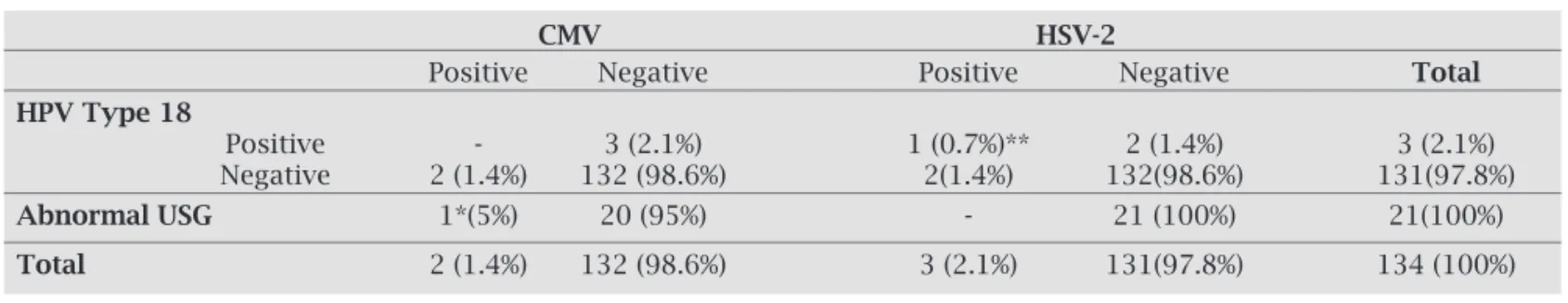
Table 2. One of two CMV positive pregnants had abnormal ultrasonographic finding. CMV DNA was negative for HPV type 18 positive 3 pregnants, while one of 3 was detected positive for HSV-2
CMV HSV-2
Positive Negative Positive Negative Total
HPV Type 18
Positive - 3 (2.1%) 1 (0.7%)** 2 (1.4%) 3 (2.1%)
Negative 2 (1.4%) 132 (98.6%) 2(1.4%) 132(98.6%) 131(97.8%)
AbnormalUSG 1*(5%) 20 (95%) - 21 (100%) 21(100%)
Total 2 (1.4%) 132 (98.6%) 3 (2.1%) 131(97.8%) 134 (100%)
*: abnormal USG findings
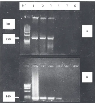
Three (3/134; 2.2%) of pregnant women were found to be positive for only HPV 18. No other types of HPV were detected in any of the pregnants (Figure 1). In control group, there was no HPV DNA positivity.
Autopsy and CMV PCR from these materials could not be performed since the family did not give permission. The other CMV DNA positive pregnant woman gave a healthy birth and no problems occurred with the mother and baby during follow-up.
Previous studies have shown that the incidence of CMV excretion from cervix increases while pregnancy proceeds.15
In our study group, the patients were at their second trimes-ter. In Turkey, Koksal et al.,16 Kaleli et al.17 and Satilmis et al.18
reported the prevalence of CMV IgG in pregnant women as 79%, 94.7% and 98.5%, respectively. CMV IgM positivity in pregnancy was reported as 0.66% by Kaleli et al..17 It is
determined that virus spread with asymptomatic infection is frequent among pregnant women and virus can be iso-lated from cervical mucus and urine of 2%-28% of pregnant women.19 There are several potential routes of infection of
the placenta during pregnancy, including ascending infec-tion from the genital tract as well as hematogenous trans-mission. As CMV secretion from cervix increases during pregnancy, a large spectrum of cells of the fetus is infected by CMV. The major target fetal organs for CMV infection are the lungs, pancreas, kidneys and the liver but comparing with these organs, CMV DNA level determined in uterine tissue and cervical smear is higher.20
In our study, 134 pregnant women were screened for HSV-2 DNA. Three (2.2%) pregnant women and 2 (2%) women in control group were found to be positive in fa-vour of HSV-2. In our country, HSV-2 is rarely seen due to the monogamy in the sexual relations. Genital herpes resulting from HSV-2 infection of the urogenital tract is a worldwide public health problem. European investiga-tions of pregnant women have shown overall prevalence of HSV-2 to range between 9% and 33%.21 HSV-2 DNA
prevalence was reported as 2% in Turkish pregnants re-cently.22 Transmission of HSV is usually via direct contact
with lesion while HSV-2 is classically transmitted sexually.6
The women most at risk for mother to child transmission are the ones who are previously uninfected. New onset HSV infection in late pregnancy carries a 30%-50% risk of neo-natal infection since there is not an adequate time for anti-bodies to develop and suppress the viral replication before labor, while infection in early pregnancy carries a risk of less than 1%.6,23 Fortunately the risk of fetal infection in
recur-rent episodes is nearly 2%.24 Risk for baby usually happens
during delivery via contact with genital lesions with recur-rent episodes of viral shedding; therefore, caesarean section is recommended in these cases.25 In pregnant population,
real-time PCR has some advantages, making results avail-able at a much earlier time, thus signifi cantly reducing the need for empirical treatment.23,26 Dual infection with HPV
and HSV positivity was found in one patient in our study. Any gynecological examination fi ndings were not found out in favor of HPV and HSV in any of the patients. Of the three
Figure 1: A; (HPV) M: fX174 DNA / Hae III marker. Lanes 1, 2, 3: HPV positive patients. Lanes 4, 5: Negative control, B; (HPV type 18), M: fX174 DNA / Hae III marker. Lanes 1, 2, 3: HPV type 18 positive patients. Lane 4: Positive control. Lane 5: Negative control.
Three (3/134; 2.2%) of the samples from the pregnant women were found to be positive for HSV-2. Dual infection with HPV and HSV positivity was found in one patient. Any gynecological examination fi ndings were not found out in favor of HPV and HSV in any of the patients (Table 2).
None of the pregnants and the control group had triple infection with CMV, HSV-2 and HPV. While there was no positivity for both CMV and HPV, HSV was found to be positive in 2 (2%) samples in control group.
DISCUSSION
In this study, of the 134 pregnant women, CMV DNA was found to be positive in two (1.5%) patients, while no CMV DNA positivity was found by real-time PCR in control group. In one of the CMV positive pregnant women, there were abnormal USG fi ndings (oligohydroamnios and in utero
growth retardation). She was followed for IUGR and
in utero exitus happened during the 20th week of pregnancy.
M 1 2 3 4 5 6
bp
140
A B A
HSV-2 DNA positive patients, all were HSV-2 IgM positive with no physical symptoms. These patients did not have a history of condom use before pregnancy and their fi rst sex-ual intercourse were after the age of 20. Although they were asymptomatic, caesarean section was recommended for HSV positive three patients in our hospital and no problems occurred during the follow up of the mothers and babies.
Studies about HPV show that its transmission mode re-mains unclear. HPV is positive in the airways of neonates and in maternal genital condylomas, which suggests that it is transmitted in the birth canal, from mothers with geni-tal condylomas.27 Since the serologic diagnosis of HPV is
inadequate, molecular diagnosis has been more popular in recent years.28 Epidemiological studies revealed
differ-ent results in pregnant women. Smith et al.,29 Gajewski et
al.30 and Takakuwa et al.31 have found out 29%, 26% and
12.5% of HPV positivity respectively in pregnant women taking routine obstetric care. In a study performed in our country, HPV DNA was found as 9.5% by PCR. One pa-tient was found type 16/30, the others were untyped.32 We
have found that 2.2% of the patients were positive for HPV 18 in our study where Smith et al.29 found that 2.6% of the
patients were positive in favor of HPV 18. The association between HSV-2 and HPV 18 was found in one of the preg-nant women. This association may be due to the similar transmission route of both viruses. During the neonatal follow-up of baby HPV DNA was not found in the laryn-geal swab by Real Time PCR.
CONCLUSION
Since HPV is considered as an infection predisposed to ma-lignancies, HPV detection with molecular methods should be considered as a routine screening test for women applied to obstetrics and gynecology clinics. This screening policy should also contain CMV and HSV infections because of their poten-tial transmission risks for the baby. We suggest that our assays provide useful information about CMV, HSV, HPV infections in the fetus and the neonate; and obstetricians have to make informed decisions about the management of pregnancies complicated by CMV, HSV, and HPV infections.
REFERENCES
1. Azam AZ, Yvan V, Fawer CL et al. Prenatal diagnosis of
con-genital cytomegalovirus infection. Obst and Gynecol 2001; 97:443-8.
2. Yamamoto AY, Mussi-Pinhata MM, Pinto PCG et al.
Useful-ness of blood and urine samples collected on fi lter paper in detecting cytomegalovirus by the polymerase chain reaction technique. J Virol Meth 2001; 97:159-64.
3. Schleiss MR, Bourne N, Bravo FJ et al.
Quantitative-com-petitive PCR monitoring of viral load following experimen-tal guinea pig cytomegalovirus infection. J Virol Meth 2003; 108:103-10.
4. Gindes L, Teperberg-Oikawa M, Sherman D et al. Congenital
cytomegalovirus infection following primary maternal infec-tion in the third trimester. BJOG 2008; 115:830-5.
5. Eskild A, Jeansson S, Hagen JA et al. Herpes simplex virus
type-2 antibodies in pregnant women: the impact of the stage of pregnancy. Epidemiol Infect 2000; 125:685-92.
6. Kriebs JM. Understanding herpes simplex virus: transmission, diagnosis and considerations in pregnancy management. J Midwifery Womens Health 2008; 53:202-8.
7. Armbruster-Moraes E, Ioshimoto LM, Leao E, Zugaib M. Prevalence of high risk human papillomavirus in the lower genital tract of Brazilian gravidas. Intern J Gynecol and Obst 2000; 69:223-7.
8. Chan PK, Chang AR, Tam WH et al. Prevalence and Genotype
Distribution of Cervical Human Papillomavirus Infection: Comparison Between Pregnant Women and Non-Pregnant Controls. Journal of Medical Virology 2002; 67:583-8. 9. Mayeaux EJ. Reducing the economic burden of HPV-related
diseases. J Am Osteopath Assoc 2008; 108:52-7.
10. De Sanjose S, Boch XF, Munoz N et al. Screening for genital
human papillomavirus: results from an international valida-tion study on human papillomavirus sampling techniques. Diag Mol Path 1999; 8:26-31.
11. Maniatis T, Fritsch EF. Molecular Cloning. In: J Sambrook ed. A Laboratory Manual. New York: 1990.
12. Baay MFD, Quint WGV, Koudstaal J et al. Comprehensive study
of several general and type-specifi c primer pairs for detection of human papillomavirus DNA by PCR in paraffi n-embedded cervical carcinomas. J Clin Microbiol. 1996; 34:745-7. 13. Tuncer S, Ustaçelebi S. Detection of Human Papillomavirus
Types 16 and 18 by Polymerase Chain Reaction in Cervical Bi-opsy Specimens. Flora 1996; 1:40-4.
14. Poljak M, Seme K. Rapid detection and typing of human papillo-maviruses by consensus polymerase chain reaction and enzyme-linked immunosorbent assay. J Virol Meth 1996; 56:231-8. 15. Chandler SH, Alexander ER, Holmes KK. Epidemiology of
cytomegalovirus infection in a heterogeneous population of pregnant women. J Infec Dis 1985; 152:249-56.
16. Koksal I, Alnaci M, Kardes OB, Aydemir V. Toxoplasma, rubel-la and cytomegalovirus seropositivity ratios among adults in Eastern Black Sea Region. Turk Bullet Microb 1994; 28:58-61. 17. Kaleli B, Kaleli I, Yurdakul B, Aksit F. Rubella and
Cytomegalo-virus Infection in Pregnancy. Turk J Infect. 1997; 11:325-7.
18. Satilmis A, Gura A, Ongun H et al. CMV seroconversion in
pregnants and the incidence of congenital CMV infection. Turk J Pediatr. 2007; 49(1):30-6.
19. Gibbs RS, Sweet RL. Maternal and fetal infectious disorders. In: Creasy RK, Resnik R eds. Maternal Fetal Medicine. Phila-delphia: W.B. Saunders, 1994.
20. Fisher S, Genbacev O, Maidji E, Pereira L. Human
cytomegalo-virus infection of cytotrophoblasts in vitro and in utero:
Impli-cations for transmission and pathogenesis. Journal of Virology 2000; 74(15):6808-20.
21. Gaytant MA, Steegers EAP, Van Laere M et al. Seroprevalence
of herpes simplex virus type 1 and type 2 among pregnant women in the Netherland. Sex Transm Dis. 2002; 29:710-4. 22. Rota S, Bozdayi G, Dogan B, Dinc B. Investigation of herpes
simplex virus DNA by real-time polymerase chain reaction in the clinical samples. Bulletin of Microbiology 2004; 38:233-9. 23. Perkins D, Chong H, Irvine B, Domagalski J. Genital co-
24. Poeran J, Wildshut H, Gaytant M et al. The incidence of neonatal herpes in The Netherlands. J Clin Virol 2008; 42 (4):321-5.
25. Moss NJ, Harper CC, Ahrens K et al. Predictors of incident
herpes simplex virus type 2 infections in young women at risk for unintended pregnancy in San Fransisco. BMC Infect Dis 2007; 7:113.
26. Gouarin S, Gault E, Vabret A et al. Real-Time PCR quantifi
ca-tion of human cytomegalovirus DNA in amniotic fl uid sam-ples from mothers with primary infection. J Clin Microbiol 2002; 40:1767-72.
27. Smith EM, Johnson SR, Pignatari S et al. Perinatal vertical
transmission of human papillomavirus and subsequent devel-opment of respiratory tract papillomatosis. Ann Otol Rhinol Laryngol 1991; 100:479-83.
28. Collier L, Oxford J Papovaviridea. In: Collier L. ed. Human Vi-rology. New York: Oxford University Press, 2000.
29. Smith EM, Ritchie JM, Yankowitz J et al. HPV prevalence and
concordance in the cervix and oral cavity of pregnant women. Infect Dis Obstet Gynecol 2004; 12:45-56.
30. Gajewski M, Wielgos M, Kami ski P et al. The occurance of
genital types of Human papillomavirus in normal pregnancy and in pregnant renal transplant recipients. Neuro Endocrinol Lett 2006; 27:529-34.
31. Takakuwa K, Mitsui T, Iwashita M et al. Studies on the
preva-lence of Human papillomavirus in pregnant women in Japan. J Perinat Med 2006; 34:77-9.
32. Guney AI, Ince U, Kullu S et al. Detection and typing of human