Hepatic Osteodystrophy: The Mechanism of Bone
Loss in Hepatocellular Disease and the Effects of
Pamidronate Treatment
Adriano L. Spirlandeli,IIngrid Dick-de-Paula,IAriane Zamarioli,IIVanda Jorgetti,IIILeandra N.Z. Ramalho,IV Marcello H. Nogueira-Barbosa,IJose B. Volpon,IIAlceu A. Jorda˜o,IFernando Q. Cunha,V Sandra Y. Fukada,VI Francisco J.A. de PaulaI,*
IDepartamento de Medicina Interna, Faculdade de Medicina de Ribeira˜o Preto, Universidade de Sa˜o Paulo, Ribeira˜o Preto, SP, BR.IIDepartamento de
Biomecaˆnica, Medicina e Rehabilitac¸a˜o do Aparelho Locomotor, Faculdade de Medicina de Ribeira˜o Preto, Ribeira˜o Preto, SP, BR.IIIDepartamento de Nefrologia, Faculdade de Medicina, Universidade de Sa˜o Paulo, Sa˜o Paulo, SP, BR.IVDepartamento de Patologia, Faculdade de Medicina de Ribeira˜o Preto, Universidade de Sa˜o Paulo, Ribeira˜o Preto, SP, BR.VDepartamento de Farmacologia, Faculdade de Medicina de Ribeira˜o Preto, Universidade de Sa˜o Paulo, Ribeira˜o Preto, SP, BR.VIDepartamento de Fı´sica e Quı´mica, Faculdade de Cieˆncias Farmaceˆuticas de Ribeira˜o Preto, Universidade de Sa˜o Paulo, Ribeira˜o Preto, SP, BR.
OBJECTIVES: The present study was designed to evaluate the bone phenotypes and mechanisms involved in bone disorders associated with hepatic osteodystrophy. Hepatocellular disease was induced by carbon tetra-chloride (CCl4). In addition, the effects of disodium pamidronate on bone tissue were evaluated.
METHODS:The study included 4 groups of 15 mice: a) C = mice subjected to vehicle injections; b) C+P = mice subjected to vehicle and pamidronate injections; c) CCl4+V = mice subjected to CCl4and vehicle injections; and
d) CCl4+P = mice subjected to CCl4and pamidronate injections. CCl4or vehicle was administered for 8 weeks, while
pamidronate or vehicle was injected at the end of the fourth week. Bone histomorphometry and biomechanical analysis were performed in tibiae, while femora were used for micro-computed tomography and gene expression. RESULTS:CCl4mice exhibited decreased bone volume/trabecular volume and trabecular numbers, as well as
increased trabecular separation, as determined by bone histomorphometry and micro-computed tomography, but these changes were not detected in the group treated with pamidronate. CCl4mice showed increased
numbers of osteoclasts and resorption surface. High serum levels of receptor activator of nuclear factor-kB ligand and the increased expression of tartrate-resistant acid phosphatase in the bones of CCl4mice supported
the enhancement of bone resorption in these mice.
CONCLUSION:Taken together, these results suggest that bone resorption is the main mechanism of bone loss in chronic hepatocellular disease in mice.
KEYWORDS: Hepatic Osteodystrophy; Osteoporosis; Mice; Bone Remodeling; Ccirrhosis.
Spirlandeli AL, Dick-de-Paula I, Zamarioli A, Jorgetti V, Ramalho LN, Nogueira-Barbosa MH, et al. Hepatic Osteodystrophy: The Mechanism of Bone Loss in Hepatocellular Disease and the Effects of Pamidronate Treatment. Clinics. 2017;72(4):231-237
Received for publication onNovember 29, 2016;First review completed onDecember 19, 2016;Accepted for publication onFebruary 14, 2017
*Corresponding author. E-mail: [email protected]
’ INTRODUCTION
The liver is a multifunctional organ that occupies a key position in the modulation of carbohydrate, protein, and lipid metabolism. At the same time, it plays a critical role in mineral metabolism and growth. As expected, chronic hepatic disorders are included in the list of diseases
con-sidered to be potential causes of osteoporosis (1). Hepatic osteodystrophy (HOD) is the generic name for the hetero-geneous bone diseases associated with chronic liver disease, and, HOD combines components of osteoporosis and osteo-malacia (2, 3).
In the literature, there has been a convergence of evidence suggesting that bone loss is the primordial bone disorder during the early stages of hepatic disease (4). Vitamin D deficiency appears later, with osteomalacia being an advanced manifestation (5). Disagreement persists regarding the pat-tern of bone loss. Some studies have shown that decreased bone formation is the predominant impairment in bone remodeling (6), while others have shown that increased bone resorption is the main cause of bone loss in HOD (7). Moreover, cholestasis could be an additional component
DOI:10.6061/clinics/2017(04)07
Copyright&2017CLINICS–This is an Open Access article distributed under the terms of the Creative Commons License (http://creativecommons.org/licenses/by/ 4.0/) which permits unrestricted use, distribution, and reproduction in any medium or format, provided the original work is properly cited.
that may contribute to the differences between previously obtained results (8, 9).
Cross-sectional (10) and longitudinal (11) clinical studies have shown that cholestasis impairs bone mass development in children and adolescents with non-severe cholestatic disease. In these studies, the serum levels of endocrine IGF-I were reduced. The importance of endocrine and autocrine/ paracrine IGF-I, as well as reduced bone formation, for the generation of osteopenia in cholestasis was also emphasized in rats subjected to bile duct ligation (12). In addition to the reduction of bone volume (BV/TV), these animals exhibited low serum levels of IGF-I and decreased expression of growth hormone receptors and IGF-I in bone (12). In these studies, there were no indications of increased bone turnover or osteo-clast activity, but a lower rate of mineral apposition indicated low osteoblastic activity (12).
Bisphosphonates, including disodium pamidronate, are analogs of inorganic pyrophosphates that exhibit carbon linked to phosphate (P-C-P) instead of to oxygen. The main pharmacological effect of bisphosphonates is that they inhibit bone resorption. Previous experimental and clinical studies have shown the benefits of pamidronate treatment in HOD (2).
The present study was designed to evaluate the mechan-isms involved in the emergence of HOD in non-cholestatic liver disease. A pharmacological model of cirrhosis was used, with C57BL/6 mice subjected to the administration of carbon tetrachloride (CCl4). The effects of disodium
pami-dronate on bone were also evaluated. Bone remodeling and microstructure were assessed by histomorphometry in tibiae and by micro-computed tomography (micro-CT) in femora, respectively. There are no data in the literature concerning the expression of genes directly or indirectly involved in bone formation and bone resorption in the bones of cirrhotic mice. Therefore, the final target of the present study was to evaluate the gene expression of RUNX-2, receptor activator of nuclear factor-kB ligand (RANKL), osteoprotegerin (OPG) (Applied Biosystems, Foster City, CA, USA), and tartrate-resistant acid phosphatase (TRAP) in the bones of control and cirrhotic mice submitted or not to disodium pamidronate therapy.
’ MATERIALS AND METHODS
The experimental protocol was approved by the Institu-tional Animal Care and Use Committee of the Ribeirão Preto Medical School, University of São Paulo (protocol no. 111/ 2011). The animal experiments were performed in strict accordance with international guidelines, as recommended in the Guide for the Care and Use of Laboratory Animals of the Ribeirão Preto Medical School, USP, Animal Research Committee. The mice were divided into 4 groups of 15 ani-mals each as follows: a) C = mice received intraperitoneal (IP) injections of vehicle for 8 weeks and one IP injection of saline solution at the end of the fourth week; b) C+P = mice received IP injections of vehicle and one IP injection of disodium pamidronate; c) CCl4= mice received IP injections
of CCl4for 8 weeks and one IP injection of saline solution;
and d) CCl4+P = mice received, twice per week, IP
injec-tions of CCl4for 8 weeks and one IP injection of disodium
pamidronate at the end of the fourth week. The clinical conditions of the animals were routinely evaluated once per day in the morning to avoid unnecessary additional stress. There was a mortality rate of 25% in the groups of mice subjected to the IP injection of CCl4, and this rate was not
influenced by the additional administration of disodium pamidronate.
The central animal facility of the Ribeirão Preto Medical School, USP, provided all of the animals used during the study. Five-week-old male mice weighing approximately 18 g were housed in cages in a room with controlled humidity and temperature (23±1o
C) conditions and with an artificial light/dark cycle of 12 hours (lights on: 06:00 AM-06:00 PM). The animals had free access to tap water and pellet chow.
To induce the cirrhosis model, the mice were treated with CCl4(1 mL/kg body weight) dissolved in olive oil 1:4 (v:v)
administered by intraperitoneal injection twice per week, as adapted from previous studies in this line of investiga-tion (13, 14). At the end of the 8-week period of CCl4
injec-tion, a two-week interval was allowed to elapse to allow for the consolidation of hepatic disease. Mice in the C+P and CCl4+P groups were treated with an IP injection
of disodium pamidronate (Eurofarma, Ribeirão Preto, SP, Brazil) dissolved in 0.9% NaCl at 1.25 mg/kg at the end of the fourth week (15). At the end of the tenth week, the mice were sacrificed by cervical dislocation, and the blood was immediately collected.
Biochemical assessment
Serum samples were stored after the end of the experi-mental protocol for future determinations. Aspartate tran-saminase (AST) and alanine trantran-saminase (ALT) activities were measured by colorimetry (Labtest, Lagoa Santa, MG, Brazil) with a spectrophotometer (SpectraMax M-5, Mole-cular Devices, Biocompare, San Francisco, CA, USA). RANKL and OPG were determined by ELISA, as indicated by the manufacturer (R&D Systems, USA). The intra-assay coeffi-cients of variation were 7.2% and 5.3% for RANKL and OPG, respectively.
Image evaluation
The left femora were scanned using a micro-CT instrument (1172; SkyScan, Kontisch, Belgium), as previously described (16). The trabecular bone volume fraction and micro-architecture of the secondary spongiosa were assessed in the distal portion of the right femora starting at 0.25 mm proximal to the distal growth plate and covering proximally a total length of 1 mm. The bones were scanned at low resolution, with an energy level of 55 kVp and intensity of 145 mA. The equipment underwent a weekly programmed calibration with phantoms presenting with densities of 0.25 and 0.75 mg/cm3. The CT-analyzer software, version 1.13.2.1, was used for quantitative assessment. The results are expressed according to standardized nomenclature (17).
Bone histomorphometry
eroded surface (ES/BS, %), trabecular separation (Tb.Sp,mm), trabecular number (Tb.N, /mm), and trabecular thickness (Tb.Th,mm).
RNA isolation and quantitative real-time PCR Total femoral RNA was prepared using a standard TRIzol (Sigma-Aldrich) method, according to the manufacturer’s instructions. RNA quantity and purity were assessed using a nanophotometer, and a sample with an A260/A280 ratio of 1.9 - 2.1 was used in the present study. cDNA was gener-ated from 1 mg of total RNA using a reverse transcriptase kit (Pre-Improm II, Promega, Madison, WI, USA), accord-ing to the manufacturer’s instructions. Subsequently, cDNA was used to assess mRNA expression using the TaqMan Fast Advanced Master Mix kit (Applied Biosystems, Foster City, CA, USA) with a thermal cycler StepOnePlust Real Time PCR System (Applied Biosystems, Foster City, CA, USA). Relative mRNA expression was determined after normaliza-tion forb-actin. RANKL (Mm00441906), OPG (Mm01205928), RUNX-2 (Mm00501584_m1), and TRAP (Mm00475698_m1) inventoried primers were purchased from Applied Biosys-tems. The expression of target genes was determined by the threshold cycle (CT) values of each sample, which were normalized to the housekeeping genes (2-DCT).
Biomechanical testing
Frozen left tibiae were thawed at room temperature for 2 h before testing. Mechanical resistance was assessed on intact tibiae using a destructive three-point bending proce-dure. Bones were positioned supine on two round bars separated by a distance of 15 mm in a mechanical testing machine (10,000 N, EMIC DL, São José dos Pinhais, PR, Brazil) and were deflected by a notched bar on the opposite side of the bone. The descending speed of the notched bar was 1 mm/min. Maximal force was determined from force/ deflection plots.
Statistical analysis
The results obtained for the four groups were compared using a Kruskal-Wallis one-way analysis of variance fol-lowed by Dunn’s post-test. Additionally, Spearman’s test
was used to determine the correlation between two para-meters. The statistical analysis was performed using Graph-Pad Instat software, version 5.0. Differences were accepted as significant atpo0.05.
’ RESULTS
Body weight gain and mouse final weight were not signif-icantly different between the groups (Table 1). In addition, the liver weights of the CCl4and CCl4+P groups were
sig-nificantly higher than those of the C and C+P groups (po0.05; Table 1).
All of the mice injected with CCl4 developed hepatic
cirrhosis, which was confirmed by the structural derange-ments of liver parenchyma in the anatomopathological exam (data not shown), as well as by biochemical changes (Table 1). The mice in the CCl4 and CCl4+P groups showed higher
serum levels of ALT and AST than the other two groups (Table 1). In addition, there was no difference in serum levels of ALT or AST between the two group pairs, i.e., CvsC+P and CCl4vsCCl4+P.
The serum levels of RANKL were significantly higher in the CCl4 group (0.087±0.01 pg/mL) than in the C group
(0.059±0.02 pg/mL) (po0.05). The cirrhotic group treated
with pamidronate (CCl4+P = 0.047±0.01) showed serum
levels of RANKL similar to those in the control group. The circulatory rates of OPG in the four groups were not significantly different (C = 9.88±4.6vs C+P = 6.2±3.0 vs CCl4= 7.52±2.7vsCCl4+P = 6.0±3.7 pg/mL).
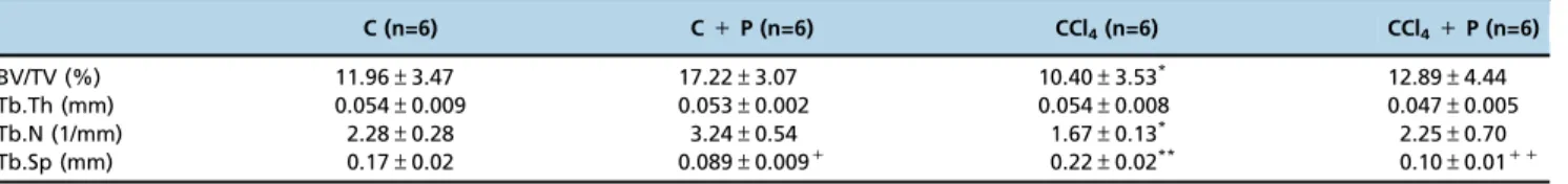
Table 2 shows the structural characteristics of femoral trabecular bone as evaluated bymCT from the four groups of mice. The CCl4 group exhibited lower BV/TV and Tb.N
values and higher Tb.Sp values than the C+P group. The CCl4+P group showed improved BV/TV, Tb.N, and Tb.Sp
values compared with the CCl4group. The trabecular
micro-structure of the CCl4+P group did not differ from that of
the C and C+P groups.
Consistent with the results obtained with femoral micro-CT, a bone histomorphometric assessment of the tibiae showed lower values of BV/TV and Tb.N and higher values of Tb.Sp in the CCl4group than in the C+P group (Table 3).
In addition, the CCl4 group exhibited higher Oc.S/BS and
Table 1-Clinical and biochemical phenotypes.
C C+P CCl4 CCl4+P
Weight (g) 29.0±1.4 28.7±3.5 29.1±1.7 26.7±2.8
Liver weight (mg/g) 51.3±4.6 47.8±3.0 56.4±5.4* 59.6±3.6**
ALT (U/L) 47.9±7.2 43.3±27.8 137.7±33.3* 133.9±22.9**
AST (U/L) 32.3±16.2 36.9±37.3 155.8±19.6* 168.8±17.8**
RANKL(pg/mL) 0.059±0.02 0.061±0.02 0.087±0.01+ 0.047±0.01++
OPG (pg/mL) 9.88±4.6 6.2±3.0 7.52±2.7 6.0±3.7
*4C and C+P; **4C and C+P;+4C;++oCCl4;po0.05
ALT: alanine transaminase; AST: aspartate transaminase; RANKL: receptor activator of nuclear factor-kB ligand; and OPG: osteoprotegerin.
Table 2-Micro-CT assessment in the femora of the four groups.
C (n=6) C+P (n=6) CCl4(n=6) CCl4+P (n=6)
BV/TV (%) 11.96±3.47 17.22±3.07 10.40±3.53* 12.89±4.44
Tb.Th (mm) 0.054±0.009 0.053±0.002 0.054±0.008 0.047±0.005
Tb.N (1/mm) 2.28±0.28 3.24±0.54 1.67±0.13* 2.25±0.70
Tb.Sp (mm) 0.17±0.02 0.089±0.009+ 0.22±0.02** 0.10±0.01++
ES/BS values than did the C+P group, whereas the CCl4+P
group showed lower Oc.S/BS and ES/BS values than the CCl4group.
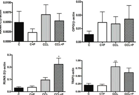
In bone, the expression of RANKL and OPG, as well as the RANKL/OPG ratio, was not significantly higher in the CCl4 group than in the C group. The expression of TRAP
was higher in the CCl4group than in the C and C+P groups.
The expression of RUNX-2 was significantly higher in bone from the CCl4+P group than in bone from the other three
groups (Figure 1).
The force needed to fracture the tibiae was significantly lower in the CCl4group (10.5±3.2 N) than in the C group
(14.7±1.7 N) (po0.05; Figure 1). The figure also shows that
pamidronate treatment efficiently improved bone resistance in cirrhotic mice. The force needed to fracture bone in the CCl4+P group (13.1±3.2 N) was similar to that in the
C group but was lower than that in the C+P group (17.3± 2.1 N) (Figure 2).
’ DISCUSSION
Bone remodeling is a crucial process that enables the skel-eton to reconcile two apparently paradoxical properties, i.e., lightness and strength. The sequential activities of bone resorption and formation, when limited to areas with pre-existing micro-damage, allow the hard tissue to renew itself, Table 3-Bone histomorphometry in the tibiae of the four groups.
C (n=7) C+P (n=6) CCl4(n=7) CCl4+P (n=7)
BV/TV (%) 10.4±2.9 12.6±2.1 7.9±1.4* 10.6±3.4
OV/BV (%) 1.84±1.42 2.39±0.42 1.32±0.77 2.74±1.46 Tb.Th (lm) 49.0±5.3 54.3±9.1 56.7±16.6 46.8±9.8 Tb.N (/mm) 2.1±0.3 2.3±0.3 1.4±02* 2.2±0.3+
Tb.Sp (lm) 439.1±95.4 376.9±58.3 657.7±149.7** 410±80.2++ Oc.S/BS (%) 1.4±0.9 0.2±0.3 2.8±2.2** 0.5±0.6++
ES/BS (%) 1.6±0.6 0.3±0.4 4.6±2.4** 0.9±0.7++
Ob.S/BS (%) 7.3±3.4 8.4±2.6 7.0±4.6 7.7±3.1 OS/BS (%) 18.9±9.7 23.08±6.5 14.79±5.2 19.4±7.1
*oC+P; **4C+P;+4CCl4;++oCCl4;po0.05
Figure 1 - Bone mRNA levels of RANKL, osteoprotegerin, RUNX-2, and TRAP in the four groups (C, C+P, CCl4, and CCl4+P). All
expression levels are presented relative tob-actin expression (RT-PCR). N=6 per genotype.* CCl4+P4C, C+P, and CCl4; ** CCl44C and
C+P,po0.05.
preventing the emergence of bone fragility. The equilib-rium of these interdependent activities is complex, involving a close interaction of genetic, nutritional, hormonal, neural, and environmental factors. A deleterious imbalance can result from the excessive activation of osteoclasts or impairment of osteoblast activity. Disease- and drug-induced osteoporosis are associated with one of these two mechanisms or their combination. For example, in glucocorticoid-induced osteo-porosis, two distinct phases were identified (18). In the first phase, increased osteoclast resorption prevails, whereas after six months, osteoblast and osteocyte dysfunction predomina-tes. Therefore, in heterogeneous diseases, it can be presumed that either mechanism may exist, depending on the cir-cumstances. Corroborating previous studies that showed the impact of chronic hepatic disorder on the skeleton, the present study contributes to advancements in this line of investigation. Based on the previous experience of our group in the study of bone disturbances in cholestasis (clinical and experimental) (10-12, 19), we observed conspicuous differ-ences in bone remodeling using an experimental model of cirrhosis. While the predominant bone impairment seems to be low osteoblastic activity in cholestasis, in hepatocellular disease, increased osteoclastic action appears to be the major mechanism of bone loss. This hypothesis was substantiated by histomorphometric analysis (i.e., increased Oc.S/BS in the CCl4 group, while there was no difference in Ob.S/BS
between groups) and by biochemical markers of bone remodeling and gene expression in bone.
Previous studies have emphasized that osteoporosis is a major complication of chronic hepatic diseases. Additionally, it is well accepted that the prevalence and severity of bone loss are increased in cholestasis (2). In a previous cross-sectional study, we demonstrated that children with congenital cholestasis had low bone mass (10). In another longitudinal study, we documented impairment of bone mass acquisi-tion (11). In these studies, the serum levels of PTH and 25-hydroxyvitamin D were normal, whereas decreased IGF-I was an early biochemical change in cholestasis (10, 11). Bone loss was an early event in hepatic cholestasis, as 85% of the children were classified as Child-Pugh A and the other 15% were classified as Child-Pugh B. After 3 years, more consistent incremental increases in the serum levels of osteocalcin were observed in the control group (76%) than in children with cholestasis (31%) (11). Moreover, Klein et al. (2002) reported decreased serum levels of osteocalcin and type I collagen telopeptide (ICTP) in children showing advanced cholestatic liver disease (6). Disturbances in bone formation were also observed in rats subjected to bile duct ligation (12). In contrast, in a group predominantly consisting of patients with hepatic liver disease, Crosbie et al. found that bone resorption was the most important cause of bone loss in hepatic osteodystrophy (5). Based on the present findings and previous reports, we hypothesize that cholestatic and hepatocytic disease leads to bone loss by diverse mechanisms. While the impairment of bone formation is the hallmark of cholestatic disorders, bone resorption prevails in hepatocytic diseases. Nevertheless, both disorders are associated with osteoporosis. In the present study, we detected decreased bone volume and trabecular number, as well as increased trabecular separation, in the tibiae and femora of mice with cirrhosis induced by CCl4. These
results agree with those recently obtained by Naussler et al. (2014) (20). In addition, the present study demonstrated bone weakness by biomechanical tests and showed that pami-dronate efficiently reversed bone loss and improved bone
strength. The present study reinforced the positive effect of disodium pamidronate on the skeleton, improving bone mass as well as bone strength. It should be emphasized that disodium pamidronate, an antiresorptive drug, not only increased bone mass in diseases associated with increased bone resorption but also improved bone mass in disorders characterized by low bone formation.
RANKL, its cellular receptor RANK, and the decoy recep-tor OPG are cytokines involved in the crosstalk between osteoblasts and osteoclasts. RANKL produced by osteoblasts dictates osteoclast differentiation and activity through RANK activation in the membranes of osteoclast precursors. OPG produced by osteoblasts deactivates RANKL, thus hamper-ing osteoclastogenesis. In mice subjected to CCl4 injection,
the serum levels of RANKL were higher than those in the control group. Additionally, pamidronate decreased the cir-culatory levels of RANKL. These results are consistent with data obtained in the ATP-binding cassette transporter B4 knockout mouse (Abcb4 / ), which is a well-established experimental model of hepatic disease. In addition to the compatible results regarding bone phenotypes obtained by micro-CT analysis, the present study also revealed similar results related to the serum levels of RANKL (21). In addi-tion, our results showed that cirrhotic mice treated with pamidronate had significantly lower serum levels of RANKL. Moreover, we showed that RANKL expression in bone was slightly increased in mice with hepatic osteodystrophy. The increased expression of TRAP in bone was additional evi-dence of enhanced osteoclast action in the bones of mice exhibiting hepatic osteodystrophy. To the best of our knowl-edge, the present study is the first to evaluate TRAP, a marker of osteoclast expression, in bones from cirrhotic mice. The increased expression of TRAP in the CCl4 group was
consistent with the higher numbers of osteoclasts on the trabecular surface and the deteriorated bone structure of the same group. Moreover, these results indirectly agreed with a previous study showing higher levels of biochemical markers of bone resorption in the serum of cirrhotic individuals, par-ticularly in those with synthetic dysfunction (22).
pamidronate treatment, the significant increase in the expres-sion of RUNX2 in bone was restricted to animals treated with CCl4. There is no clear explanation for this occurrence, but in
a previous study, it was observed that RUNX-2 expression was elevated in the livers of cirrhotic mice previously treated with CCl4(29). The study called attention to RUNX-2
regu-lating collagen synthesis, demonstrating that myofibroblasts, including hepatic stellate cells and portal fibroblasts, express RUNX-2 (29). This insightful study indicated that CCl4could
up-regulate RUNX-2 in mesenchymal cells, unlike in oste-oblasts (29). No previous study has evaluated the effects of disodium pamidronate in mice previously indirectly subjected to CCl4by injection. The present study encourages
further studies to address the mechanism of action of diso-dium pamidronate in HOD.
This study has some limitations. The dynamic parameters of bone histomorphometry were not evaluated. However, the present findings are important preliminary data for a more comprehensive study examining alterations in bone remodeling, namely, increased bone resorption as the main mechanism of bone loss in cirrhosis predominantly due to hepatocyte disorders.
The present study showed that liver disease provoked by CCl4-induced hepatocyte damage profoundly affected bone
microstructure and strength. Biochemical, molecular, and histomorphometric evidence suggested that increased bone resorption is the main mechanism involved in the process of bone loss. Based on previous data obtained in our laboratory, these results strikingly differed from bone disturbances associated with hepatic cholestasis, which mainly presents as changes in bone formation.
’ ACKNOWLEDGMENTS
This research was supported by the Conselho Nacional de Apoio Científico e Tecnológico-CNPq (470279/2013-3), NAP-DIN (11.1.21625.01.0) and the Fundac¸ão de Apoio ao Ensino e Pesquisa (FAEPA-HCFMRP); FJAP
receivedfinancial support from CNPq (307901/2014-9). The authors are grateful to Marta T. N. Maibashi for her efficient technical assistance.
’ AUTHOR CONTRIBUTIONS
de Paula FJ was responsible for the research proposal and the design of the study, discussed all of the results, and wrote the manuscript. Spirlandeli AL participated in the design of the study, collection of all of the data, analy-sis of the results, and preparation of the manuscript. Dick-de-Paula I, Zamarioli A, Jorgetti V, Ramalho LN, Nogueira-Barbosa MH, Volpon JB, Jordão AA, Cunha FQ, and Fukada SY participated in the design of the study and the collection of the data and contributed to the preparation of the manuscript.
’ REFERENCES
1. Bours SP, van den Bergh JP, van Geel TA, Geusens PP. Secondary osteo-porosis and metabolic bone disease in patients 50 years and older with osteoporosis or with a recent clinical fracture: a clinical perspective. Curr Opin Rheumatol. 2014;26(4):430-9, http://dx.doi.org/10.1097/BOR.0000 000000000074.
2. Gasser RW. Cholestasis and metabolic bone disease - a clinical review. Wien Med Wochenschr. 2008;158(19-20):553-7, http://dx.doi.org/10.1007/ s10354-008-0594-z.
3. Miroliaee A, Nasiri-Toosi M, Khalilzadeh O, Esteghamati A, Abdollahi A, Mazloumi M. Disturbances of parathyroid hormone-vitamin D axis in non-cholestatic chronic liver disease: a cross-sectional study. Hepatol Int. 2010;4(3):634-40, http://dx.doi.org/10.1007/s12072-010-9194-2. 4. Orsini LG, Pinheiro MM, Castro CH, Silva AE, Szejnfeld VL. Bone mineral
density measurements, bone markers and serum vitamin D concentrations in men with chronic non-cirrhotic untreated hepatitis C. PloS one. 2013; 8(11):e81652, http://dx.doi.org/10.1371/journal.pone.0081652.
5. Crosbie OM, Freaney R, McKenna MJ, Hegarty JE. Bone density, vitamin D status, and disordered bone remodeling in end-stage chronic liver disease. Calcif Tissue Int. 1999;64(4):295-300, http://dx.doi.org/10.1007/ s002239900622.
6. Klein GL, Soriano H, Shulman RJ, Levy M, Jones G, Langman CB. Hepatic osteodystrophy in chronic cholestasis: evidence for a multifactorial etiol-ogy. Pediatr Transplant. 2002;6(2):136-40, http://dx.doi.org/10.1034/ j.1399-3046.2002.01060.x.
7. Chen CC, Wang SS, Jeng FS, Lee SD. Metabolic bone disease of liver cirrhosis: is it parallel to the clinical severity of cirrhosis? J Gastroenterol Hepatol. 1996;11(5):417-21, http://dx.doi.org/10.1111/j.1440-1746.1996. tb00284.x.
8. Janes CH, Dickson ER, Okazaki R, Bonde S, McDonagh AF, Riggs BL. Role of hyperbilirubinemia in the impairment of osteoblast proliferation associated with cholestatic jaundice. J Clin Invest. 1995;95(6):2581-6, http://dx.doi.org/10.1172/JCI117959.
9. Angulo P. Strengthening the bones in primary biliary cirrhosis. Hepatol-ogy. 2013;58(6):1871-3, http://dx.doi.org/10.1002/hep.26696.
10. de Albuquerque Taveira AT, Fernandes MI, Galvão LC, Sawamura R, de Mello Vieira E, de Paula FJ. Impairment of bone mass development in children with chronic cholestatic liver disease. Clin Endocrinol. 2007; 66(4):518-23.
11. Taveira AT, Pereira FA, Fernandes MI, Sawamura R, Nogueira-Barbosa MH, Paula FJ. Longitudinal evaluation of hepatic osteodystrophy in children and adolescents with chronic cholestatic liver disease. Braz J Med Biol Res. 2010;43(11):1127-34, http://dx.doi.org/10.1590/S0100-879X 2010007500118.
12. Pereira FA, Facincani I, Jorgetti V, Ramalho LN, Volpon JB, Dos Reis LM, et al. Etiopathogenesis of hepatic osteodystrophy in Wistar rats with cholestatic liver disease. Calcif Tissue Int. 2009;85(1):75-83, http://dx.doi. org/10.1007/s00223-009-9249-3.
13. Abe W, Ikejima K, Lang T, Okumura K, Enomoto N, Kitamura T, et al. Low molecular weight heparin prevents hepatic fibrogenesis caused by carbon tetrachloride in the rat. J Hepatol. 2007;46(2):286-94, http://dx.doi. org/10.1016/j.jhep.2006.08.023.
14. Moreno M, Chaves JF, Sancho-Bru P, Ramalho F, Ramalho LN, Mansego ML, et al. Ghrelin attenuates hepatocellular injury and liver fibrogenesis in rodents and influences fibrosis progression in humans. Hepatology. 2010;51(3):974-85, http://dx.doi.org/10.1002/hep.23421.
15. Rao SH, Evans KD, Oberbauer AM, Martin RB. Bisphosphonate treatment in the oim mouse model alters bone modeling during growth. J Biomech. 2008;41(16):3371-6, http://dx.doi.org/10.1016/j.jbiomech.2008.09.028. 16. de Paula FJ, Dick-de-Paula I, Bornstein S, Rostama B, Le P, Lotinun S, et al.
VDR haploinsufficiency impacts body composition and skeletal acquisi-tion in a gender-specific manner. Calcif Tissue Int. 2011;89(3):179-91, http://dx.doi.org/10.1007/s00223-011-9505-1.
17. Bouxsein ML, Boyd SK, Christiansen BA, Guldberg RE, Jepsen KJ, Muller R. Guidelines for assessment of bone microstructure in rodents using micro-computed tomography. J Bone Miner Res. 2010;25(7):1468-86, http://dx.doi.org/10.1002/jbmr.141.
18. Frenkel B, White W, Tuckermann J. Glucocorticoid-Induced Osteoporosis. Adv Exp Med Biol. 2015;872:179-215, http://dx.doi.org/10.1007/978-1-4939-2895-8.
19. Pereira FA, Mattar R, Facincani I, Defino HL, Ramalho LN, Jorgetti V, et al. Pamidronate for the treatment of osteoporosis secondary to chronic cholestatic liver disease in Wistar rats. Braz J Med Biol Res. 2012;45(12): 1255-61, http://dx.doi.org/10.1590/S0100-879X2012007500143. 20. Nussler AK, Wildemann B, Freude T, Litzka C, Soldo P, Friess H, et al.
Chronic CCl4 intoxication causes liver and bone damage similar to the human pathology of hepatic osteodystrophy: a mouse model to analyse the liver-bone axis. Arch Toxicol. 2014;88(4):997-1006, http://dx.doi.org/ 10.1007/s00204-013-1191-5.
21. Hochrath K, Ehnert S, Ackert-Bicknell CL, Lau Y, Schmid A, Krawczyk M, et al. Modeling hepatic osteodystrophy in Abcb4 deficient mice. Bone. 2013;55(2):501-11, http://dx.doi.org/10.1016/j.bone.2013.03.012. 22. Lai JC, Bikle DD, Lizaola B, Hayssen H, Terrault NA, Schwartz JB. Total
25(OH) vitamin D, free 25(OH) vitamin D and markers of bone turnover in cirrhotics with and without synthetic dysfunction. Liver Int. 2015; 35(10):2294-300, http://dx.doi.org/10.1111/liv.12819.
23. Idris AI, Rojas J, Greig IR, Van’t Hof RJ, Ralston SH.
Aminobispho-sphonates cause osteoblast apoptosis and inhibit bone nodule formation in vitro. Calcif Tissue Int. 2008;82(3):191-201, http://dx.doi.org/10.1007/ s00223-008-9104-y.
24. Im GI, Qureshi SA, Kenney J, Rubash HE, Shanbhag AS. Osteoblast proliferation and maturation by bisphosphonates. Biomaterials. 2004; 25(18):4105-15, http://dx.doi.org/10.1016/j.biomaterials.2003.11.024. 25. Casado-Diaz A, Santiago-Mora R, Dorado G, Quesada-Gomez JM.
Rise-dronate positively affects osteogenic differentiation of human mesench-ymal stromal cells. Arch Med Res. 2013;44(5):325-34, http://dx.doi.org/ 10.1016/j.arcmed.2013.05.002.
27. Duque G, Li W, Adams M, Xu S, Phipps R. Effects of risedronate on bone marrow adipocytes in postmenopausal women. Osteoporos Int. 2011;22(5): 1547-53, http://dx.doi.org/10.1007/s00198-010-1353-8.
28. Koch FP, Merkel C, Al-Nawas B, Smeets R, Ziebart T, Walter C, et al. Zoledronate, ibandronate and clodronate enhance osteoblast differentia-tion in a dose dependent manner--a quantitative in vitro gene expression
analysis of Dlx5, Runx2, OCN, MSX1 and MSX2. J Craniomaxillofac Surg. 2011;39(8):562-9, http://dx.doi.org/10.1016/j.jcms.2010.10.007. 29. Hattori S, Dhar DK, Hara N, Tonomoto Y, Onoda T, Ono T, et al.