Attenuation and immunogenicity of
recombinant yellow fever 17D-dengue
type 2 virus for rhesus monkeys
1Departamento de Bioquímica e Biologia Molecular,
Instituto Oswaldo Cruz, FIOCRUZ, Rio de Janeiro, RJ, Brasil
2Departamento de Qualidade,
3Departamento de Desenvolvimento Tecnológico,
Instituto de Tecnologia em Imunobiológicos, Rio de Janeiro, RJ, Brasil
4Escola Nacional de Saúde Pública, Rio de Janeiro, RJ, Brasil
R. Galler1, R.S. Marchevsky2,
E. Caride3, L.F.C. Almeida3,
A.M.Y. Yamamura3,
A.V. Jabor3, M.C.A. Motta3,
M.C. Bonaldo1,
E.S.F. Coutinho4
and M.S. Freire3
Abstract
A chimeric yellow fever (YF)-dengue serotype 2 (dengue 2) virus was constructed by replacing the premembrane and envelope genes of the YF 17D virus with those from dengue 2 virus strains of Southeast Asian genotype. The virus grew to high titers in Vero cells and, after passage 2, was used for immunogenicity and attenuation studies in rhesus monkeys. Subcutaneous immunization of naive rhesus mon-keys with the 17D-D2 chimeric virus induced a neutralizing antibody response associated with the protection of 6 of 7 monkeys against viremia by wild-type dengue 2 virus. Neutralizing antibody titers to dengue 2 were significantly lower in immune animals than in YF-naive monkeys and protection against challenge with wild-type den-gue 2 virus was observed in only 2 of 11 YF-immune monkeys. An anamnestic response to dengue 2, indicated by a sharp increase of neutralizing antibody titers, was observed in the majority of the monkeys after challenge with wild-type virus. Virus attenuation was demonstrated using the standard monkey neurovirulence test. The 17D-D2 chimera caused significantly fewer histological lesions than the YF 17DD virus. The attenuated phenotype could also be inferred from the limited viremias compared to the YF 17DD vaccine. Overall, these results provide further support for the use of chimeric viruses for the development of a new live tetravalent dengue vaccine.
Correspondence
R. Galler
Departamento de Bioquímica e Biologia Molecular
Instituto Oswaldo Cruz, FIOCRUZ 21045-900 Rio de Janeiro, RJ Brasil
Fax: +55-21-2590-3495 E-mail: rgaller@ioc.fiocruz.br
Research supported by PADCT (No. 0442-98), CNPq (Nos. 50.1526/ 2003-0 and 472809/2003-2), PDTIS/ FIOCRUZ, and PAPES/FIOCRUZ (No. 0250.250.156).
Received May 18, 2005 Accepted August 13, 2005
Key words
•YF 17D virus recombinants •Dengue vaccine
•Immunogenicity •Attenuation •Rhesus monkeys
Introduction
The genus Flavivirus consists of 70 mem-bers, several of which cause human illnesses with the most important being yellow fever (YF), Japanese encephalitis, tick-borne en-cephalitis, and dengue type 1-4 viruses. Den-gue viruses have spread throughout the
vac-cine candidates for dengue viruses have been developed using serial passages in cultured vertebrate cells and tested in humans (4-8), but so far none has been licensed.
Since the establishment of the prototype flavivirus genome structure and expression (9), recombinant DNA technology has been an alternative for flavivirus vaccine devel-opment (for a review, see Ref. 10). In par-ticular, a strategy first developed by Bray and Lai (11) has been used by several groups to create new chimeric flaviviruses through the exchange of the viral envelope proteins (for a review, see Ref. 12). In this context, the YF 17D virus, one of the most effective and safest vaccines available and therefore very attractive as a live carrier, was used for insertion of the premembrane/envelope (prM/ E) genes of several dengue viruses (13-17), resulting in attenuated and immunogenic chimeras. Here, we describe the attenuation, immunogenicity and protective ability of a chimeric 17D-dengue serotype 2 virus in rhesus monkeys.
Material and Methods
Cells and viruses
Vero cells(ATCC, CCL 81) were main-tained in 199 medium with Earle’s salts (E199) buffered with sodium bicarbonate and supplemented with 5% fetal bovine se-rum and antibiotics.
The wild-type dengue 2 (D2) 44-2 strain has been described elsewhere (13). It origi-nated from a human case of dengue fever and belongs to the group of D2 Southeast Asian genotype viruses more recently intro-duced in the Americas (18). YF 17DD is a live attenuated virus used in the YF vaccine manufactured by Bio-Manguinhos, Oswaldo Cruz Foundation (FIOCRUZ), Rio de Janei-ro, RJ, Brazil.
The 17D-D2 virus was obtained after transfection of Vero cells with in vitro tran-scribed full-length RNA (13). The
superna-tant resulting from the transfected culture was harvested when a cytopathic effect was evident. This stock was titrated and used to infect two T175 flasks of Vero cells at a multiplicity of infection of 0.002. The super-natant was harvested 7 days later when cyto-pathic effect was pronounced, supplemented with 10% sorbitol as a stabilizer, aliquoted and frozen at -70ºC. This virus was used for all experiments described below at passage 2. All virus stocks or monkey serum samples were titrated by plaque formation on Vero cell monolayers using 6-well plates and car-boxymethylcellulose as overlay (13).
RT/PCR and sequencing
Viral suspensions were used for RNA extraction with Trizol LS (Gibco-BRL, Life Technologies, Gaithersburg, MD, USA). The extracted RNA was used as template for cDNA synthesis with YF or Den-specific synthetic oligonucleotide primers (GeneAmp RNA PCR Core Kit, Perkin-Elmer, Boston, MA, USA). Primers were designed on the basis of YF 17D strain sequence (Gene bank accession number X03700) and the D2 NGC strain sequence (M29095). PCR products were gel purified (Qiaquick gel extraction kit, Qiagen, Hilden, NDRW, Germany) and sequenced using the ABI PRISM dye termi-nator cycle sequencing core kit and an ABI 3100 instrument (Applied Biosystems, Fos-ter City, CA, USA).
Monkeys
rela-tive humidity of ~60% and 12 h of artificial light and 12 h of darkness). Animals were fed twice daily with monkey chow supple-mented with fresh fruits and were allowed water ad libitum.
Studies were carried out according to a protocol approved by the Institutional Com-mittee for Experimentation and Care of Re-search Animals (CEUA-FIOCRUZ: P0112/ 02).
Immunogenicity studies: Experiment 1
A total of 12 rhesus monkeys, 2 females and 10 males weighing 3,400 to 7,240 g, were divided into 4 groups of 3 animals each and used in the experiment. The overall design of immunogenicity experiment 1 is given in Table 1. Group 1 received a subcu-taneous (sc) dose of the chimeric 17D-D2 virus at day 30 and was challenged with 4.85 log10 plaque-forming units (PFU) of D2
44-2 virus by the sc route at day 60. Group 2 received an sc dose of YF 17DD vaccine (4.3 log10 PFU) at day 0 and a similar dose of
chimeric 17D-D2 virus 30 days later (day 30). The animals were challenged as above on day 60. Group 3 received the YF 17DD vaccine only at day 30 and was challenged with D2 44-2 virus on day 60. Group 4 received solely the challenge virus at day 60. Samples for viremia measurements were collected on days 1-8 after inoculation of YF 17DD virus, 1-8 days after 17D-D2 virus
and 1-10 days after D2 44-2 virus. All vire-mias were assayed by plating serum samples onto Vero cells (undiluted and at 1:30 and 1:300 dilutions). Blood samples were taken at days 0, 15, 30, 45, 60, 75, and 90 for measuring the antibody response to D2 and YF by the PRNT assay.
Immunogenicity studies: Experiment 2
This experiment was designed to extend our observations on the immunogenicity of the 17D-D2 virus and the influence of YF pre-immunity on the protective efficacy of the chimeric virus. A total of 12 male rhesus monkeys weighing 2,110 to 3,260 g were divided into 3 groups of 4 animals each (Table 2). Group 1 was immunized with YF 17DD at day 0, re-immunized with 17D-D2 virus at day 30 and challenged with D2 44-2 virus 60 days later (day 90). Group 2 was immunized with YF 17DD at day 0, re-immunized with 17D-D2 virus at day 120 and challenged with D2 44-2 virus at day 180. Group 3 was immunized with 17D-D2 virus at day 0 and challenged with D2 44-2 virus at day 60. Blood samples for the analy-sis of seroconversion by PRNT were taken on day 0 (all pre-immune samples), day 30 (group 1 with YF 17DD and groups 1-3 after challenge inoculation with 44-2 virus) or day 60 (groups 1-3 with 17D-D2) after the respective viral inoculation. For group 2, antibody to YF was also measured at day
Table 1. Design of Experiment 1 on the immunogenicity of 17D-D2 virus in naive and YF-immune rhesus monkeys.
1st inoculation (0-30 days) 2nd inoculation (31-60 days) 3rd inoculation (61-90 days)
Group Virus Viremia Seroconversion Virus Viremia Seroconversion Virus Viremia Seroconversion
(days)a (days)b (days)a (days)b (days)a (days)b
1 - - - 17D-D2 1-8 45, 60 D2 44-2 1-10 75, 90
2 17DD 1-8 0, 15, 30 17D-D2 1-8 45, 60 D2 44-2 1-10 75, 90
3 - - - 17DD 1-8 45, 60 D2 44-2 1-10 75, 90
4 - - - D2 44-2 1-10 75, 90
aSerial bleedings from days 1-8 (17DD) or 1-10 (17D-D2 and D2 44-2) after inoculation; bdays from the beginning of experiment; YF 17DD virus
120 prior to the administration of the 17D-D2 virus. For all groups the interval between immunization with 17D-D2 virus and chal-lenge was 60 days. Viremia samples were collected on days 2, 4, and 6 after YF 17DD immunization, days 1-8 after 17D-D2 and days 1-10 after D2 44-2 challenge.
Viral neutralization
PRNT assays were carried out in Vero cells in 6-well plates as described elsewhere (19). The neutralizing antibody titer or 50% PRNT (PRNT50) was identified as the
high-est serum dilution that reduced the number of virus plaques by 50% or more. The chal-lenge viruses used in the PRNT were YF 17DD and D2 44-2.
Monkey neurovirulence test
This test was performed in two groups of 10 captive-bred healthy rhesus monkeys composed of 12 males and 8 females (2,430 to 3,600 g). Methods for intracerebral virus inoculation (5.05 log10 PFU/mL),
measure-ments of viremia and seroconversion, clini-cal observations, autopsy, and histologiclini-cal examination have been described (20,21).
Statistical analysis
Means and standard deviations were cal-culated for clinical and combined histologi-cal scores. When there was a suggestion that
the data were asymmetrical or variances were not homogeneous, the Kruskal-Wallis non-parametric test was performed. Differences were considered to be statistically signifi-cant when P was 0.05 or less. Statistical analyses were done using the software Stata 7.0 (Stata Corporation, College Station, TX, USA, 2002).
Results
Establishing the wild-type dengue 2 challenge model
In this preliminary experiment, we ana-lyzed the infectivity of a wild-type D2 virus to be used to challenge monkeys. Five rhesus monkeys were inoculated with a single sc
dose of 5.0 log10 PFU of D2 44-2 virus.
Monkeys were bled at day 0 and thereafter for 10 consecutive days. Viremia, assayed by plaque titration in Vero cell monolayers, was detectable from days 1 to 8 in 3 animals and on days 1 through 6 in the remaining two. The magnitude of viremia ranged from 0.88 to 2.87 log10 PFU/mL (data not shown).
Given the consistency of the D2 44-2 virus in generating measurable viremias in all ani-mals we reasoned this would be an appropri-ate virus to be used to challenge monkeys after immunization with the 17D-D2 virus.
Immunogenicity of the 17D-D2 virus
In order to examine the immunogenicity
Table 2. Design of Experiment 2 on the immunogenicity of 17D-D2 virus in naive and YF-immune rhesus monkeys.
1st inoculation 2nd inoculation 3rd inoculation
Group Virus Viremia Seroconversion Virus Viremia Seroconversion Virus Viremia Seroconversion
(days)a (days)b (days)a (days)b (days)a (days)b
1 17DD 2, 4, 6 0, 30 17D-D2 1-8 90 D2 44-2 1-10 120
2 17DD 2, 4, 6 0, 120 17D-D2 1-8 180 D2 44-2 1-10 210
3 - - - 17D-D2 1-8 60 D2 44-2 1-10 90
aBleedings at days 2, 4, and 6 after YF 17DD innoculation or serial bleedings from days 1 through 8 or 1 through 10 after inoculation with 17D-D2
or D2 44-2 viruses, respectively; bdays from the beginning of experiment; YF 17DD virus dose: 4.78 log10 PFU; 17D-D2 virus dose: 5.1 log10 PFU;
of the chimeric virus we carried out two separate experiments, both including groups of flavivirus-naive and YF-immune rhesus monkeys.
In experiment 1, 12 rhesus monkeys were divided into 4 groups of 3 each. Groups 2 and 3 were given one human dose of YF 17DD vaccine (at day 0 and day 30, respec-tively; Table 1). Thirty days later, groups 1 (naive) and 2 were given an equivalent dose of 17D-D2 virus. At day 60 all 12 animals were challenged sc with 4.87 log10 PFU of
D2 44-2 virus. The data on seroconversion and viremia after challenge are shown in Table 3.
In group 1, 2 of 3 flavivirus-naive ani-mals that received the 17D-D2 virus were protected from challenge. Animal 151 had a lower neutralizing antibody response to D2 (1:108 and 1:63, at 15 and 30 days post-infection (pi), respectively) compared to the other 2 animals, and was not protected (Table 3). Animals L18 and M22 had higher PRNT (1:731 and 1:1310, respectively) and were protected, as evidenced by the absence of viremia (Table 3). The 3 YF-immune mon-keys (group 2) developed measurable neu-tralizing antibodies to D2 after immuniza-tion with the 17D-D2 virus but only one monkey (151A), which had a relatively high
PRNT of 1:407, did not show viremia after challenge with 44-2 virus. All 6 animals in groups 1 and 2 showed a boost type response after challenge with wild-type D2 virus (Table 3).
As expected, the group 3 YF-immune animals had no antibodies to D2 until after challenge with D2 44-2 virus and accord-ingly all 3 animals developed measurable post-challenge viremia of over 3 log10 PFU/
mL (Table 3). The group 4 naive monkeys showed no antibodies to YF or D2 viruses and developed viremia after challenge with wild-type D2 virus (Table 3).
In the second experiment we expanded the number of flavivirus-naive and YF-immune monkeys. Three groups of 4 monkeys each
were used. None of the monkeys showed any titer of neutralizing antibodies to YF or D2 at day 0 (Table 4). Groups 1 and 2 received one
Table 3. Immunogenicity of 17D-D2 virus in naive and YF-immune rhesus monkeys (Experiment 1).
Group Monkey PRNT50 Challenge
0a YFb D2c Viremiad PRNT50 to D2 (day 30)
1 L18 1:6 <1:5 1:731 0/0 1:4677
M22 1:6 <1:5 1:1310 0/0 1:2005
151 <1:5 <1:5 1:63 2.93/4 1:3236
2 R33 1:6 1: 25 1:105 3.78/4 1:3099
R41 1:5 1:158 1:81 3.97/5 1:5662
151A 1:5 1:126 1:407 0/0 1:620
3 S51 1:7 1:316 <1:5 3.09/6 1:1422
R59 1:10 1:501 <1:5 3.02/5 1:904 165 1:5 1:630 <1:5 3.74/5 1:747
4 S19 1:5 <1:5 <1:5 3.28/4 1:1135
R55 <1:5 <1:5 <1:5 2.2/4 1:1010 185 <1:6 <1:5 <1:5 2.31/4 1:686
aPRNT for dengue 2 (D2) and yellow fever (YF) prior to any inoculation; bPRNT for YF
at day 30 after YF vaccination; cPRNT for D2 virus at day 30 after inoculation of
17D-D2 virus; dpeak viremia titer (log
10 PFU/mL)/total number of days of viremia. PFU =
plaque-forming units; PRNT = plaque reduction neutralization titer; PRNT50 =
neutral-izing antibody titer or 50% PRNT.
Table 4. Immunogenicity of 17D-D2 virus in naive and YF-immune rhesus monkeys (Experiment 2).
Group Monkey PRNT50 Challenge
Prea YF D2b Viremiae PRNT50 to D2 (day 30)
1 U11 <1/5 >1/630c 188 2.54/4 25988
U45 <1/5 1/501 126 3.91/5 29854
U59 <1/5 1/257 2131 1.40/3 45748
U67 1/5 1/135 51 3.20/5 28454
2 U23 1/5 1/240d 71 4.54/5 14894
U63 <1/5 1/389 2641 <0.7/0 4769 U69 <1/5 >1/630 96 4.22/6 24618
U75 <1/5 1/288 65 3.35/5 51531
3 U47 <1/5 <1/5 288 <0.7/0 57361 U55 <1/5 <1/5 8925 <0.7/0 3099 U57 <1/5 <1/5 4624 <0.7/0 4266 U65 <1/5 <1/5 6090 <0.7/0 3393
aPRNT
50 for dengue 2 (D2) and yellow fever (YF) prior to any inoculation; bPRNT for
D2 virus 60 days after inoculation with 17D-D2 virus, prior to challenge; cPRNT values
at day 30 after YF vaccination, prior to 17D-D2 inoculation; dPRNT values at day 120
after YF vaccination, prior to 17D-D2 inoculation; ePeak viremia titer/total number of
mia caused by the YF 17DD vaccine strain and the 17D-D2 chimeric virus after sc in-oculation is a sensitive indicator of attenua-tion reflecting viral viscerotropism. In ex-periment 1, 4 of 6 animals inoculated with YF 17DD virus developed measurable vire-mia with titers ranging from 1.2 to 2.43 log10
PFU/mL on 1-6 days pi. Viremia was not detectable after the 6th day pi. Of the 6 monkeys that received the 17D-D2 virus, 2 showed minimal detectable titers of viremia (1.2 log10 PFU/mL) for 1 day only in the
10-day interval when blood samples were ob-tained (Table 5). One of these animals was YF-immune, and the other, YF-non-immune. In experiment 2, of 8 animals inoculated with the YF 17DD vaccine, four animals had detectable viremia on the 4th day with a peak titer of 1.9 log10 PFU/mL and one
ani-mal (U63), on the 6th day (2.33 log10 PFU/
mL). Only 2 of 12 animals that received the 17D-D2 virus developed detectable viremia (1.2 log10 PFU/mL). One of these was
YF-immune (monkey U75 viremic on two con-secutive days) and the other, non-immune (Table 5).
In both experiments it was evident that upon sc inoculation of similar doses, the 17D-D2 virus caused significantly lower lev-els of viremia compared to the YF 17DD vaccine virus. There was no apparent effect of prior YF-immunity on 17D-D2 viremia.
Low neurovirulence of the 17D-D2 chimera
Further evidence for the attenuation of the 17D-D2 virus was obtained by neuro-virulence testing in monkeys using the YF 17DD vaccine as control. A total of 10 mon-keys for each virus were inoculated intra-cerebrally (ic) and the animals were ob-served for 30 days for clinical signs, after which they were sacrificed and their central nervous system (CNS) and several other organs were removed for histological analy-ses. Individual scores for each of the 20 monkeys used in the test are shown in Table
Table 5. Viremia of 17DD and 17D-D2 viruses after subcutaneous inoculation of rhesus monkeys.
Viremia (17 DD) Viremia (17D-D2)
Range Total number of Range Total number of (Log10 PFU/mL) viremic days (Log10 PFU/mL) viremic days
Experiment 1
Group 1 - - 1.2 1
Group 2 1.2-2.43 8 1.2 1
Group 3 1.2 3 -
-Group 4 - - -
-Experiment 2
Group 1 0.6 3 <0.7
-Group 2 1.6-2.33 2 1.2 2
Group 3 - - 1.2 1
PFU = plaque-forming units.
human dose of YF 17DD virus sc and all developed antibodies to YF at 30 days pi. Group 1 received the 17D-D2 virus 30 days after immunization with YF 17DD virus and was challenged with D2 44-2 virus 60 days after receiving the 17D-D2 dose. None of the animals were protected, all of them showing high-post-challenge viremia titers. Monkey U59 showed a very high antibody titer to D2 (1:2131) but was not protected. However, peak viremia caused by the challenge virus was lower compared to the other monkeys in this group (Table 4). Group 2 was given the 17D-D2 virus 120 days after YF immunization and was challenged 60 days later. Only one animal (U63), which showed the highest titer of neu-tralizing antibodies to D2 in the group, was protected from viremia after challenge. In group 3, the YF-non-immune animals had measurable antibody titers to D2 60 days after immunization with the 17D-D2 chimera (Table 4). All animals were protected from challenge with D2 44-2. Animal U47 showed a lower neutralizing antibody titer (1:286) compared to the other animals but was also protected (Table 4).
Attenuation of 17D-D2 virus
vire-6. Only 2 of the 20 monkeys displayed signs of encephalitis (V9 and V75), both inocu-lated with the YF 17DD virus. For the 17D-D2 virus, only 2 monkeys (V08 and V51) displayed minor clinical signs (anorexia and a rough coat) resulting in an overall clinical score of 0.03 for the virus. The YF 17DD virus induced somewhat more intense clini-cal signs in 3 monkeys (V9, V11 and V75) with a fourth monkey showing minor clini-cal signs (V79). The cliniclini-cal score for the chimeric virus was about one third that for the reference vaccine virus but the differ-ence was not statistically significant (P = 0.65, Kruskal-Wallis test).
None of the 20 animals displayed any anatomical or histological abnormality in any of the extra-neural organs analyzed which included tongue, salivary mandibular gland, heart, lung, liver, kidney, urinary bladder, mesenteric lymph node, axillary lymph node, spleen, stomach, duodenum, and colon. De-spite intracerebral inoculation with the 17D-D2 virus, the monkeys did not show any evidence of clinical dengue infection nor were any specific lesions produced either at the site of injection or in the spinal cord, brain stem, thalamus, and frontal, parietal, temporal and occipital cortex. The appear-ance of specific microscopic lesions after inoculation of the YF 17DD virus indicated involvement of the CNS.
None of the 10 animals inoculated with the 17D-D2 virus showed lesions of grade 2 or higher in any of the 7 CNS areas studied and only 3 had grade 1 lesions in the sub-stantia nigra. Another animal had grade 1 in the putamen. For the YF 17DD virus, grade 3 lesions were observed in two animals (V75 and V79) in the nucleus caudatus. Grade 2 lesions were noted in the substantia nigra of 7 animals and in one or more of the 7 dis-criminatory areas of 8 of 10 monkeys.
Only 3 monkeys inoculated with the 17D-D2 virus (V39, V55 and V89) displayed minor neuronal involvement in the target area (substantia nigra), yielding a mean score
Table 6. Clinical and histological scores for the neurovirulence test in rhesus monkeys.
Virus Monkey Clinical Discriminatory Target Combined score areas area histological score
17D-D2 V08 0.1 0.04 0 0.02
V33 0 0 0 0
V39 0 0 0.5 0.25
V47 0 0 0 0
V51 0.2 0 0 0
V55 0 0 0.5 0.25
V61 0 0.07 0 0.04
V65 0 0 0 0
V77 0 0 0 0
V89 0 0 0.5 0.25
Mean ± SD 0.03 ± 0.06a 0.011 ± 0.02 0.15 ± 0.02 0.081 ± 0.12b
17DD V09 0 0.54 2 1.27
V11 0.3 0.5 1.5 1
V43 0 0.07 0.5 0.16
V45 0 0.43 1 0.71
V67 0 1.12 2 1.56
V75 0.5 1.05 2 1.52
V79 0.07 1.36 1.5 1.43
V81 0 0.2 1 0.6
V85 0 0.5 1.5 1
V91 0 0.92 2 1.46
Mean ± SD 0.087 ± 0.17a 0.67 ± 0.42 1.5 ± 0.52 1.07 ± 0.46b
aP = 0.65 for 17D-D2 vs 17DD (Kruskal-Wallis test); bP = 0.0004 for 17D-D2 vs 17DD
(Kruskal-Wallis test).
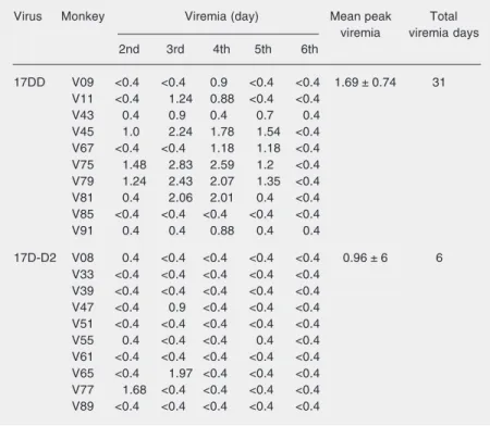
these animals on days 2 through 6 pi for analysis of viremia. As described above for viremia after sc inoculation, the viremia lev-els of the 17D-D2 virus were clearly lower (peak titer of 0.96 log10 PFU/mL and mean
duration of 0.6 days) compared to YF 17DD (mean peak titer of 1.69 log10 PFU/mL and
mean duration of 3.1 days) after ic inocula-tion (Table 7). Taken together, the viremia data after both sc and ic inoculation of mon-keys demonstrated limited viral replication, particularly for the chimera.
Nucleotide sequence analysis of the 17D-D2 virus
The 17D-D2 virus at passage 2 had its genome entirely sequenced. The extreme 5' and 3' ends were sequenced on one strand only. The genome consists of 10,874 nucleo-tides. We detected 3 nucleotide changes at passage 2 virus genome as compared to the cDNA sequence in the full-length infectious clone at positions 1765 (G→A), 2170 (C→T) and 10,467 (G→A), all of which were silent. Given that our 17D-D2 virus and the ChimeriVax-D2 virus (16) have their prM/E genes derived from different D2 strains, it was relevant to compare their respective amino acid sequences (Table 8). The D2 moiety in the FIOCRUZ 17D-D2 viral ge-nome consists of D2 New Guinea C (NGC) sequences from nucleotide 432 (numbering according to the D2 NGC sequence, GenBank accession number M29095) to nucleotide 1715 where an NsiI site was created for fragment exchange. From this position to nucleotide 2414 (the signalase cleavage site between E and NS1), the cDNA used for the construction came from a Brazilian isolate of D2 virus (13). Throughout the prM/E region, the 17D-D2 virus differs from ChimeriVax™-D2 at 89 nucleotide positions, leading to 4 amino acid substitutions (Table 8).
In the YF moiety, the 17D-D2 virus dif-fers at 4 nucleotide positions in relation to
Table 8. Differences in amino acid sequence between ChimeriVax-D2 and 17D-D2 viruses.
Gene (amino acid) Virus
17D-D2 ChimeriVax-D2a
prM/M 125b Threonine Isoleucine
E 141 Isoleucine Valine
E 164 Isoleucine Valine
E 308 Isoleucine Valine
NS5 391c Serine Asparagine
NS5 657 Asparagine Aspartic acid
aThe ChimeriVax-D2 sequence was derived by splicing together the prM/E genes from
dengue-2 (D2) type virus PUO 218 strain (GenBank accession number D00345) and the yellow fever backbone based on the YF 17D sequence of Rice et al. (9) (GenBank accession number X03700) and introducing the nucleotide sequence differences reported by Guirakhoo et al. (16). bNumbering based on D2 virus NGC strain (GenBank
accession number M29095). cNumbering based on YF 17D sequence (GenBank
accession number X03700).
to be highly significant (P = 0.0004), further confirming the attenuated nature of our 17D-D2 chimeric virus (Table 6).
Blood samples were also collected from
Table 7. Viremia in monkeys inoculated intracerebrally with 17DD and 17D-D2 vi-ruses.
Virus Monkey Viremia (day) Mean peak Total
viremia viremia days 2nd 3rd 4th 5th 6th
17DD V09 <0.4 <0.4 0.9 <0.4 <0.4 1.69 ± 0.74 31 V11 <0.4 1.24 0.88 <0.4 <0.4
V43 0.4 0.9 0.4 0.7 0.4
V45 1.0 2.24 1.78 1.54 <0.4 V67 <0.4 <0.4 1.18 1.18 <0.4 V75 1.48 2.83 2.59 1.2 <0.4 V79 1.24 2.43 2.07 1.35 <0.4 V81 0.4 2.06 2.01 0.4 <0.4 V85 <0.4 <0.4 <0.4 <0.4 <0.4 V91 0.4 0.4 0.88 0.4 0.4
17D-D2 V08 0.4 <0.4 <0.4 <0.4 <0.4 0.96 ± 6 6 V33 <0.4 <0.4 <0.4 <0.4 <0.4
ChimeriVax-D2: 6898 (NS4B, silent), 8656 (NS5, silent), 8808 (NS5 391/S→N), and 9605 (NS5 657/N→D; Table 8). No hetero-geneous positions were noted in the YF moi-ety of the chimeric virus.
Discussion
In a previous report we described the construction of a chimeric 17D-D2 virus through the exchange of the prM/E genes. This virus was characterized in terms of in vitro growth, immunogenicity and attenua-tion for mice (13). Here, we have further extended these studies to non-human pri-mates.
We examined the ability of the 17D-D2 virus to induce protective immune responses in flavivirus-naive rhesus monkeys. Alto-gether, 6 of 7 naive monkeys immunized with 17D-D2 (in both experiments 1 and 2) developed neutralizing antibodies to D2 and were protected from challenge. The neutral-izing titers to D2 significantly increased af-ter challenge, suggesting an anamnestic re-sponse. In contrast, animals that received the YF 17DD vaccine or medium only showed consistent viremia after wild-type D2 virus challenge and seroconverted to D2 thereaf-ter.
The question of pre-immunity to YF is of importance in validating the use of YF-based recombinant vaccines in areas with intense vaccination for YF, as is the case for many South American countries. In experiment 1, 3 animals received a human dose of YF 17DD vaccine by the sc route followed by a single dose of 17D-D2 virus 30 days later. All 3 animals seroconverted to YF. Only animal 151A had a significant neutralizing
antibody titer to D2 and was protected. The other two animals (R33 and R41) had lower anti-D2 antibody levels prior to challenge and were not protected suggesting that pre-immunity to YF interfered with immuniza-tion by the 17D-D2 virus. Guirakhoo et al. (16) reported that anti-YF 17D immunity
did not preclude induction of immunity and protection by the ChimeriVax-D2 virus. These investigators used an interval of 120 days between YF and chimeric virus vacci-nations. It is possible that the interference in experiment 1 was due to a shorter interval (30 days) between the YF and 17D-D2 im-munizations. This possibility was addressed in experiment 2. Two groups of 4 monkeys received the YF 17DD vaccine 30 or 120 days prior to inoculation with the 17D-D2 virus. A third group received the chimera only. Neutralizing antibody titers to D2 were generally lower in the YF pre-immune groups as compared to the YF-naive group, with the exception of monkeys U59 and U63. The 6 animals that did not have high anti-D2 neu-tralizing antibody titers were not protected from challenge, while monkeys U59 and U63 were partially or completely protected (Table 4). All 4 naive monkeys given the chimera only were fully protected. No dif-ference was observed for the time intervals of 30 and 120 days between YF 17DD and 17D-D2 virus vaccination. The YF-Vax and FIOCRUZ YF vaccines used for pre-immu-nization of monkeys in the two studies (16) and in this study are based on different substrains of the 17D virus, 17D-204 and YF 17DD, respectively. These substrains have been shown to differ in monkey neuroviru-lence (21), but whether they elicit somewhat different anti-YF immune responses in mon-keys that would explain the differences in results from the two groups is unclear.
immu-nized with the YF 17D virus. The neutraliz-ing antibody titers elicited by the 17D-D2 virus in the YF-non-immune monkeys (geo-metric mean titer 1234) were significantly higher than in YF-immune animals (geo-metric mean titer 188; P = 0.05, Kruskal-Wallis test). The apparent limited replicative capability of our 17D-D2 virus could pro-vide an explanation for its inability to solidly immunize monkeys against D2 in the pres-ence of immunity to YF.
Most flavivirus proteins elicit some type of immune responses, either humoral or cel-lular (22). There is evidence in the literature that immunity to YF NS1 protein protects mice (23,24) and monkeys (25) from an otherwise fatal challenge. Studies on human cytotoxic T lymphocyte responses to live attenuated YF 17D vaccine led to the identi-fication of HLA-B35-restricted cytotoxic T lymphocyte epitopes on nonstructural pro-teins NS1, NS2B and NS3 (26). van der Most et al. (27) have shown that the YF 17D virus envelope and NS3 proteins are major targets of the antiviral T cell response in mice. Since the chimeric virus contains eight of the YF virus proteins it is conceivable that immunity to any of those may compromise chimeric virus replication in the YF immune host, thereby reducing the response to the dengue proteins and resulting in the lower level of protection observed in experiments 1 and 2.
One of the hallmarks of the YF 17D vaccine is its extremely low incidence of adverse events. A total of 21 cases of neuro-logic disease have been reported (2) among millions of vaccinees and encephalitic reac-tions were also observed in the early devel-opment of the YF 17D virus (28). Since neurotropism is a concern with YF 17D vaccines, a standardized neurovirulence test in monkeys was established to ensure the attenuation of any YF 17D virus (29,30). Although dengue in general is known to be non-neurovirulent (31) recent reports have indicated cases of encephalitis after natural
dengue infection (32-34). Because YF 17D-dengue chimeric viruses contain the replica-tive machinery of YF 17D virus and the envelope genes of dengue viruses, their safety should be assessed by the formal neuroviru-lence test. Other attenuated dengue candi-date vaccine viruses have also been tested in the same manner (35-39) before entering clinical trials. Here we demonstrated that the 17D-D2 virus caused lower viremia of shorter duration after ic inoculation as compared to the YF 17DD vaccine control. The viremias of both viruses were within the WHO limit of less than 100 LD50/0.03 mL of serum. The
peak viremia of our 17D-D2 chimera was lower than that observed for ChimeriVax-D2 virus inoculated into the CNS of rhesus monkeys in a similar test but using a mixture of all 4 17D-dengue chimeric viruses (ChimeriVax-dengue types 1 through 4 (15). Vaccine-related clinical signs were ob-served in 4 of 10 monkeys that received YF 17DD virus beginning at day 8 pi and in-cluded encephalitis (monkeys V9 and V75). Clinical signs were observed in only 1 mon-key of 10 that received the 17D-D2 virus and were minor. Accordingly, the clinical score for the 17D-D2 group was lower than that for the YF 17DD group. Likewise, the histo-logical lesions in both target and discrimina-tory areas of monkeys inoculated with the 17D-D2 virus were less frequent, with fewer neuronal changes and yielding a combined histological score significantly lower than that observed for the control YF 17DD vac-cine virus (P < 0.0004). These data, together with the observed limited viremia, demon-strate the high attenuation of our 17D-D2 virus, similar to that of the recently described tetravalent ChimeriVax-dengue vaccine (15). A total of 93 nucleotide sequence differ-ences and 6 amino acid substitutions have been detected between our 17D-D2 and ChimeriVax-D2 viruses. Although they seem to replicate with the same efficiency in Vero cells, reaching 7.0 log10 PFU/mL, the
be a burden for the former virus to replicate in rhesus monkeys.
It is generally accepted that a dengue vaccine should be tetravalent. The attenua-tion profile for monkeys as well as the im-munogenicity and protective efficacy of the 17D-D2 virus lend further support to the development of a tetravalent dengue vac-cine using chimeric 17D-dengue viruses. However, the data shown here suggest that the prM/E sequences used for the construc-tion of chimeras may influence vaccine per-formance. In this regard a more immuno-genic 17D-D2 variant may need to be pro-duced, which replicates better in the primate model, and then tested in a tetravalent vac-cine formulation.
Acknowledgments
The authors are grateful to Instituto de Tecnologia em Imunobiológicos (Bio-Man-guinhos), Fundação Oswaldo Cruz, Rio de Janeiro, RJ, Brazil, for providing the YF 17DD virus and for all the support for the tests. We are also indebted to Dr. Antônio M. Marinho (Centro de Criação de Animais de Laboratório, Fundação Oswaldo Cruz) for providing the rhesus monkeys used in all experiments. The technical assistance of José M. da Silva, Idevaldo I. Ferreira, Mauro F. da Silva, and Edney do Monte is gratefully acknowledged.
References
1. Gubler DJ (2002). Epidemic dengue/dengue hemorrhagic fever as a public health, social and economic problem in the 21st century.
Trends in Microbiology, 10: 100-103.
2. Monath T (2003). Yellow Fever Vaccine. 4th edn. W.B. Saunders Company, Philadelphia, PA, USA.
3. Monath TP & Heinz FX (1996). Flaviviruses. In: Fields BN (Editor),
Fields Virology. 3rd edn. Lippincott-Raven Publishers, Philadelphia/ New York, 961-1035.
4. Bhamarapravati N & Sutee Y (2000). Live attenuated tetravalent dengue vaccine. Vaccine, 18: 44-47.
5. Edelman R, Wasserman SS, Bodison SA et al. (2003). Phase I trial of 16 formulations of a tetravalent live-attenuated dengue vaccine.
American Journal of Tropical Medicine and Hygiene, 69: 48-60. 6. Sabchareon A, Lang J, Chanthavanich P et al. (2004). Safety and
immunogenicity of a three dose regimen of two tetravalent live-attenuated dengue vaccines in five- to twelve-year-old Thai chil-dren. Pediatric Infectious Disease Journal, 23: 99-109.
7. Sabchareon A, Lang J, Chanthavanich P et al. (2002). Safety and immunogenicity of tetravalent live-attenuated dengue vaccines in Thai adult volunteers: role of serotype concentration, ratio, and multiple doses. American Journal of Tropical Medicine and Hygiene, 66: 264-272.
8. Sun W, Edelman R, Kanesa-Thasan N et al. (2003). Vaccination of human volunteers with monovalent and tetravalent live-attenuated dengue vaccine candidates. American Journal of Tropical Medicine and Hygiene, 69: 24-31.
9. Rice CM, Lenches EM, Eddy SR et al. (1985). Nucleotide sequence of yellow fever virus: implications for flavivirus gene expression and evolution. Science, 229: 726-733.
10. Pugachev KV, Guirakhoo F, Trent DW et al. (2003). Traditional and novel approaches to flavivirus vaccines. International Journal of Parasitology, 33: 567-582.
11. Bray M & Lai CJ (1991). Construction of intertypic chimeric dengue viruses by substitution of structural protein genes. Proceedings of
the National Academy of Sciences, USA, 88: 10342-10346. 12. Lai CJ & Monath TP (2003). Chimeric flaviviruses: novel vaccines
against dengue fever, tick-borne encephalitis, and Japanese en-cephalitis. Advances in Virus Research, 61: 469-509.
13. Caufour PS, Motta MC, Yamamura AM et al. (2001). Construction, characterization and immunogenicity of recombinant yellow fever 17D-dengue type 2 viruses. Virus Research, 79: 1-14.
14. Chambers TJ, Liang Y, Droll DA et al. (2003). Yellow fever virus/ dengue-2 virus and yellow fever virus/dengue-4 virus chimeras: biological characterization, immunogenicity, and protection against dengue encephalitis in the mouse model. Journal of Virology, 77: 3655-3668.
15. Guirakhoo F, Pugachev K, Zhang Z et al. (2004). Safety and efficacy of chimeric yellow fever-dengue virus tetravalent vaccine formula-tions in nonhuman primates. Journal of Virology, 78: 4761-4775. 16. Guirakhoo F, Weltzin R, Chambers TJ et al. (2000). Recombinant
chimeric yellow fever-dengue type 2 virus is immunogenic and protective in nonhuman primates. Journal of Virology, 74: 5477-5485.
17. van Der Most RG, Murali-Krishna K, Ahmed R et al. (2000). Chi-meric yellow fever/dengue virus as a candidate dengue vaccine: quantitation of the dengue virus-specific CD8 T-cell response. Jour-nal of Virology, 74: 8094-8101.
18. Rico-Hesse R, Harrison LM, Salas RA et al. (1997). Origins of dengue type 2 viruses associated with increased pathogenicity in the Americas. Virology, 230: 244-251.
19. Stefano I, Sato HK, Pannuti CS et al. (1999). Recent immunization against measles does not interfere with the sero-response to yellow fever vaccine. Vaccine, 17: 1042-1046.
20. Galler R, Pugachev KV, Santos CL et al. (2001). Phenotypic and molecular analyses of yellow fever 17DD vaccine viruses associ-ated with serious adverse events in Brazil. Virology, 290: 309-319. 21. Marchevsky RS, Freire MS, Coutinho ES et al. (2003).
Virol-ogy, 316: 55-63.
22. Rothman AL (2004). Dengue: defining protective versus pathologic immunity. Journal of Clinical Investigation, 113: 946-951.
23. Schlesinger JJ, Brandriss MW & Walsh EE (1985). Protection against 17D yellow fever encephalitis in mice by passive transfer of monoclonal antibodies to the nonstructural glycoprotein gp48 and by active immunization with gp48. Journal of Immunology, 135: 2805-2809.
24. Schlesinger JJ, Foltzer M & Chapman S (1993). The Fc portion of antibody to yellow fever virus NS1 is a determinant of protection against YF encephalitis in mice. Virology, 192: 132-141.
25. Schlesinger JJ, Brandriss MW, Cropp CB et al. (1986). Protection against yellow fever in monkeys by immunization with yellow fever virus nonstructural protein NS1. Journal of Virology, 60: 1153-1155. 26. Co MD, Terajima M, Cruz J et al. (2002). Human cytotoxic T lympho-cyte responses to live attenuated 17D yellow fever vaccine: identifi-cation of HLA-B35-restricted CTL epitopes on nonstructural proteins NS1, NS2b, NS3, and the structural protein E. Virology, 293: 151-163.
27. van der Most RG, Harrington LE, Giuggio V et al. (2002). Yellow fever virus 17D envelope and NS3 proteins are major targets of the antiviral T cell response in mice. Virology, 296: 117-124.
28. Fox JP, Manso C & Souza Aguiar JR (1942). Encephalitis in man following vaccination with 17D yellow fever virus. American Journal of Hygiene, 36: 117-142.
29. Levenbook IS, Pelleu LJ & Elisberg BL (1987). The monkey safety test for neurovirulence of yellow fever vaccines: the utility of quanti-tative clinical evaluation and histological examination. Journal of Biological Standardization, 15: 305-313.
30. World Health Organization (1998). Requirements for yellow fever vaccine. WHO Technical Report Series, 872: 31-68.
31. Nathanson N, Davis M, Thind IS et al. (1966). Histological studies of the monkey neurovirulence of group B arboviruses. II. Selection of indicator centers. American Journal of Epidemiology, 84: 524-540. 32. Hommel D, Talarmin A, Deubel V et al. (1998). Dengue encephalitis
in French Guiana. Research in Virology, 149: 235-238.
33. Leao RN, Oikawa T, Rosa ES et al. (2002). Isolation of dengue 2 virus from a patient with central nervous system involvement (trans-verse myelitis). Revista da Sociedade Brasileira de Medicina Tropi-cal, 35: 401-404.
34. Lum LC, Lam SK, Choy YS et al. (1996). Dengue encephalitis: a true entity? American Journal of Tropical Medicine and Hygiene, 54: 256-259.
35. Angsubhakorn S, Moe JB, Latendresse JR et al. (1986). The neuro-virulence of flaviviruses in crab-eating monkeys (Macaca fascicula-ris). Southeast Asian Journal of Tropical Medicine and Public Health, 17: 604-612.
36. Angsubhakorn S, Moe JB, Marchette NJ et al. (1987). Neuroviru-lence detection of dengue virus using rhesus and cynomolgus mon-keys. Journal of Virological Methods, 18: 13-24.
37. Angsubhakorn S, Moe JB, Marchette NJ et al. (1987). Neuroviru-lence effects of dengue-2 viruses on the rhesus (Macaca mulatta) brain and spinal cord. Southeast Asian Journal of Tropical Medicine and Public Health, 18: 52-55.
38. Angsubhakorn S, Yoksan S, Bhamarapravati N et al. (1988). Den-gue-4 vaccine: neurovirulence, viraemia and immune responses in rhesus and cynomolgus monkeys. Transactions of the Royal Socie-ty of Tropical Medicine and Hygiene, 82: 746-749.