Association of Activating KIR Copy Number Variation of
NK Cells with Containment of SIV Replication in Rhesus
Monkeys
Ina Hellmann1, So-Yon Lim1, Rebecca S. Gelman2, Norman L. Letvin1*
1Division of Viral Pathogenesis, Beth Israel Deaconess Medical Center, Harvard Medical School, Boston, Massachusetts, United States of America,2Department of Biostatistics, Dana Farber Cancer Institute and Harvard School of Public Health, Boston, Massachusetts, United States of America
Abstract
While the contribution of CD8+cytotoxic T lymphocytes to early containment of HIV-1 spread is well established, a role for
NK cells in controlling HIV-1 replication during primary infection has been uncertain. The highly polymorphic family of KIR molecules expressed on NK cells can inhibit or activate these effector cells and might therefore modulate their activity against HIV-1-infected cells. In the present study, we investigated copy number variation inKIR3DHloci encoding the only
activating KIR receptor family in rhesus monkeys and its effect on simian immunodeficiency virus (SIV) replication during primary infection in rhesus monkeys. We observed an association between copy numbers ofKIR3DHgenes and control of SIV replication inMamu-A*01–rhesus monkeys that express restrictiveTRIM5alleles. These findings provide further evidence for an association between NK cells and the early containment of SIV replication, and underscore the potential importance of activating KIRs in stimulating NK cell responses to control SIV spread.
Citation:Hellmann I, Lim S-Y, Gelman RS, Letvin NL (2011) Association of Activating KIR Copy Number Variation of NK Cells with Containment of SIV Replication in Rhesus Monkeys. PLoS Pathog 7(12): e1002436. doi:10.1371/journal.ppat.1002436
Editor:Guido Silvestri, Emory University, United States of America
ReceivedJuly 5, 2011;AcceptedOctober 30, 2011;PublishedDecember 15, 2011
Copyright:ß2011 Hellmann et al. This is an open-access article distributed under the terms of the Creative Commons Attribution License, which permits unrestricted use, distribution, and reproduction in any medium, provided the original author and source are credited.
Funding:This work was supported by the NIAID Center for HIV/AIDS Vaccine Immunology grant AI067854. The funders had no role in study design, data collection and analysis, decision to publish, or preparation of the manuscript.
Competing Interests:The authors have declared that no competing interests exist. * E-mail: [email protected]
Introduction
Natural killer (NK) cells are the primary effector cells of the innate immune system, representing a first line of defense against viruses through their ability to lyse virally infected cells without prior antigen sensitization [1–3]. NK cells express a complicated set of activating and inhibitory receptors on their cell surfaces that recognize specific ligands on target cells [4]. Inhibitory receptors transmit inhibitory signals to NK cells that protect healthy cells from destruction by NK cell-mediated cytotoxicity, whereas activating NK cell receptors transmit activating signals to these effector cells. It is the balance of these opposing signals that determines the activation state of an NK cell and, in so doing, regulates NK cell-mediated killing and cytokine production [5–7]. Among these receptor families expressed by NK cells are the inhibitory and activating killer cell immunoglobulin-like receptors (KIR). The highly polymorphic KIRs recognize MHC class I molecules as ligands [8,9], and the coincident expression of certain KIRs and MHC class I molecules in an individual influences the outcome of a number of viral infections [10,11].
Recent studies have shown that activating KIRs and their MHC class I ligands can affect AIDS pathogenesis. The expression of
KIR3DS1, an activating KIR receptor, has been shown to delay AIDS progression when its ligand, HLA-B Bw4 alleles with an isoleucine at position 80 (HLA-B Bw4-80Ile), is coexpressed in an individual [12]. Consistent with this finding, anin vitrofunctional analysis showed that KIR3DS1+NK cells are able to inhibit
HIV-1 replication in HLA-B Bw4-80Ile+ target cells [13]. Further,
KIR3DS1+ NK cells selectively expand during acute HIV-1
infection in the presence ofHLA-B Bw4-80Ile[14]. In addition to these findings, others have reported an association between the expression of certain inhibitoryKIR3DL1allotypes and protection against HIV-1 disease progression, when the KIR3DL1 ligand,
HLA-B Bw4alleles, is also expressed in an individual [15]. Studies of the contributions of NK cells to HIV-1 control have been limited by the difficulties associated with finding individuals who can be evaluated during the earliest phase of the infection. The SIV-infected rhesus monkey therefore provides a critical model for exploring NK cell biology in the setting of an AIDS virus infection [16]. We have previously shown that there are five KIR receptor families in rhesus monkeys [17]. KIR3DH is the only activating KIR family in this nonhuman primate species, and this family of molecules is highly polymorphic [18–21]. An understanding of this KIR gene family of rhesus monkeys provides an important basis for exploring the contributions of KIR receptors and NK cells in early AIDS pathogenesis in the SIV/macaque model. In the present study, we evaluated the copy number variation (CNV) of activating KIRs in rhesus monkeys and demonstrated an association between the extent of this CNV and SIV control during primary SIV infection in a cohort of Mamu-A*01– rhesus monkeys that were homozygous for the restrictiveTRIM5alleles.
Results
Establishment and validation of a qPCR assay to determineKIR3DHCNV
contribution to the control of virus replication during the acute phase of SIV infection. To date, only one activating KIR receptor family, KIR3DH, also termed KIR3DS in recent publications to be consistent with the nomenclature used in describing human KIRs [19,21], has been identified in rhesus monkeys. Interestingly, the KIR3DH receptor family in this macaque species comprises genes that display extensive polymorphism. To determine the copy number of activating KIR genes in rhesus monkeys, we developed a quantitative real-time PCR assay (qPCR) using a primer/probe set that binds to a conserved region ofKIR3DHgenes that encodes the transmembrane domain of the KIR3DH proteins. This primer/probe set was designed to amplify 23 previously described
KIR3DH alleles (GenBank accession numbers MmKIR3DH1-4 (AF334648-AF334651),MmKIR3DH7-21 (EU702453-EU702473),
MmKIR3DH-like1-4 (AY505479-82)) [17,18]. We confirmed that this primer/probe set does not bind inhibitory KIR genes by sequencing the qPCR amplicons (data not shown).
Genomic DNA derived from B-lymphoblastoid cell lines (B-LCLs) or peripheral blood mononuclear cells (PBMCs) of 77 rhesus monkeys was extracted, andKIR3DHcopy numbers were determined for each monkey by qPCR using serial dilutions of a plasmid containing the amplicon of theKIR3DHqPCR reaction.
STAT6, since it is present at two copies per diploid genome (pdg), was used as a reference gene in these studies [22]. Intra-experimental reproducibility of the qPCR assay was confirmed
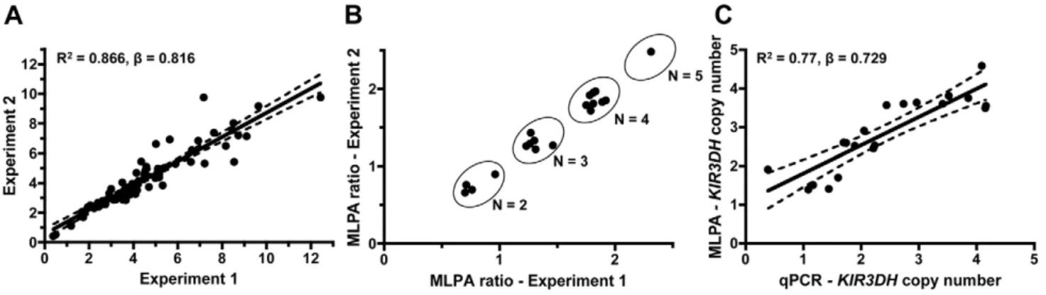
by analyzing triplicate samples for each of the 77 monkeys in two separate experiments, determiningKIR3DHcopy numbers based on the means of the triplicate values (R2 = 0.866, b = 0.816) (Figure 1A).
To validate the accuracy of theKIR3DHcopy number qPCR assay, we developed a multiplex ligation-dependent probe amplification assay (MLPA). Twenty genomic DNA samples were analyzed using one oligonucleotide set for theKIR3DHgenes and, for reference genes, two oligonucleotide sets forEP300encoding the E1A binding protein p300 and one set forCREBBPencoding the CREB-binding protein.EP300andCREBBPare each present at two copies pdg in the rhesus monkey genome. Because of the extensive polymorphism at the KIR3DH loci, only one MLPA oligonucleotide set could be designed that bound to the same
KIR3DH genes amplified in the qPCR assay. Relative MLPA signals forKIR3DH,normalized against the control gene signals, were assessed in two MLPA experiments using the same DNA samples. These signals clustered into groups corresponding to copy numbers 2, 3, 4 and 5 (Figure 1B). The KIR3DH copy numbers determined by MLPA were correlated with KIR3DH
copy numbers determined by qPCR (R2 = 0.77, b = 0.729) (Figure 1C).
KIR3DHcopy numbers vary extensively in Indian-origin rhesus monkeys
Using this assay, we determinedKIR3DH copy numbers in a cohort of 77 Indian-origin rhesus monkeys (Figure 2A).KIR3DH
copy numbers varied extensively in this cohort of monkeys, ranging from 0 to 12 copies pdg (median = 4.11 copies pdg). We rejected that this distribution had a Gaussian distribution by the skewness test (P = 0.001; i.e., the distribution was not symmetric) as well as by the kurtosis test (P = 0.005; i.e., the distribution is more sharply peaked than the Gaussian), the chi-squared test (P = 0.0006), and the Shapiro-Wilks test (P = 0.003) (data not shown). Since the aim of this study was to determine the association of activating KIR copy numbers on early control of SIV replication, we first sought to determine whether activating KIR copy numbers in this species co-stratified with other alleles already implicated in SIV control. Therefore, we assessed whether
KIR3DHcopy numbers in this cohort of animals were associated with theirMamu-A*01orMamu-B*17status or their expression of particularTRIM5alleles. The expression of certain MHC class I
Author Summary
NK cells are effector cells of the innate immune system that contribute to protection against virus infections through their ability to lyse virus-infected cells without prior antigen sensitization. Their role in controlling HIV-1 replication during primary infection has been uncertain. NK cell activation is regulated by inhibitory and activating KIRs that recognize MHC class I molecules expressed by target cells. In the present study, we identify an association between the copy number of activating KIR genes in rhesus monkeys and the control of SIV replication during primary infection in Mamu-A*01– rhesus monkeys that express restrictive TRIM5alleles. This observation under-scores the potential importance of activated NK cells in the control of SIV spread during the early stages of infection.
Figure 1. Intra-run reproducibility inKIR3DHcopy number determination and validation of quantitative real-time PCR estimates of KIR3DHcopy numbers by MLPA (multiplex ligation-dependent probe amplification).(A)KIR3DHcopy numbers were determined using triplicates of each DNA sample from 77 rhesus monkeys in two separate experiments to validate intra-experiment reproducibility (R2= 0.87,b=
0.816). (B) Comparison of signal ratios of two MLPA experiments. Pairs of ratios (20 samples) cluster around groups corresponding toKIR3DHcopy numbers of 2, 3, 4 and 5. (C) Relationship betweenKIR3DHcopy numbers determined by qPCR and MLPA (R2= 0.77,b= 0.729). The 95% confidence
interval is shown by dashed lines. doi:10.1371/journal.ppat.1002436.g001
alleles, particularly Mamu-A*01 and Mamu-B*17, has been associated with control of virus replication and delayed disease progression following SIV infection of Indian-origin rhesus monkeys [23–27]. We have also recently demonstrated the role of rhesus monkeyTRIM5 alleles 1–5 in restricting SIV infection and the impact of this restriction on the clinical outcome of SIV infectionin vivo[28]. Although MHC class I,TRIM5andKIR3DH
genes are encoded on different chromosomes in rhesus monkeys, contributions to SIV control by one allele could be a surrogate for the effects of another allele, as seen forCCL3Land Mamu-A*01
[29]. We found thatKIR3DHcopy numbers were not significantly different betweenMamu-A*01+andMamu-A*01–
rhesus monkeys (Mann-Whitney,P = 0.31) (Figure 2B). Moreover,KIR3DHcopy numbers were also not significantly different betweenMamu-B*17–
and Mamu-B*17+ monkeys (Mann-Whitney, P = 0.49). There
were insufficient Mamu-B*08+ animals (n = 3) in this cohort to
assess the associations of this allele withKIR3DHcopy numbers. Finally, we found that KIR3DH copy numbers were not significantly different in rhesus monkeys expressing onlyTRIM5
alleles 1–5 and rhesus monkeys expressing at least one of the
permissiveTRIM5 alleles 6–11 (Mann-Whitney, P = 0.75), nor were they significantly different when considering the four subsets ofMamu-A*01andTRIM5(Kruskal-Wallis,P = 0.19).
HighKIR3DHcopy numbers are associated with lower peak SIV RNA levels inMamu-A*01–rhesus monkeys
To explore whether copy number variation of activating
KIR3DH alleles in rhesus monkeys might be associated with protection against SIV replication, we evaluated a cohort of 57 rhesus monkeys infected with SIVmac251. SIV plasma RNA levels were measured in these monkeys at peak and set-point of the infection, days 14 and 70 post-SIV challenge, respectively, since these measures of viremia have been shown to be predictors of SIV disease progression in rhesus monkeys [30,31]. No association of
KIR3DHcopy numbers and viral load at peak (Figure 3A) or set-point (data not shown) was observed. To explore further a possible relationship between activating KIR copy numbers and control of SIV replicationin vivo, we divided this cohort of rhesus monkeys into two groups based on their expression or lack of expression of the MHC class I allele Mamu-A*01. Since its expression can
Figure 2. Distribution ofKIR3DHcopy numbers in Indian-origin rhesus monkeys.Copy numbers ofKIR3DHgenes were determined using quantitative real-time PCR on genomic DNA of 77 Indian-origin rhesus monkeys. (A) Distribution ofKIR3DHcopy numbers in the entire cohort of monkeys. (B) Boxplots ofKIR3DHcopy number distribution in rhesus monkeys divided into three cohorts:Mamu-A*01–andMamu-A*01+rhesus
monkeys,Mamu-B*17–
andMamu-B*17+rhesus monkeys and rhesus monkeys expressing onlyTRIM5alleles 1–5 or expressing at least oneTRIM5
allele from the group 6–11. (C) The median, mean and standard deviation (SD) ofKIR3DHcopy numbers are shown for various subgroups of rhesus monkeys.Pvalues were calculated using the Mann-Whitney U test (two-tailed).
contribute to SIV control, we reasoned that the expression of
Mamu-A*01might mask effects of other host alleles on controlling viral replication during SIV infection. On the basis of similar reasoning, the Mamu-A*01+ and Mamu-A*01–
rhesus monkeys were further divided into monkeys expressing only the TRIM5
alleles 1–5 and monkeys expressing at least oneTRIM5allele from the group 6–11. Finally, the monkeys were further subdivided into those havingKIR3DHcopy numbers above the median ($4.111 copies pdg) and those having KIR3DHcopy numbers below the median (,4.111 copies pdg). As expected, the Mamu-A*01+
monkeys had lower viral load values at peak and set-point than the
Mamu-A*01– monkeys (data not shown). In Mamu-A*01+ rhesus
monkeys, even when those monkeys were divided according to theirTRIM5 allele expression, there was no association between
KIR3DHcopy numbers and peak (Figure 3B) or set-point (data not shown) viral load. However, there were too few monkeys with
TRIM5 alleles 1–5 to determine whether there was such an association. Strikingly, in the Mamu-A*01– rhesus monkeys, we observed a very strong negative trend toward an association betweenKIR3DHcopy numbers and peak plasma SIV RNA levels (P = 0.08) (Figure 3C). The association between KIR3DHcopy numbers and peak plasma SIV RNA levels was even more pronounced inMamu-A*01–rhesus monkeys expressing only the
TRIM5 alleles 1–5. In this group of monkeys, animals having
KIR3DHcopy numbers above the median had significantly lower peak SIV RNA levels than animals havingKIR3DHcopy numbers
below the median (Mann-Whitney, P = 0.02), with a 0.4 log median difference. We also analyzed the association between
KIR3DHcopy number and peak plasma viral load by fitting the data to a parabola or a linear spline with a fixed knot at aKIR3DH
copy number of 5. The fixed knot of 5 was chosen because of previous reports that an individual NK cell usually expresses 3–5 KIRs that are randomly selected on their surface [32]. We hypothesized thatKIR3DHcopy numbers above 5 might not be associated with a linear increase in surface expression ofKIR3DH
molecules. In theMamu-A*01–,TRIM5 1–5 expressing monkeys, we observed comparable significance for the coefficient of the linear term in the parabola and for the slope of the spline for copy number#5 (P = 0.015 and P = 0.016), the squared term in the parabola and the slope of the second spline were not significant (P = 0.074 and P = 0.53) (Supplementary Figure S2 in Text S1). Interestingly, this association was no longer apparent by the time viral set-point was reached on day 70 post-infection (data not shown). These results suggest that NK cells from this subset of monkeys expressing higher numbers of KIR3DH copies may contribute more to control of SIV replication than NK cells from monkeys expressing lower numbers of activating KIR copies.
KIR3DHCNV is not associated with other clinical sequelae of SIV infection
We then assessed some of the clinical consequences of SIV infection inMamu-A*01–rhesus monkeys. The loss of peripheral
Figure 3. Association ofKIR3DHcopy numbers and peak plasma SIV RNA levels in Indian-origin rhesus monkeys.KIR3DHcopy numbers were determined by quantitative real-time PCR using genomic DNA from rhesus monkeys. Peak plasma SIV RNA levels were quantified on day 14 post-SIVmac251-infection. Scatter plots represent the relationship between SIV peak viral load andKIR3DHcopy numbers in the entire cohort of rhesus monkeys (P= 0.70) (A), inMamu-A*01+rhesus monkeys (P= 0.24) (B) and inMamu-A*01–rhesus monkeys (P= 0.08) (C). TheMamu-A*01+
andMamu-A*01–rhesus monkeys were subdivided into two groups: one group havingKIR3DHcopy numbers below the median (black) and the other
group havingKIR3DHcopy numbers above the median (blue). These groups were further subdivided into monkeys expressing onlyTRIM5alleles 1–5 or expressing at least oneTRIM5allele of the group 6–11.Pvalues were determined using the Mann-Whitney U test (two-tailed).
doi:10.1371/journal.ppat.1002436.g003
blood CD4+T cells, and more importantly, central memory (CM)
CD4+T cells following SIV infection have been shown to predict
survival of the infected rhesus monkeys [31]. Therefore, we evaluated the loss of peripheral blood CD4+T cells and central
memory (CM) CD4+T cells on days 14 (peak) and 70 (set-point)
following SIVmac251 infection in these monkeys. Data were assessed by dividing theMamu-A*01–rhesus monkeys into those having KIR3DHcopy numbers above the median ($4.11 copies pdg) and those havingKIR3DHcopy numbers below the median (,4.11 copies pdg). Neither loss of total CD4+T cells nor loss of
CM CD4+T cells was significantly associated withKIR3DHcopy
numbers in these monkeys (Figure 4A, B). We also evaluated survival as a clinical indicator of long-term protection following SIV infection in the Mamu-A*01– rhesus monkeys. Assessing the percentage of monkeys that had died by day 283 following SIV infection (day 283 was chosen ahead of time; after 283 days some monkeys were euthanized and some monkeys were used in other experiments), we observed no association betweenKIR3DHcopy numbers and survival following infection (data not shown). Therefore, peak viral load was the only clinical correlate of
KIR3DHcopy numbers in this cohort of monkeys.
KIR3DHCNV is not associated with the expression of particular KIR3DHalleles
Since some reports by other investigators had suggested that particular KIR alleles were associated with high plasma viral RNA levels in SIV-infected rhesus monkeys [20,33], we investigated whether particular KIR3DHalleles contributed to the association betweenKIR3DH copy numbers and control of peak SIV RNA levels in these monkeys. Full-length KIR3DH cDNA clones generated from 8 unrelated, Mamu-A*01– rhesus monkeys with different KIR3DH copy numbers ranging from 1–10 copies pdg were acquired by PCR. Two of the isolated KIR cDNA sequences were identical to rhesus monkey KIR alleles that had previously been reported (Mamu-KIR3DS10-JHB-HQ (GU112262) and
Mamu-KIR3DS05-JHB-HH (GU112301)) [19]. The majority of isolated KIR cDNA clones differed in their sequences from previously reported rhesus monkey KIR sequences. These novel sequences have been assigned the GenBank accession numbers JN613291-JN613300. The predicted amino acid sequences of all
KIR3DHalleles were aligned (Supplementary Figure S1 in Text S1). While someKIR3DHcDNA sequences were only observed in individual monkeys (eg. JN613291), other KIR3DH alleles were observed in multiple monkeys (eg. JN613292) (Figure 5). Impor-tantly, there was no apparent trend toward certainKIR3DHalleles being expressed in rhesus monkeys with low or highKIR3DHcopy numbers. Therefore, there was no evidence that a particular
KIR3DHallele was responsible for the observed effect on early SIV control.
HigherKIR3DHcopy numbers associate with high and stableKIR3DHtranscript levels
To assess how activating KIR CNV might affect early SIV containment, we first determined the association between the number ofKIR3DHcopies in a cell and the expression ofKIR3DH
genes by that cell. We utilized the qPCR assay that we developed for determining KIR3DH CNV to measure KIR3DH mRNA expression levels in peripheral blood CD16+NK cells of 28 naı¨ve
rhesus monkeys. Relative KIR3DH mRNA expression was sig-nificantly associated withKIR3DHcopy numbers in the evaluated cell populations, as determined by linear regression analysis (P = ,0.001, R2 = 0.51) (Figure 6A). To determine if these differences inKIR3DHRNA expression levels persisted over time, we sampled 15 of these naı¨ve rhesus monkeys one month later, andKIR3DHRNA expression levels at both sampling dates were measured in the same qRT-PCR run. Relative mRNA expression levels of KIR3DH genes from the first and second sampling associated positively, reaching statistical significance as determined by linear regression analysis (P = 0.03, R2 = 0.33) (Figure 6B). These findings suggest that increased KIR3DH copy numbers associate with high, stableKIR3DHmRNA expression levels.
KIR3DHCNV and NK cell frequencies on day 28 following SIV infection
We next investigated the relative representation of various subpopulations of NK cells in rhesus monkeys expressing different numbers of KIR3DH copies. Rhesus monkey NK cells were defined as CD3– CD8a+ NKG2A+, and CD16 and CD56
expression were used to delineate three NK cell subsets: CD16+,
Figure 4. Lack of association betweenKIR3DHcopy numbers and clinical course following SIVmac251 infection in Mamu-A*01–
rhesus monkeys.Peripheral blood CD4+T cell and central memory (CM) CD4+T cell counts were measured by flow cytometry and complete blood
counting. Loss of CD4+T cells (A) and CM CD4+T cells (B) were determined on days 14 and 70 post-challenge (pc). TheMamu-A*01–
rhesus monkeys were divided into two groups: monkeys havingKIR3DHcopy numbers below (black) or above (blue) the median.
CD56+ and double-negative (DN) NK cells (Figure 7A). Since
these subsets have been previously shown to mediate different effector functions, an expansion or contraction might only be expected to occur in certain NK cell subsets. The relative representation of NK cells, as a percentage of total circulating lymphocytes, and of NK cell subsets, as a percentage of total NK cells, were determined by flow cytometric analysis of PBMCs of naı¨ve monkeys and of monkeys sampled on day 28 following SIVmac251 infection (Figure 7B). Data were displayed by
grouping the monkeys into those havingKIR3DHcopy numbers below and those havingKIR3DHcopy numbers above the median. In the naı¨ve rhesus monkeys, the relative representation of circulating NK cells did not associate with differences inKIR3DH
copy numbers. We observed a modest increase in the relative representation of NK cells in monkeys harboring a greater number ofKIR3DH copies than the median on day 28 post-SIVmac251 infection (median, 9.27%; range, 2.15–15.82%; n = 9) compared to monkeys having KIR3DH copy numbers below the median
Figure 6.KIR3DHmRNA expression is associated withKIR3DHcopy numbers in CD14–CD16+NK cells.CD14–CD16+NK cells were isolated
from PBMCs of naı¨ve rhesus monkeys using immunomagnetic beads. RNA was extracted from these isolated cells andKIR3DHmRNA expression levels were determined by quantitative RT-PCR. Relative RNA expression was calculated using the 22DDCPmethod. (A) Scatter plots show the association
betweenKIR3DHcopy numbers andKIR3DHmRNA expression levels (P= ,0.001, R2= 0.51). (B)KIR3DHmRNA expression levels were measured a second time in the same animals after a one month interval (P = 0.03, R2 = 0.33).
doi:10.1371/journal.ppat.1002436.g006
Figure 5. Expression of particularKIR3DHalleles is not associated withKIR3DHcopy numbers.KIR3DHcDNA clones of 8 unrelated rhesus monkeys withKIR3DHcopy numbers ranging from 1–10 copies pdg were obtained using a PCR method described by Blokhuiset al.[18]. Two previously reported KIR alleles (GU112262, GU112301) and ten novelKIR3DHalleles (JN613291-JN613300) were identified.
doi:10.1371/journal.ppat.1002436.g005
(median, 6.02%; range, 1.34%–9.04%; n = 5). However, this difference in circulating NK cells did not reach statistical significance (Mann-Whitney,P = 0.24). Since there is no available antibody for staining KIR3DH molecules, NK cells that express KIR3DH receptors on their surface can’t be distinguished from those that do not.
Cytokine production of stimulated NK cell
subpopulations and expression of NK cell activation molecules does not associate withKIR3DHCNV
We finally assessed some aspects of the functionality of NK cells from rhesus monkeys harboring different copy numbers of
KIR3DHloci. We evaluated cytokine secretion by NK cells from naı¨ve and SIVmac251-infected monkeys in response to in vitro
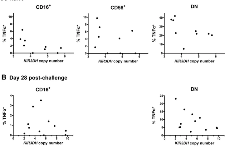
stimulation. PBMCs were stimulated with K562 cells and the intracellular expression of two cytokines produced by NK cells – tumor necrosis factor a (TNFa) and interferon c (IFNc) – was measured in the three primary NK cell subsets: CD16+, CD56+
and DN NK cells. As expected, the CD16+NK cells secreted little
cytokine [34], while the other NK cell subpopulations did secrete cytokines upon stimulation (Figure 8). There was, however, no obvious association between TNFa or IFNc secretion upon stimulation and KIR3DHcopy numbers by the CD56+and DN
NK cell subpopulations. This was seen in NK cells sampled from both naı¨ve and recently infected monkeys (Figure 8A, B and data not shown).
We also assessed whether the surface expression of the activation-associated molecules CD69, HLA-DR and NKp46 on NK cell subsets was associated with KIR3DH copy numbers in
naı¨ve rhesus monkeys and in the same cohort of monkeys at day 35 and at set-point following SIV infection. The intracellular levels of the proliferation-associated Ki67 molecule in NK cells during primary infection were also assessed. No association was observed between KIR3DH copy number and the expression of these molecules in CD16+, DN or CD56+NK cells pre- or post-infection
(Supplementary Figure S3 in Text S1 and data not shown).
Discussion
The protective effects of particular KIRs for HIV-1 infections in humans have, for the most part, been shown in epidemiologic studies [12,35–37]. Functional studies of NK cells expressing specific KIRs have been difficult to carry out in the early phases of HIV-1 infections because the timing of HIV-1 acquisition cannot be precisely determined from the clinical histories of patients. The SIV-infected rhesus monkey therefore represents a potentially important model for studying KIR receptors expressed on the surface of NK cells and the effects of these cells on the control of viral replication during primary infection. However, the ligands of rhesus monkey KIRs are not well understood. The interaction between a particular KIR and its ligand might, however, be crucial for that KIR to modify disease outcome in HIV-1/SIV infections. In fact, some reports suggest that associations between particular KIR receptors and clinical sequelae of HIV-1 infections are only observed when the ligands of those KIRs are considered [12,36,38]. For example, coexpression ofKIR3DS1and its ligand
HLA-B Bw4-80Ilealleles was associated with a delayed progression to AIDS in HIV-1-infected individuals, whereas the expression of
KIR3DS1in the absence of theHLA-B Bw4-80Ilealleles was not
Figure 7. Distribution of peripheral blood NK cell subsets inMamu-A*01– rhesus monkeys.(A) Representative flow cytometric plots defining NK cells. Rhesus monkey NK cells were defined as CD3–CD8a+NKG2A+, and CD16 and CD56 expression was used to delineate three primary
NK cell subsets: CD16+, CD56+and double negative (DN) NK cells. (B) Percentages of circulating total NK cells as well as CD16+, CD56+and
double-negative (DN) NK cells were compared in naı¨ve rhesus monkeys and SIVmac251-infected rhesus monkeys on day 28 post-challenge. The rhesus monkeys were divided into animals havingKIR3DHcopy numbers below (black) or above (blue) the median.
associated with a delayed clinical progression following HIV-1 infection [12]. In contrast to this observation, other studies in humans have demonstrated that the expression of particular KIR molecules was associated with a more favorable HIV-1 disease outcome or decreased risk of HIV-1 acquisition irrespective of the expression of the KIR ligands [37,39,40]. Also, studies of SIV-infected rhesus monkeys showed that the expression of particular inhibitoryKIR3DLandKIR3DHmolecules was associated with high levels of SIV replication without consideration of the ligands of those KIR molecules [20,33]. Consistent with the findings of these latter studies, we observed an association betweenKIR3DHcopy numbers and peak SIV RNA levels during primary infection in monkeys that did not expressMamu-A*01and expressed the restrictive TRIM5
alleles 1-5 without considering the contribution of specific KIR ligands. It is possible however, that the observed effect might be more profound if KIR3DH ligands were taken into consideration. The characterization of KIR3DH-expressing NK cell subpop-ulations could be important for clarifying the role of activating KIRs in modulating SIV replication. There is, however, no monoclonal antibody that recognizes rhesus monkey KIR3DH, and, therefore, surface expression of KIR3DH on monkey NK cells cannot be monitored. Without such an antibody, NK cells expressing high levels of KIR3DH molecules on their surface and high frequencies of KIR3DH+NK cells cannot be distinguished.
In the present studies, we used KIR3DH RNA expression as a surrogate for KIR3DH cell surface expression. The primer/probe
set used in the assay to quantifyKIR3DHRNA expression binds to a conserved region of the KIR3DH genes that encodes the transmembrane domain of the KIR3DH proteins. Therefore, truncated KIR proteins that have lost their transmembrane domains due to frameshift deletions and are not anchored in the cell membrane, as seen for allotypes of the humanKIR2DS4and
KIR2DL4[41,42], would not be detected. We assume that higher
KIR3DH copy numbers, and the resulting higher KIR3DH
transcript levels, indicate an increased surface expression of activating KIR receptors on subpopulations of NK cells.
The MHC class I allele Mamu-A*01 and TRIM5 alleles are genetic determinants of the control of SIV replication in rhesus monkeys [25,29,43]. A link was also reported between CCL3L
CNV and AIDS progression in SIV-infected rhesus monkeys [22]. We, however, showed thatCCL3LCNV was serving as a surrogate for the expression ofMamu-A*01, and the relatively benign clinical course observed following SIV infection in certain monkeys was actually a consequence ofMamu-A*01expression by those animals [29]. Because of these findings, it was important in the present studies to show thatKIR3DHcopy numbers were not acting as a surrogate marker for the Mamu-A*01 and TRIM5 status of the monkeys. In our data, CNV ofKIR3DHwas not associated with the expression of eitherMamu-A*01, Mamu-B*17or the restrictive
TRIM5alleles. In addition, when Mamu-A*01 and TRIM5 were both included in models, they did not eliminate the relationship of
KIR3DHcopy number and peak viral load.
Figure 8. Secretion of tumor necrosis factora(TNFa) in NK cell subsets following stimulation with K562 cells.PBMCs were stimulated
with K562 cells at an effector-to-target-ratio of 10:1. Percentages of TNFa+cells above background in CD16+, CD56+and DN NK cells were measured
in peripheral blood of naı¨ve rhesus monkeys (A) and SIVmac251-infected rhesus monkeys on day 28 post-challenge (B). For the day 28 post-challenge timepoint, insufficient CD56+NK cells were acquired to allow an analysis.
doi:10.1371/journal.ppat.1002436.g008
Very few studies have evaluated CNV in KIR loci. One study showed thatKIR2DS2copies ranged from 0 to 2 copies in humans [38]. In that study,KIR2DS2copy numbers were estimated using KIR typing rather than single gene analysis. Pelak et al.
documented up to 3 copies ofKIR3DS1and 3 copies ofKIR3DL1
in humans using a qPCR-based assay that was similar to the quantitative assay we used in the present studies [Pelak et al., personal communication]. We, however, observed a wider range ofKIR3DHcopies in rhesus monkeys with one monkey having 12
KIR3DH copies per cell. We defined copy numbers of the KIR3DH family without evaluating individual KIR3DH genes. These results are consistent with a recent study in rhesus monkeys showing 0-4KIR3DSgenes per haplotype [19].
Both Gaudieriet al.and Pelak et al.investigated the effects of copy number variation of KIR2DS2 and KIR3DS1 on HIV-1 disease outcome. Higher copy numbers of the activatingKIR2DS2
were associated with greater CD4+ T cell loss and rapid
progression to AIDS [38]. Since these investigators only indirectly determinedKIR2DS2copy numbers and there is a strong linkage disequilibrium betweenKIR2DS2andKIR2DL2, the rapid HIV-1 disease progression in these individuals might not be attributable to the expression ofKIR2DS2. In the study byPelak et al., higher numbers of activatingKIR3DS1copies were associated with lower plasma virus RNA levels at set-point in HIV-1 infected individuals that express the KIR3DS1-ligandHLA-B Bw4-80Ilealleles [Pelak
et al., personal communication]. The findings in the present study are in line with these observations.
The effect of activating KIR copy numbers on SIV replication may be more modest than the effects on SIV control mediated by the MHC class I moleculeMamu-A*01and the restrictiveTRIM5alleles [25,28,43]. Since theKIR3DHcopy number effect was only seen in the Mamu-A*01– monkeys that were homozygous for restrictive
TRIM5alleles, it is likely that a strongerMamu-A*01-associated effect may obscure this NK cell contribution to SIV control. It is not immediately obvious why theKIR3DHcopy number effect was only observed in the monkeys expressing the restrictiveTRIM5alleles.
In the present studies we showed that higher numbers of
KIR3DH copies were associated with lower peak viral load following SIV infection, but we did not observe associations between KIR3DH copy numbers and other clinical sequelae of SIV infection. The contributions of NK cells to the control of SIV replication may be manifested during the early stages of SIV infection. Then, during the course of SIV-infection, virus-specific CD8+T cells expand that maintain control over viral replication
throughout the chronic phase of infection [44–48]. The contribu-tions of adaptive CD8+T cells to viral replication are likely much
greater than those mediated by NK cells, and these effects may simply obscure those of KIR3DH-expressing NK cells on SIV control during the later stages of infection. Further, the NK cells may become too dysfunctional later in the course of infections to mediate an antiviral effect [49–51].
Materials and Methods
Ethics statement
All of the animals used in this present study were Indian-origin rhesus macaques. All monkeys were housed in accordance with the guidelines of theNIH Guide for the Care and Use of Laboratory Animalsand with the approval of the Institutional Animal Care and Use Committee of Harvard Medical School and the National Institutes of Health.
Animals and SIV infections
SIV-challenged monkeys were either infected intravenously or intrarectally with an uncloned SIVmac251 inoculum [31]. The
expression of Mamu-A*01, Mamu-B*08, Mamu-B*17 and the expression ofTRIM5alleles were assessed by PCR [28,29].
Plasma viral load assay and CD4+T lymphocyte counts
Plasma viral RNA levels were measured using an ultra-sensitive branched DNA amplification assay (Bayer Diagnostics, Berkeley, CA). Counts of total peripheral blood CD4+T lymphocytes and
central memory CD4+ T lymphocytes were calculated by
multiplying the total lymphocyte count by the percentage of CD3+CD4+T cells, times the percentage of CD95+CD28+T cells
for counts of central memory CD4+ T cells, determined by
monoclonal antibody staining and flow cytometric analysis [52].
Extraction of genomic DNA
Total genomic DNA was extracted from peripheral blood mononuclear cells (PBMCs) or H.papio-immortalized B-lympho-blastoid cell lines (B-LCLs) using the DNeasy Blood and Tissue Kit (Qiagen, Valencia, CA). The purity of the isolated genomic DNA was verified by A260/A280 ratio: average 1.88 (range 1.74–2.00).
The DNA samples were stored at220uC until use. DNA integrity was verified by gel-electrophoresis of selected samples.
Determination ofKIR3DH copy numbers using real-time PCR
Activating KIR copy number determinations were performed by quantitative real-time PCR (qPCR) using the 7300 Real-time PCR System (Applied Biosystems, Foster City, CA). With KIR3DH being the only activating KIR receptor family in rhesus monkeys, aKIR3DHprimer andTaqManprobe set for qPCR was designed to specifically amplify previously identified Mm-KIR3DH
alleles (GenBank accession numbersMmKIR3DH1-4 (AF334648-AF334651), MmKIR3DH7-21 (EU702453-EU702473), and
MmKIR3DH-like1-4 (AY505479-82)) [17,18], thereby avoiding recognition of any inhibitory KIR genes.KIR3DH-specific primer sequences were: Forward: 59 -CACCAGACACCTGCCTATTG-TGA-39; Reverse: 59 -GAGTCTCTTTTTGTCGGAGCACCA-39; Probe: 59 -FAM-TAGGTACTCGGTGGCCACCATCAT-BHQ-39. qPCR products were sequenced to confirm the spe-cificity of the amplification. TheSTAT6gene, present in a single copy per haploid rhesus genome [22], was used as an endogenous reference gene: Forward: 59 -AACCTAAAGAGAATGGGAG-TGT-39; Reverse: 59-GAATATAGTCACAACCCTGGATC-39; Probe: 59-FAM-CTCTGCCCTTCTCCTGCCTCCC-BHQ-39. Primers were purchased from Invitrogen and probes were purchased from Biosearch Technologies. Per qPCR reaction, 12.5 ng of total genomic DNA were added toTaqManUniversal PCR Master Mix (Applied Biosystems) and the specific primers and probe. All samples were run in triplicate. Thermal cycling conditions were as follows: 2 min at 50uC, 10 min at 95uC, followed by 40 cycles of a two-step PCR of 15 s at 95uC and 1 min at 60uC. qPCR results were analyzed using the SDS v1.4.0 software (Applied Biosystems).
Absolute quantification ofKIR3DHcopy numbers using plasmid standard
To determine absolute KIR3DHcopy numbers, plasmid DNA standards for KIR3DHand STAT6 were created. The plasmids contained the specific sequence amplified in the qPCR reaction.
First-strand cDNA was synthesized from RNA isolated from rhesus PBMCs using an oligo(dT)20primer and the SuperScript III
First-Strand Synthesis System for RT-PCR (Invitrogen, Carlsbad, CA), following the manufacturer’s protocol. Then, PCR was performed using the Platinum PCR SuperMix High Fidelity (Invitrogen). Amplification conditions were: 5min at 94uC; 35 cycles of 30 s at 94uC, 30 s at 55uC, and 45 s at 68uC; and 20 min at 68uC. PCR products were resolved by gel electrophoresis, excised and purified with the QIAquick Gel Extraction Kit (Qiagen). Purified PCR products were ligated into the pGEM-T Easy Vector (Promega, Madison, WI), resulting in pKIR3DH and pSTAT6. Clones containing the correct insert were verified by sequencing. Plasmids were isolated and purified using the EndoFree Plasmid Maxi Kit (Qiagen), and the plasmid DNA concentration was measured using a NanoDrop ND-1000 spectrophotometer (Thermo Scientific, Wilmington, DE). Six serial log dilutions (108– 103copies) of pKIR3DH and pSTAT6 plasmid DNA were used to generate standard curves by plotting CTvalues versus log copies of each qPCR plate. An R2value of a
standard curve of less than 99% was considered imprecise and the corresponding qPCR plate (96 wells) of DNA samples was repeated. Absolute copy numbers were calculated by determining the number ofKIR3DHcopies per sample from the standard curve and then by normalizing against the number ofSTAT6copies in the same sample. Values were multiplied by 2 to obtain copy numbers per diploid genome.
Confirmation of qPCRKIR3DH copy number estimates
To validate that theKIR3DHabsolute copy numbers calculated from qPCR were accurate, we determinedKIR3DHcopy numbers using a multiplex ligation-dependent probe amplification assay (MLPA). Two adjacent oligonucleotides per locus – one set for the
KIR3DH loci and, as reference loci, two sets for EP300 (E1A binding protein p300) and one set for CREBBP (CREB-binding protein) – were designed to contain the same primer binding sequences for later amplification. Only one KIR3DH MLPA oligonucleotide set, recognizing the sameKIR3DHalleles as in the qPCR assay, could be designed due to the polymorphic nature of theKIR3DHloci. The oligonucleotide sequences are listed in the Supplementary Table S1 in Text S1 and were purchased from IDT (IDT, Coralville, IA). All MLPA reagents were purchased from MRC Holland (MRC-Holland, Amsterdam, Netherlands). The MLPA reaction was carried out according to the manufac-turer’s protocol using 100 ng of genomic DNA. For each DNA sample, the oligonucleotide sets for the KIR3DH genes and the reference loci were ligated to the DNA in a multiplex PCR and only ligated oligonucleotides were amplified using the FAM-labeled universal primer pair. Amplified PCR products differed in length and could therefore be distinguished. The amplification products were analyzed on an ABI 3130XL capillary DNA analyzer (Applied Biosystems). The results were analyzed using GeneMapper Software (Applied Biosystems). The final analysis of the MLPA data was carried out using Microsoft Excel software. For each sample, peak signal values forKIR3DHwere normalized by the average signal of the reference probes to determine relative MLPA signals [53]. Relative MLPA signals from two different experiments using the same DNA samples formed discrete clusters corresponding to copy numbers. Absolute copy numbers of individual clusters were determined by assuming that the distance between successive clusters corresponded to 1 copy and that the copy number genotype of the first cluster corresponded to the distance from 0 divided by the average distance of successive clusters.
RNA isolation
PBMCs were sorted for CD14–CD16+NK cells using magnetic
cell sorting (MACS Microbeads by Miltenyi Biotec). Total RNA was isolated from the CD14–CD16+NK cells using the RNeasy
Mini Kit (Qiagen). RNA samples were stored at280uC until use.
Quantification ofKIR3DH RNA expression
KIR3DH RNA expression in CD14–CD16+ NK cells was
determined by performing real-time quantitative reverse tran-scription PCR (qRT-PCR) using the sameKIR3DHand STAT6
primers and probes used for copy number determination of DNA. Per qRT-PCR reaction, 50 ng of total RNA was added to the
TaqmanOne-Step RT-PCR Master Mix (Applied Biosystems) and theKIR3DH-andSTAT6-specific primers and probes. All samples were run in triplicates. Thermal cycling conditions were as follows: 30 min at 48uC, 10 min at 95uC, followed by 40 cycles of a two-step PCR of 15 s at 95uC and 1 min at 60uC. qRT-PCR results were analyzed using the SDS v1.4.0 software (Applied Biosystems). Relative RNA expression was determined using the 22DDCt method (Applied Biosystems). Briefly, for each sample, CTvalues
ofKIR3DHwere first normalized against the CTvalues ofSTAT6.
Normalized KIR3DH CT values for each monkey were then
divided by the normalizedKIR3DH CT values of one reference
monkey that was evaluated in each qPCR run to determine relativeKIR3DHRNA expression.
Discrimination ofKIR3DHalleles
Total RNA, isolated from rhesus CD14–CD16+NK cells, was
used to synthesize first-strand cDNA using an oligo(dT)20primer
and the SuperScript III First-Strand Synthesis System for RT-PCR (Invitrogen), following the manufacturer’s protocol. RT-PCR amplification was performed using the Platinum PCR SuperMix High Fidelity (Invitrogen). The primer sequences were as described by Blokhuiset al. [18]. Amplification conditions were as follows: 5 min at 94uC; 35 cycles of 30 s at 94uC, 30 s at 66uC, and 90 s at 68uC; and 20 min at 68uC. PCR products were subjected to gel electrophoresis, excised and purified with a QIAquick Gel Extraction Kit (Qiagen) following the manufactur-er’s protocol. Purified PCR products were ligated into the pGEM-T Easy vector (Promega), according to the manufacturer’s instructions. Briefly, ligation reactions were incubated at room temperature for one hour and then transformed into JM109 High Efficiency competent cells (Promega). Between 23 and 28 insert-containing colonies per sample were sequenced and analyzed using the Sequencher Software (Gene Codes, Ann Arbor, MI). Corresponding amino acid sequences were aligned using Clus-talW2 Multiple Sequence Alignment [54]. Only KIR sequences that were obtained from more than one clone are reported. The novel rhesus monkey KIR cDNA sequences have been assigned the GenBank accession numbers JN613291-JN613300.
Monoclonal antibodies (MAbs) and immunophenotyping of NK cells
The antibodies used in this study were anti-CD8a-Peridinium Chlorophyll Protein-Cy5.5 (SK1), anti-CD56-Phycoerythrin-Cy7 (N901, Beckman Coulter, Brea, CA), anti-CD3-Pacific Blue (SP34.2), anti-CD159 (NKG2A)-Allophycocyanin (Z199, Beckman Coulter), anti-CD16-Allophycocyanin-Cy7 (3G8), anti-TNFa -Fluo-rescein Isothiocyanate (MAb11), anti-IFNc-Alexa Fluor 700 (B27) anti-CD335 (NKp46)-Phycoerythrin (BAB281, Beckman Coulter), CD69-energy-coupled dye (TP1.55.3, Beckman Coulter), anti-HLA-DR-Phycoerythrin-Cy7 (L243 (G46-6), anti-Ki67-Alexa Fluor 488 (B56), and anti-CD56-Peridinium Chlorophyll Protein-Cy5.5
(B159). All antibodies unless otherwise indicated were purchased from BD Biosciences. The LIVE/DEAD Fixable Aqua Dead Cell stain kit (Invitrogen) was used as a viability marker to distinguish live cells from dead cells in all flow cytometric analyses. All acquisitions were made on a LSR II flow cytometer (BD Biosciences) and analyzed using FlowJo software (TreeStar Inc., Ashland, OR). PBMCs were isolated from EDTA-anticoagulated blood by Ficoll-Paque (GE Healthcare, Piscataway, NJ) gradient separation and either stained immediately or cryopreserved in the vapor phase of liquid nitrogen. Later, cryopreserved cells were thawed and rested at 37uC in a 5% CO2environment for 6 hrs. The viability of these cells
was . 90%. PBMCs were stained with anti-surface MAbs to delineate NK cells (CD3, CD8, NKG2A, CD56, and CD16) and anti-surface molecules CD69, HLA-DR and NKp46. Cells were then fixed and permeabilized with Cytofix/Cytoperm solution (BD Biosciences) and stained with antibodies specific for Ki67. Labeled cells were fixed in 1% formaldehyde-PBS.
PBMC stimulation and intracellular cytokine staining
To determine cytokine production of NK cells in response to K562 cells, PBMC were incubated for 6 hours in the presence of RPMI 1640/10% fetal calf serum alone (unstimulated), with K562 at an effector-to-target ratio of 10:1, or with phorbol myristate acetate (PMA) as a positive control. All samples contained Monensin (GolgiStop, BD Biosciences) and Brefeldin (Golgi Plug, BD Biosciences). Cells were next stained with antibodies specific for cell surface molecules CD3, CD8, NKG2A, CD56, and CD16. Cells were then fixed and permeabilized with Cytofix/Cytoperm solution (BD Biosciences) and stained with antibodies specific for TNFaand IFNc. Labeled cells were fixed in 1% formaldehyde-PBS. All data are reported after background correction.
Statistical analyses
All statistical analyses and graphic analyses were conducted using GraphPad Prism (GraphPad Prism Software, La Jolla, CA) and STATA (StataCorp LP, College Station, Texas). Normality was assessed using the skewness test, the kurtosis test, the chi-squared-test and the Shapiro-Wilks-test. The non-parametric Mann-Whitney-test was used for the comparison of two groups and the Kruskal-Wallis test was used for comparing more than two groups. Linear regression models were done for the relationship of peak viral load and KIR3DH copy number either overall (and incorporating covariates forMamu-A*01andTRIM5groupings) or in subgroups. Subsequently, two other regression models (para-bolic, i.e. second degree polynomial, and two linear splines with a fixed knot at 5) were used because of previous reports that an individual NK cell usually expressed at most 5 KIRs [32]. AllP
values are two-sided and none are corrected for multiple comparisons.Pvalues of,0.05 were considered significant.
Supporting Information
Text S1 Supplementary figures and tables.
(PDF)
Acknowledgments
We thank Yue Sun, Mary Carrington, David B. Goldstein and Piotr Kozlowski for helpful discussions.
Author Contributions
Conceived and designed the experiments: IH NLL. Performed the experiments: IH SYL. Analyzed the data: IH SYL RSG. Wrote the paper: IH NLL RSG.
References
1. Biron CA (1999) Initial and innate responses to viral infections--pattern setting in immunity or disease. Curr Opin Microbiol 2: 374–381.
2. Cerwenka A, Lanier LL (2001) Natural killer cells, viruses and cancer. Nat Rev Immunol 1: 41–49.
3. Orange JS (2002) Human natural killer cell deficiencies and susceptibility to infection. Microbes Infect 4: 1545–1558.
4. Lanier LL (2005) NK cell recognition. Annu Rev Immunol 23: 225–274. 5. Trinchieri G (1989) Biology of natural killer cells. Adv Immunol 47: 187–376. 6. Moretta A, Bottino C, Mingari MC, Biassoni R, Moretta L (2002) What is a
natural killer cell? Nat Immunol 3: 6–8.
7. Vivier E, Nunes JA, Vely F (2004) Natural killer cell signaling pathways. Science 306: 1517–1519.
8. Moretta A, Sivori S, Vitale M, Pende D, Morelli L, et al. (1995) Existence of both inhibitory (p58) and activatory (p50) receptors for HLA-C molecules in human natural killer cells. J Exp Med 182: 875–884.
9. Pende D, Biassoni R, Cantoni C, Verdiani S, Falco M, et al. (1996) The natural killer cell receptor specific for HLA-A allotypes: a novel member of the p58/p70 family of inhibitory receptors that is characterized by three immunoglobulin-like domains and is expressed as a 140-kD disulphide-linked dimer. J Exp Med 184: 505–518.
10. Parham P (2005) MHC class I molecules and KIRs in human history, health and survival. Nat Rev Immunol 5: 201–214.
11. Martin MP, Carrington M (2005) Immunogenetics of viral infections. Curr Opin Immunol 17: 510–516.
12. Martin MP, Gao X, Lee JH, Nelson GW, Detels R, et al. (2002) Epistatic interaction between KIR3DS1 and HLA-B delays the progression to AIDS. Nat Genet 31: 429–434.
13. Alter G, Martin MP, Teigen N, Carr WH, Suscovich TJ, et al. (2007) Differential natural killer cell-mediated inhibition of HIV-1 replication based on distinct KIR/HLA subtypes. J Exp Med 204: 3027–3036.
14. Alter G, Rihn S, Walter K, Nolting A, Martin M, et al. (2009) HLA class I subtype-dependent expansion of KIR3DS1+and KIR3DL1+NK cells during acute human immunodeficiency virus type 1 infection. J Virol 83: 6798–6805. 15. Martin MP, Pascal V, Yeager M, Phair J, Kirk GD, et al. (2007) A mutation in KIR3DS1 that results in truncation and lack of cell surface expression. Immunogenetics 59: 823–829.
16. Haigwood NL (2004) Predictive value of primate models for AIDS. AIDS Rev 6: 187–198.
17. Hershberger KL, Shyam R, Miura A, Letvin NL (2001) Diversity of the killer cell Ig-like receptors of rhesus monkeys. J Immunol 166: 4380–4390. 18. Blokhuis JH, Doxiadis GG, Bontrop RE (2009) A splice site mutation converts
an inhibitory killer cell Ig-like receptor into an activating one. Mol Immunol 46: 640–648.
19. Blokhuis JH, van der Wiel MK, Doxiadis GG, Bontrop RE (2010) The mosaic of KIR haplotypes in rhesus macaques. Immunogenetics 62: 295–306. 20. Chaichompoo P, Bostik P, Stephenson S, Udompunturuk S, Kobkitjaroen J,
et al. (2010) Multiple KIR gene polymorphisms are associated with plasma viral loads in SIV-infected rhesus macaques. Cell Immunol 263: 176–187. 21. Kruse PH, Rosner C, Walter L (2010) Characterization of rhesus macaque KIR
genotypes and haplotypes. Immunogenetics 62: 281–293.
22. Degenhardt JD, de Candia P, Chabot A, Schwartz S, Henderson L, et al. (2009) Copy number variation of CCL3-like genes affects rate of progression to simian-AIDS in Rhesus Macaques (Macaca mulatta). PLoS Genet 5: e1000346. 23. Yasutomi Y, Reimann KA, Lord CI, Miller MD, Letvin NL (1993) Simian
immunodeficiency virus-specific CD8+lymphocyte response in acutely infected rhesus monkeys. J Virol 67: 1707–1711.
24. Pal R, Venzon D, Letvin NL, Santra S, Montefiori DC, et al. (2002) ALVAC-SIV-gag-pol-env-based vaccination and macaque major histocompatibility complex class I (A*01) delay simian immunodeficiency virus SIVmac-induced immunodeficiency. J Virol 76: 292–302.
25. Mothe BR, Weinfurter J, Wang C, Rehrauer W, Wilson N, et al. (2003) Expression of the major histocompatibility complex class I molecule Mamu-A*01 is associated with control of simian immunodeficiency virus SIVmac239 replication. J Virol 77: 2736–2740.
26. Loffredo JT, Maxwell J, Qi Y, Glidden CE, Borchardt GJ, et al. (2007) Mamu-B*08-positive macaques control simian immunodeficiency virus replication. J Virol 81: 8827–8832.
27. Yant LJ, Friedrich TC, Johnson RC, May GE, Maness NJ, et al. (2006) The high-frequency major histocompatibility complex class I allele Mamu-B*17 is associated with control of simian immunodeficiency virus SIVmac239 replication. J Virol 80: 5074–5077.
28. Lim SY, Rogers T, Chan T, Whitney JB, Kim J, et al. (2010) TRIM5alpha Modulates Immunodeficiency Virus Control in Rhesus Monkeys. PLoS Pathog 6: e1000738.
CCL3L copy number variation, to the control of SIVmac251 replication in Indian-origin rhesus monkeys. PLoS Genet 6: e1000997.
30. Lifson JD, Nowak MA, Goldstein S, Rossio JL, Kinter A, et al. (1997) The extent of early viral replication is a critical determinant of the natural history of simian immunodeficiency virus infection. J Virol 71: 9508–9514.
31. Letvin NL, Mascola JR, Sun Y, Gorgone DA, Buzby AP, et al. (2006) Preserved CD4+ central memory T cells and survival in vaccinated SIV-challenged monkeys. Science 312: 1530–1533.
32. Valiante NM, Uhrberg M, Shilling HG, Lienert-Weidenbach K, Arnett KL, et al. (1997) Functionally and structurally distinct NK cell receptor repertoires in the peripheral blood of two human donors. Immunity 7: 739–751.
33. Bostik P, Kobkitjaroen J, Tang W, Villinger F, Pereira LE, et al. (2009) Decreased NK cell frequency and function is associated with increased risk of KIR3DL allele polymorphism in simian immunodeficiency virus-infected rhesus macaques with high viral loads. J Immunol 182: 3638–3649.
34. Reeves RK, Gillis J, Wong FE, Yu Y, Connole M, et al. (2010) CD16- natural killer cells: enrichment in mucosal and secondary lymphoid tissues and altered function during chronic SIV infection. Blood 115: 4439–4446.
35. Martin MP, Qi Y, Gao X, Yamada E, Martin JN, et al. (2007) Innate partnership of HLA-B and KIR3DL1 subtypes against HIV-1. Nat Genet 39: 733–740.
36. Qi Y, Martin MP, Gao X, Jacobson L, Goedert JJ, et al. (2006) KIR/HLA pleiotropism: protection against both HIV and opportunistic infections. PLoS Pathog 2: e79.
37. Boulet S, Sharafi S, Simic N, Bruneau J, Routy JP, et al. (2008) Increased proportion of KIR3DS1 homozygotes in HIV-exposed uninfected individuals. Aids 22: 595–599.
38. Gaudieri S, DeSantis D, McKinnon E, Moore C, Nolan D, et al. (2005) Killer immunoglobulin-like receptors and HLA act both independently and synergis-tically to modify HIV disease progression. Genes Immun 6: 683–690. 39. Jennes W, Verheyden S, Demanet C, Adje-Toure CA, Vuylsteke B, et al. (2006)
Cutting edge: resistance to HIV-1 infection among African female sex workers is associated with inhibitory KIR in the absence of their HLA ligands. J Immunol 177: 6588–6592.
40. Ravet S, Scott-Algara D, Bonnet E, Tran HK, Tran T, et al. (2007) Distinctive NK-cell receptor repertoires sustain high-level constitutive NK-cell activation in HIV-exposed uninfected individuals. Blood 109: 4296–4305.
41. Middleton D, Gonzalez A, Gilmore PM (2007) Studies on the expression of the deleted KIR2DS4*003 gene product and distribution of KIR2DS4 deleted and nondeleted versions in different populations. Hum Immunol 68: 128–134.
42. Goodridge JP, Lathbury LJ, Steiner NK, Shulse CN, Pullikotil P, et al. (2007) Three common alleles of KIR2DL4 (CD158d) encode constitutively expressed, inducible and secreted receptors in NK cells. Eur J Immunol 37: 199–211. 43. Sauermann U, Siddiqui R, Suh YS, Platzer M, Leuchte N, et al. (2008) Mhc
class I haplotypes associated with survival time in simian immunodeficiency virus (SIV)-infected rhesus macaques. Genes Immun 9: 69–80.
44. Borrow P, Lewicki H, Hahn BH, Shaw GM, Oldstone MB (1994) Virus-specific CD8+cytotoxic T-lymphocyte activity associated with control of viremia in primary human immunodeficiency virus type 1 infection. J Virol 68: 6103–6110. 45. Koup RA (1994) Virus escape from CTL recognition. J Exp Med 180: 779–782. 46. Matano T, Shibata R, Siemon C, Connors M, Lane HC, et al. (1998) Administration of an anti-CD8 monoclonal antibody interferes with the clearance of chimeric simian/human immunodeficiency virus during primary infections of rhesus macaques. J Virol 72: 164–169.
47. Jin X, Bauer DE, Tuttleton SE, Lewin S, Gettie A, et al. (1999) Dramatic rise in plasma viremia after CD8(+) T cell depletion in simian immunodeficiency virus-infected macaques. J Exp Med 189: 991–998.
48. Schmitz JE, Kuroda MJ, Santra S, Sasseville VG, Simon MA, et al. (1999) Control of viremia in simian immunodeficiency virus infection by CD8+ lymphocytes. Science 283: 857–860.
49. Mavilio D, Lombardo G, Kinter A, Fogli M, La Sala A, et al. (2006) Characterization of the defective interaction between a subset of natural killer cells and dendritic cells in HIV-1 infection. J Exp Med 203: 2339–2350. 50. Alter G, Teigen N, Davis BT, Addo MM, Suscovich TJ, et al. (2005) Sequential
deregulation of NK cell subset distribution and function starting in acute HIV-1 infection. Blood 106: 3366–3369.
51. Mavilio D, Benjamin J, Daucher M, Lombardo G, Kottilil S, et al. (2003) Natural killer cells in HIV-1 infection: dichotomous effects of viremia on inhibitory and activating receptors and their functional correlates. Proc Natl Acad Sci U S A 100: 15011–15016.
52. Sun Y, Permar SR, Buzby AP, Letvin NL (2007) Memory CD4+T-lymphocyte loss and dysfunction during primary simian immunodeficiency virus infection. J Virol 81: 8009–8015.
53. Kozlowski P, Lin M, Meikle L, Kwiatkowski DJ (2007) Robust method for distinguishing heterozygous from homozygous transgenic alleles by multiplex ligation-dependent probe assay. Biotechniques 42: 584, 586, 588.
54. Chenna R, Sugawara H, Koike T, Lopez R, Gibson TJ, et al. (2003) Multiple sequence alignment with the Clustal series of programs. Nucleic Acids Res 31: 3497–3500.