Presence of autoantibodies against
HeLa small nuclear ribonucleoproteins
in chagasic and non-chagasic cardiac
patients
Departamento de Ciências Biológicas, Faculdade de Ciências Farmacêuticas, Universidade Estadual Paulista, Araraquara, SP, Brasil
M.C. Bosetto, M.S. Peixoto, L.I.R. de Castro and R.M.B. Cicarelli
Abstract
We detected anti-human small nuclear ribonucleoprotein (snRNP) autoantibodies in chagasic patients by different immunological meth-ods using HeLa snRNPs. ELISA with Trypanosoma cruzi total lysate antigen or HeLa human U small nuclear ribonucleoproteins (UsnRNPs) followed by incubation with sera from chronic chagasic and non-chagasic cardiac patients was used to screen and compare serum reactivity. Western blot analysis using a T. cruzi total cell extract was also performed in order to select some sera for Western blot and immunoprecipitation assays with HeLa nuclear extract. ELISA showed that 73 and 95% of chronic chagasic sera reacted with HeLa UsnRNPs and T. cruzi antigens, respectively. The Western blot assay demon-strated that non-chagasic cardiac sera reacted with high molecular weight proteins present in T. cruzi total extract, probably explaining the 31% reactivity found by ELISA. However, these sera reacted weakly with HeLa UsnRNPs, in contrast to the chagasic sera, which showed autoantibodies with human Sm (from Stefanie Smith, the first patient in whom this activity was identified) proteins (B/B’, D1, D2, D3, E, F, and G UsnRNP). Immunoprecipitation reactions using HeLa nuclear extracts confirmed the reactivity of chagasic sera and human UsnRNA/RNPs, while the other sera reacted weakly only with U1snRNP. These findings agree with previously reported data, thus supporting the idea of the presence of autoimmune antibodies in chagasic patients. Interestingly, non-chagasic cardiac sera also showed reactivity with T. cruzi antigen and HeLa UsnRNPs, which suggests that individuals with heart disease of unknown etiology may develop autoimmune antibodies at any time. The detection of UsnRNP autoan-tibodies in chagasic patients might contribute to our understanding of how they develop upon initial T. cruzi infection.
Correspondence
R.M.B. Cicarelli
Departamento de Ciências Biológicas FCF, Universidade Estadual Paulista Rod. Araraquara-Jaú, km 01 14801-902 Araraquara, SP Brasil
Fax: +55-16-3301-6840 E-mail: cicarell@fcfar.unesp.br
Research supported by FAPESP (Nos. 97/01891-1 and 99/11393-4). M.C. Bosetto was supported by CAPES and M.S. Peixoto was supported by FAPESP.
Received February 4, 2003 Accepted October 14, 2003
Key words •Chagas’ disease •Autoantibodies •Small nuclear
Introduction
In Brazil and Latin America, Chagas’ disease still represents a serious social and medical problem since this endemic disease affects about 8 millions of mostly poor in-habitants living under precarious housing conditions. The disease has three phases: acute, latent and chronic. Myocarditis is a visceral involvement present in all phases. In the acute phase, characterized by high levels of parasitemia, cases may range from asymp-tomatic to oligosympasymp-tomatic and to serious and even fatal, although death occurs in less than 3.5% of chagasic individuals. This phase is characterized by exponential parasite growth, triggering an intense immunological response. The latent phase follows the acute one and precedes the chronic one for about 10 to 20 years. At the end of the chronic phase, the worst clinical manifestations ap-pear, with the occurrence of cutaneous erup-tions, megaesophagus and megacolon, car-diomegaly, and occasionally hepatospleno-megaly.
Sera from chagasic individuals are char-acterized by the presence of anti- Trypanoso-ma cruzi antibodies accompanied in some cases by detectable autoantibodies (1-3). These sera present antibodies that react with P ribosomal proteins (4,5) and are consid-ered to be specific serological markers for systemic lupus erythematosus (SLE) (6), but without any clear correlation between indi-vidual clinical situation and the presence of circulating autoantibodies. Nonetheless, the nature of antigen targeted was never estab-lished. Many autoimmune reactions detected in chagasic individuals can be explained by the similarity between the proteins of host and parasite, which induce autoantibodies by molecular cross-reactions with cardiac and nervous tissue structures (7-13). Such anti-heart immune response might arise by molecular mimicry among some T. cruzi antigens homologous to cardiac proteins or bysecondary cardiac proteins from the
myo-carditis caused by the parasite during the acute phases (14). A human cardiac myosin heavy chain heart-specific epitope (1442-1447 residues, AAALDK) has also been detected, showing molecular mimicry with secondary epitopes (AAAGDK hexapeptide) of the B13 immunodominant recombinant protein of T. cruzi (15).
A theory proposed to explain autoimmu-nity suggests that the phenomenon may be initiated by a foreign protein, which might share epitopes with some human proteins. This may cause a primary autoimmune reac-tion with the appearance of initial autoanti-bodies. These antibodies may react with hu-man factors such as ribonucleoproteins (RNPs), thus causing a secondary reaction that leads to the appearance of anti-RNP autoantibodies. Bach-Elias et al. (16) sug-gested that the proteins of the T. cruzi para-site, destroyed by the host immune system, might be presented again, thus triggering autoantibody production. The presence of common epitopes in T. cruzi and human U small nuclear RNPs (UsnRNP) has been de-tected. Other experimental data involving molecular mimicry support the hypothesis of chronic cardiac autoimmunity pathogen-esis as well as irreversible digestive lesions. There are at least two non-mutually exclu-sive explanations for the generation of au-toimmunity: 1) the parasite infection dis-turbs immunoregulation, leading to the loss of self antigen tolerance, and 2) immune recognition of T. cruzi antigens, which cross-react with some types of mammalian anti-gens, causes autoreactivity of B or T lym-phocyte clones, which proliferate and lead to autoimmune lesions in chagasic patients (16). Since the etiology of Chagas’ disease is well known, this disease has become an impor-tant model for autoimmunity studies and may also permit the study of the onset of autoantibody production.
immunological methods. The detection of these antibodies should facilitate future un-derstanding about how these antibodies de-veloped upon initial T. cruzi infection.
Material and Methods
Chagasic and non-chagasic sera
Serum samples from chronic chagasic individuals with cardiac involvement, from non-chagasic cardiac patients and from nor-mal individuals, and SLE sera were kindly provided by the Departamento de Cardiolo-gia, Hospital das Clínicas, Faculdade de Medicina de Ribeirão Preto, Universidade de São Paulo, Ribeirão Preto, SP, Brazil. The study was approved by the Research Ethics Committee of FCF, UNESP.
Trypanosoma cruzi cell culture and mainte-nance (Y strain)
Epimastigote forms of T. cruzi (Y strain) were established and cultured in liver infusion tryptose medium at 25ºC for 15 days. Five hundred milliliters of culture (2 x 10⁹ total cells) was used to prepare the total lysate antigen for enzyme-linked immunosorbent assay (ELISA) and Western blot analysis.
HeLa nuclear extracts
HeLa nuclear extracts were prepared by the method of Dignam et al. (17), aliquoted and stored at -80ºC until the time for use. The extracts were treated with phenol/chlo-roform/isoamylalcohol (PCA, 25:24:1, v/v; GibcoBRL, Grand Island, NY, USA), vor-texed and centrifuged at 10,000 g for 5 min. The aqueous phase (snRNAs) was trans-ferred to another tube and the same volume of cold acetone was added to the phenol phase. The preparation was mixed gently, incubated overnight at -20ºC, and centri-fuged at 10,000 g for 20 min at 4ºC. The pellet was washed with 70% ethanol,
al-lowed to dry for a short time and resus-pended in electrophoresis sample buffer (50% glycerol, 10% SDS, 25% ß-mercaptoetha-nol, 100 mM Tris-HCl, pH 6.8, 0.025% bro-mophenol blue).
ELISA
Microplates were coated overnight at 4ºC with 1.0 µg of antigen per well in 0.1 M carbonate/bicarbonate buffer, pH 9.6. UsnRNPs isolated from HeLa nuclear ex-tracts were kindly provided by Dr. Montserrat Bach-Elias (Consejo Superior de Investiga-ciones Científicas, Barcelona, Spain) and also used as antigen to coat the plates. The plates were washed once with PBS, pH 7.2, containing 0.05% Tween 20 (PBS/Tw) and incubated with blocking buffer (PBS/Tw plus 0.5% low fat milk) for 1 h at 37ºC. The plates were washed again and the sera diluted 1:100 in blocking buffer were added to the wells and incubated for 1 h at room temperature. The plates were washed three times and 50 µl peroxidase-labeled anti-human immuno-globulin G (IgG) antibodies (GibcoBRL) di-luted 1:10,000 in blocking buffer was added to each well and incubated for 1 h at room temperature. The plates were washed again and the color was developed by adding 50 µl OPD to each well (GibcoBRL) (0.4 µg/ml in citrate buffer containing H2O2). After 10 min of incubation at 37ºC in the dark, absorbance at 492 nm was measured with an automated plate reader (BioRad). The cut-off values used in these assays were: high (0.16 and 0.40), medium (0.10 and 0.20) and low (0.02 and 0.01), when using HeLa UsnRNPs and T. cruzi lysate antigen, respectively. Normal human serum absorbance was below 0.001.
Western blotting
(5% stacking gel, 10% separating gel). The samples were transferred to nitrocellulose membranes (1 h, 100 V, 4ºC), wrapped in aluminum foil and stored at -20ºC until use (18).
The nitrocellulose sheets were blocked using PBS/Tw/0.5% low fat milk (blocking solution) for 1 h at room temperature under
shaking, followed by washes (three times, 5 min each) with PBS/Tw. Nitrocellulose strips (1 cm) were cut and incubated for 16 h at 4ºC with shaking with serum diluted 1:100 in blocking solution. After incubation, the strips were washed as described above and incu-bated with peroxidase-labeled anti-human IgG (Sigma, St. Louis, MO, USA) diluted 1:2000 in PBS/Tw or peroxidase-labeled pro-tein A (Sigma; diluted 1:2000) for 1 h at room temperature followed by washing with 0.05 M Tris-HCl, pH 7.6, three times for 5 min and developed for 10 min with diamino-benzidine (GibcoBRL) diluted as suggested by the manufacturer, and the reaction was blocked with distilled water.
Immunoprecipitation assays using HeLa nuclear extracts (19)
Human sera were precipitated with an equal volume of 100% saturated ammonium sulfate solution and the precipitate washed twice with a 50% saturated solution. The precipitated IgGs were bound to protein A-Sepharose (GibcoBRL) overnight at 4ºC and incubated with HeLa nuclear extract for 18 h at 4ºC under rotation. The pellet was washed and treated with proteinase K (20 mg/ml) for 15 min at 37ºC, followed by PCA extraction (v/v). The small RNAs (UsnRNAs) were precipitated with ethanol and submitted to electrophoresis on 10% denaturing urea/poly-acrylamide/TBE buffer gel followed by sil-ver staining.
Results
ELISA
Eighty-five sera from non-chagasic car-diac individuals and 133 sera from chagasic cardiac patients were analyzed by ELISA using T. cruzi antigen and then assessed for reactivity using a chagasic serum sample with a high degree of reactivity (average absorbance >0.7) as positive control. These
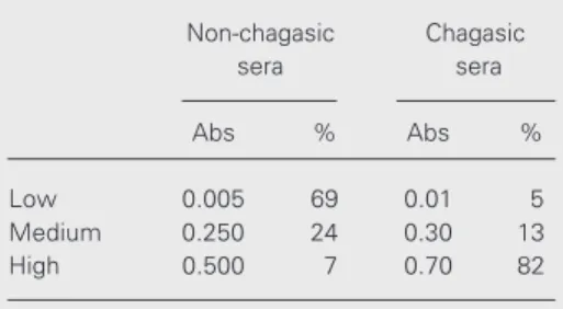
Table 1. Reactivity with Trypanosoma cruzi total extract of sera from patients with non-chagasic and chagasic heart disease.
Non-chagasic Chagasic sera sera
Abs % Abs %
Low 0.005 69 0.01 5 Medium 0.250 24 0.30 13 High 0.500 7 0.70 82
Eighty-five sera from non-chagasic patients and 133 sera from chagasic patients were tested by ELISA using T. cruzi antigen and their degree of reactivity (low, medium and high) was deter-mined. A chagasic serum with strong reactivity and normal human sera were used as positive and negative controls, respectively (data not shown). Mean absorbances (Abs) are given in the Table.
Table 2. Reactivity with HeLa human U small nuclear ribonucleoproteins (UsnRNPs) of sera from patients with non-chagasic and chagasic heart disease.
Non-chagasic Chagasic sera sera
Abs % Abs %
Low 0.04 32 0.05 27 Medium 0.12 56 0.15 68 High 0.18 12 0.25 5
results are shown in Table 1. As expected, among the non-chagasic cardiac sera, 69% showed low reactivity with T. cruzi antigen (average absorbance = 0.005), and only 7% of them showed cross-reactivity with this antigen (average absorbance = 0.5). The cut-off points for high, medium and low reactiv-ity were given in Material and Methods.
A set of 36 chagasic sera and 22 non-chagasic cardiac sera (medium and high re-activity) were analyzed by ELISA using HeLa UsnRNPs as antigen and the results are shown in Table 2. The absorbance values of non-chagasic sera were lower (see the mean ab-sorbance values annotated in the Table) than those obtained with chagasic sera, probably because infection with the parasite can raise autoantibody levels.
Similar results were previously obtained by Bach-Elias et al. (16), who reported 73% cross-reactivity of chagasic sera with HeLa UsnRNP antigens, thus supporting the idea of the presence of autoimmune antibodies in these individuals. However, it is interesting to note that non-chagasic cardiac sera, which showed high and medium reactivity with T. cruzi antigen, also reacted with HeLa UsnRNPs in 68% of cases. This may suggest that individuals with cardiac involvement of unknown etiology may somehow develop autoimmune antibodies, which could recog-nize some epitopes in the T. cruzi antigen.
Western blot analysis
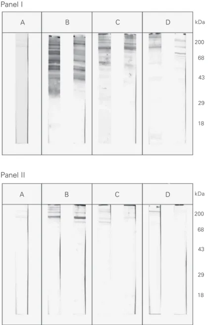
In an attempt to understand the ELISA results in Table 1 the same sera were also tested by Western blot containing T. cruzi total cell extract as antigen, which showed different patterns of reactivity. Two sera from each reactivity group were chosen to illus-trate the results presented in Figure 1, panel I (chagasic sera) and panel II (non-chagasic cardiac sera). These results demonstrate that non-chagasic cardiac sera only reacted with high molecular weight proteins, which are present in T. cruzi total extract, helping to
explain the lower absorbance values obtained by ELISA, as well as the cross-reactivity among these non-chagasic cardiac sera.
In order to determine the cross-reactivity between chagasic sera and human UsnRNPs, we analyzed all chagasic sera by Western blot using HeLa UsnRNPs as antigen and compared them with SLE serum as positive
Figure 1. Western blot of sera from patients with chagasic (Panel I) and non-chagasic (Panel II) heart disease using T. cruzi total cell extract. Two sera from each group were chosen to illustrate the results. A, Normal human serum. B, Sera with high reactivity to ELISA. C, Sera with medium reactivity to ELISA. D, Sera with low reactivity to ELISA. The molecular weights are indicated in kDa.
kDa
200
68
43
29
18 Panel I
Panel II
A B C D
A B C D kDa
200
68
43
29
(16,20). However, in the presentexperiments, the non-chagasic cardiac sera showed very faint bands similar to those for normal se-rum.
Immunoprecipitation assays
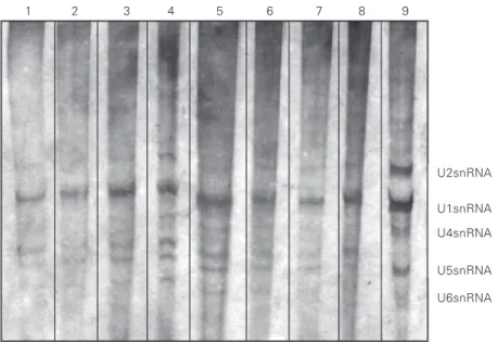
In an attempt to determine whether the previously tested antibodies were able to recognize human UsnRNA/RNPs, some sera were used in immunoprecipitation reactions with HeLa nuclear extracts, as described in Material and Methods. The results are shown in Figure 3 and demonstrate that chagasic sera, in fact, presented antibodies against different human UsnRNA/RNPs (Figure 3, lanes 3 through 6), while other sera, includ-ing the normal serum, reacted weakly with U1snRNP (Figure 3, lanes 7 and 8). The U1snRNA reactivity seems to better explain the cross-reactions obtained in ELISA using UsnRNPs as antigen and chagasic sera with higher absorbance values (Table 2) since these chagasic sera may have high affinity antibodies that can immunoprecipitate UsnRNPs. Anti-m3G antibody was used as a positive control to characterize the UsnRNPs in HeLa nuclear extracts (Figure 3, lane 9).
Discussion
Many of the specific host-parasite inter-actions in Chagas’ disease may explain the tissue lesions of the peripheral nervous sys-tem observed in acute or chronic disease (6). Many T. cruzi antigens are involved in chronic chagasic cardiopathy, such as FL-160-1, a surface protein associated with the 160 kDa flagellum which has an epitope that cross-reacts with the nervous tissue, and Gp85, a surface glycoprotein which reacts with chronic chagasic sera as well as with common glycolipid antigens present both in the mammalian nervous system and in T. cruzi trypomastigote forms (7,13,21,22). Experiments using molecular cloning have revealed that chronic chagasic cardiopathy
U2snRNA
U1snRNA
U4snRNA
U5snRNA
U6snRNA
1 2 3 4 5 6 7 8 9
Figure 3. Immunoprecipitation of HeLa nuclear extract by human sera. Human small nuclear RNAs (UsnRNAs) were analyzed in 10% urea/polyacrylamide gel/TBE [1X]. Lane 1, Normal human serum; lane 2, cardiac non-chagasic serum; lanes 3 through 8, chagasic sera; lane 9, anti-m3G antibody used as positive control to characterize the HeLa UsnRNAs.
B/B’ A
C
D3 D2
D1
1 2 3 4
Figure 2. Western blot of serum from a patient with chagasic heart disease (lane 2 - high degree of reactivity in Figure 1, Panel I) and from a patient with non-chagasic heart disease (lane 3 - high degree of reactivity in Figure 1, Panel II) using HeLa UsnRNPs. Lane 1, Serum from a patient with systemic lupus erythematosus as positive control; lane 4, normal human serum. The typical human Sm proteins (A, B/B’, C, D1, D2, D3) are indicated.
patients present a strong humoral response against C-terminal regions of four cloned P ribosomal proteins, TcP1, TcP2, TcPJL5 and TcPo, which may be important in Chagas’ disease immunopathology (10). Moreover, chronic serum antibodies recognized an an-tigen (43-45 kDa) in normal mouse heart and skeletal muscle tissue, suggesting that this glycoprotein may be a target for autoanti-bodies in Chagas’ disease (11).
Fatenejad et al. (23) showed that the au-toimmune response to snRNPs and possibly to other autoantigens in lupus is a specific reaction similar to that seen in a typical immune response to foreign antigens. The presence of autoantibodies has always indi-cated a compliindi-cated immune reaction, which cannot simply be explained by a direct par-ticipation of the target antigen. Several T. cruzi antigens present cross-reactivity with human tissues. Nevertheless, almost nothing has been reported thus far with respect to UsnRNPs. Studies carried out by Miatello and Fiorotto (24) demonstrated the exist-ence of cross-reactivity between antibodies directed against the ribosomal antigen of the parasite and normal myocardial tissue of mice and rabbits after experimental infec-tion or immunizainfec-tion with T. cruzi, thus suggesting the presence of infection-induced autoantibodies. Skeiky et al. (12) suggested that T. cruzi P proteins must contribute to the development of autoantibodies in chagasic patients and the appearance of anti-P au-toantibodies may be explained by a similar mechanism in Chagas’ disease and SLE. Solana et al. (25) demonstrated that chagasic patient sera reacted with human factors and parasite ribosomal antigens by ELISA and Western blot, although they did not present cross-reactivity with sera from patients with other infections or autoimmune diseases.
The present study confirmed the pres-ence of autoantibodies in chagasic patients against heterologous UsnRNPs, which could be a consequence of epitope spreading from the initial parasite epitope. Experiments
us-ing ELISA have indicated that it is easier to detect the presence of these autoantibodies by this methodology; however, they are dif-ficult to visualize by immunoprecipitation assays. Interestingly, non-chagasic cardiac sera showed high and medium reactivity with T. cruzi antigen (lower absorbance) and also reacted with HeLa UsnRNPs (Tables 1 and 2). This suggests that individuals with heart disease of unknown etiology may de-velop autoimmune antibodies at any time, which could cross-react with T. cruzi. The Western blotting results showed that these non-chagasic cardiac sera reacted mainly with higher molecular weight T. cruzi epi-topes (Figure 1, panel II), but presented faint bands with HeLa UsnRNPs as well as nor-mal sera (Figure 2, lanes 3 and 4). The explanation for these findings is still un-known.
Since two primary hypotheses are pro-posed to account for pathogenesis in chronic T. cruzi infections (the persistence of the parasites at specific sites in the infected hosts causes chronic inflammatory reactivity and T. cruzi infection induces immune responses which are targeted at self tissues), our results support this autoimmune etiology, defining another putative autoantigen like snRNPs, but also argue in favor of the idea that Chagas’ disease must be treated as a parasitic disease with efforts to enhance effective immune responses and reduce the parasite load. This is supported by the results with non-cardiac chagasic sera.
the ß1-adrenergic receptors. This fact was attributed to a certain homology existing between the R13 peptide of the parasite and an epitope of the second extracellular loop of the ß1-adrenergic receptor.
Bach-Elias et al. (16) analyzed in chagas-ic sera the presence of autoantibodies di-rected against some fairly known autogens, Sm and RNP (here named anti-UsnRNPs), which are typical of some au-toimmune rheumatic diseases such as SLE and mixed connective tissue disease. The main reason to use these antigens was the fact that an external agent such as a retrovirus may initially be responsible for the develop-ment of anti-UsnRNP antibodies, mainly through the activation of antiviral antibody synthesis. These antibodies cross-react with a mimicry region in host UsnRNPs and may therefore trigger the onset of an autoimmune process. Three similar characteristics be-tween Chagas’ disease and some
autoim-mune diseases such SLE may be mentioned: a) similar etiology, which means that an external agent may initiate an autoimmune process, b) presence of antibodies which recognize host proteins as foreign, and c) a chronic and generalized autoimmune pro-cess.
Then, the present results give more sup-port to the previous explanations, since the autoantibodies in chronic chagasic patients were demonstrated here by using different methodologies. Similar studies using inbred mouse strains chronically infected with T. cruzi have been done by our group (Ambrósio DL, Castro LIR and Cicarelli RMB).
Acknowledgments
We thank Dr. Montserrat Bach-Elias (CSIC, Barcelona, Spain) for providing the UsnRNPs from HeLa nuclear extracts.
References
1. Szarfman A, Terranova VP, Rennard SI, Foidart JM, de Fátima Lima M, Scheinman JI & Martin GR (1982). Antibodies to laminin in Chagas’ disease. Journal of Experimental Medicine, 155: 1161-1171.
2. Acosta AM, Sadigursky M & Santos-Buch CA (1983). Anti-striated muscle antibody activity produced by Trypanosoma cruzi. Proceed-ings of the Society for Experimental Biology and Medicine, 172: 364-369.
3. Schmunis GA (1987). Autoimmunity in Chagas’ disease. Memórias do Instituto Oswaldo Cruz, 82 (Suppl 1): 287-310.
4. Levin MJ, Mesri E, Benarous R, Levitus G, Schijman A, Levy-Yeuati P, Chiale PA, Ruiz AM, Kahn A & Rosenbaum MB (1989). Identifica-tion of a major Trypanosoma cruzi antigenic determinant in chronic Chagas’ disease. American Journal of Tropical Medicine and Hy-giene, 41: 530-538.
5. Sepulveda P, Liegeard P, Wallukat G, Levin MJ & Hontebeyrie M (2000). Modulation of cardiocyte functional activity by antibodies against Trypanosoma cruzi ribosomal P2 protein C terminus. Infec-tion and Immunity, 68: 5114-5119.
6. Bonfa E & Elkon KB (1986). Clinical and serological associations of the anti-ribosomal P protein antibody. Arthritis and Rheumatism, 29: 981-985.
7. Avila JL (1992). Molecular mimicry between Trypanosoma cruzi and host nervous tissues. Acta Cientifica Venezolana, 43: 330-340. 8. Eisen H & Kahn S (1991). Mimicry in Trypanosoma cruzi: fantasy
and reality. Current Opinion in Immunology, 3: 507-510.
9. Grauert MR, Houndayer M & Hontebeyrie-Josckowiciz M (1993).
Trypanosoma cruzi infection enhances polyreactive antibody re-sponse in an acute case of human Chagas’ disease. Clinical and Experimental Immunology, 93: 85-92.
10. Levin MJ (1993). Humoral autoimmune response in Chagas’ dis-ease: Trypanosoma cruzi ribosomal antigens as immunizing agents.
FEMS Immunology and Medical Microbiology, 7: 205-210. 11. McCormick TS & Rowland EC (1993). Trypanosoma cruzi:
recogni-tion of a 43 kDa muscle glycoprotein by antibodies present during murine infection. Experimental Parasitology, 77: 273-281. 12. Skeiky YA, Benson DR, Parsons M, Elkon KB & Reed SG (1992).
Cloning and expression of Trypanosoma cruzi ribosomal protein PO and epitope analysis of anti-PO autoantibodies in Chagas’ disease patients. Journal of Experimental Medicine, 176: 201-211. 13. van Voorhis WC, Barrett L, Koelling R & Farr AG (1993). FL-160
proteins of Trypanosoma cruzi are expressed from a multigene family and contain two distinct epitopes that mimic nervous tis-sues. Journal of Experimental Medicine, 178: 681-694.
14. Teixeira AR, Teixeira ML & Santos-Buch CA (1975). The immunol-ogy of experimental Chagas’ disease. IV. Production of lesions in rabbits similar to those of chronic Chagas’ disease in man. Ameri-can Journal of Pathology, 80: 163-180.
Try-panosoma cruzi antigen. Proceedings of the National Academy of Sciences, USA, 92: 3541-3545.
16. Bach-Elias M, Bahia D, Teixeira DC & Cicarelli RMB (1998). Pres-ence of autoantibodies against small nuclear ribonucleoprotein epi-topes in Chagas’ patients’ sera. Parasitology Research, 84: 796-799.
17. Dignam JD, Lebovitz RM & Roeder RG (1983). Accurate transcrip-tion initiatranscrip-tion by RNA polymerase II in a soluble extract from iso-lated mammalian nuclei. Nucleic Acids Research, 1: 1475-1489. 18. Towbin H, Stachelin T & Gordon J (1992). Electrophoretic transfer of
proteins from polyacrylamide gels to nitrocellulose sheets: proce-dure and some applications. Biotechnology, 24: 145-149.
19. Steitz JA (1989). Immunoprecipitation of ribonucleoproteins using autoantibodies. Methods in Enzymology, 180: 468-481.
20. Cicarelli RMB, Castro LIR, Martins EC, Cegatti MB, Bahia D & Bach-Elias M (1998). Spliceosomal factors are recognized by chagasic sera. International Proceedings Division, Monduzzi Editore - 10th International Congress of Immunology, November 1-6, 1998. New Delhi, India, 1141-1145.
21. Travassos LR & Almeida IC (1993). Carbohydrate immunity in
Ameri-can trypanosomiasis. Springer Seminars in Immunopathology, 15: 183-204.
22. Gea S, Ordonez P, Cerban F, Iosa D, Chizzolini C & Vottero-Cima E (1993). Chagas’ disease cardioneuropathy: association of anti- Try-panosoma cruzi and anti-sciatic nerve antibodies. American Journal of Tropical Medicine and Hygiene, 49: 581-588.
23. Fatenejad S, Bennett M, Moslehi J & Craft J (1998). Influence of antigen organization on the development of lupus autoantibodies.
Arthritis and Rheumatism, 41: 603-612.
24. Miatello CS & Fiorotto ER (1989). Ribosomal antibody response in rabbits and mice infected with Trypanosoma cruzi.Revista Argen-tina de Microbiologia, 21: 141-145.
25. Solana ME, Katsin AM, Umenzawa ES & Miatello CS (1995). High specificity of Trypanosoma cruzi epimastigotes ribonucleoprotein as antigen in serodiagnosis of Chagas’ disease. Journal of Clinical Microbiology, 33: 1456-1460.