Acta bot. bras. 14(1): 57-69. 2000 57
ANATONUCALANDULTRASTRUCTURALASPECTSOFLEAFGALLSIN
FICUS MICROCARPA L. F. (MORACEAE) INDUCED BY
GYNAIKOTHRIPS FICORUMMARCHAL (THYSANOPTERA)
Recebido em 21/05/1999. Aceito em 16/12/1999
Silvana Cristina P. M. de Souzal
Jane Elizabeth Kraus2 Rosy Mary S. Isaias3 Lea de Jesus Neves4
RESUMO - (Aspectos anatômicos e ultra-estruturais de galhas foliares em Ficus microcarpa L.f.
(Moraceae) causadas por Gynaikothdpsficorum MarchaI (Thysanoptera)). Galhas foliares são comuns em diversas espécies de Ficus. O objetivo deste trabalho é estudar as alterações estruturais envolvidas na formação das galhas foliares induzidas por Gynaikothrips ficorum (Thysanoptera) em Ficus microcarpa, uma planta ornamental. As galhas de folhas jovens e maduras foram separadas em dois estádios de desenvolvimento, com base na presença de lesões e no grau de dobramento da lâmina. Durante o desenvolvimento da galha formam-se intumescimentos na lâmina tanto nas folhas jovens quanto nas maduras, os quais coincidem com as áreas de hipertrofia celular e hiperplasia dos tecidos. Estes intumescimentos foram detectados em maior quantidade e mais precocemente em folhas jovens, que, por possuirem células menos diferenciadas, mostraram respostas cecidogenéticas mais rápidas. A hipertrofia e, a hiperplasia foram os processos marcantes na ontogênese desta galha, assim como ocorre em outras induzidas por tripes. Entretanto, apesar dos numerosos sítios de alimentação e a grande área de ataque, as galhas foliares de F. microcarpa podem ser consideradas rudimentares, pois não se observa a diferenciação de novos tecidos.
Palavras-chave - galha foliar, tripes, Ficlls microcarpa, Moraceae, anatomia
ABSTRACT - (Anatomical and ultrastructural aspects of leaf galls in Ficus microcarpa L.f. (Moraceae) induced by Gynaikothdps ficorum MarchaI (Thysanoptera)). Several species of Ficus present leaf galls and the goal of this research is to study the structural alterations involved in the formation of leaf galls caused by Gynaikothrips ficorum on F. microcarpa, an ornamental plant. The galls of young and mature leaves were separated into two developmental stages based on the presence of lesions on leaf lamina and
I Scholarship of the PIBIC-CNPq, e-mail: [email protected]
2 Instituto de Biociências da Universidade de São Paulo, Departamento de Botânica, C. Postal 11461, CEP 05422-970, São
Paulo, SP, Brasil, e-mail: [email protected] (requests for offprints)
3 Instituto de Ciências Biológicas da Universidade Federal de Minas Gerais, Departamento de Botânica, C. Postal 486, CEP
31270-110, Belo Horizonte, MG, Brasil, e-mail [email protected]
4 Museu Nacional da Universidade Federal do Rio de Janeiro, Departamento de Botânica, Quinta da Boa Vista, São Cristóvão,
58 Souza, Kraus, Isaias & Neves: Anatomical and ultrastructural aspects of leaf galls in Fir us lIlicrocarpa L.f.
the degree of leaf folding, Swellings of the lamina were observed in young and mature leaves during gall development which coincided with the areas of cellular hypertrophy and tissue hyperplasia. Swellings were detected in a greater amount and more precociously on young leaves when compared to mature ones . In young leaves, the cecidogenetic responses were quicker and led to further structural differences because younger cells are not completely differentiated. Cell hypertrophy and tissue hyperplasia were striking processes involved in the ontogenesis of the studied gall, similar to other galls induced by thrips. Nevertheless , in spite of the numerous sites of feeding and the wide area of attack, F. microcarpa galls can be considered rudimentary, since no new tissue differentiation was observed.
Key words - leaf gall, thrips, Ficus microcarpa, Moraceae, anatomy
Introduction
Several Ficus species present leaf galls. Condit (1969) reported t~e presence of galls in four species of Ficus, while Mani (1964) reported the existence of leaf galls in 11 such species. Both authors inc1uded Ficus microcarpa in their lists. Neves & Isaias (1987) ftrst reported the presence of gall inducing insects on the flowers of F. microcarpa in Brazil. Other studies
on Ficus inc1ude ontogenetical studies on galls
of F. religiosa (Raman & Gopinathan 1987) and
F. tomentella (Neves 1987).
Ficus microcarpa has been misidentifted
many times as F. retusaand F. benjamlÍza(Mello
Filho et al1983), probably because oftheir very similar vegetative characteristics. This species is very widely used for urban landscaping in Brazil, and infestation by Gynaikothripsftcorum (Thysanoptera) has resuted in the death of many specimens (Mello Filho et aI. 1983). Even though infestation is currently less intense, it has not been eradicated fram major urban centers, such as São Paulo, São Caetano do Sul, and severa I towns in the state ofRio de Janeiro. The development of insect galls is dependent upon the continuous presence of the insect, which through mechanical and chemical stimuli induces the plant cells to differentiate in an abnormal way (Mani 1964; Meyer & Maresquelle 1983), causing severa I degrees of alteration in the host organ. From an ecological viewpoint , galls are adaptations that have allowed galling organisms to colonize and radiate in several different habitats (Price et al
1986, 1987; Fernandes & Price 1988).
Thrip induced galls are usually leaf galls simpler than those caused by dipterans or hymenopterans (Mani 1964) and relatively are poor1y known (Ananthakrishnan 1992). The understanding of the factors that are cause plant tissues to change anatomically and morphologically may shed some light on the intimate biological association between gall inducing agents and host plants. The goal of this research is to study the structural alterations and reactions of plant tissues involved in the formation of the leaf gall induced by G. ftcorum
on F. microcarpa.
Material and methods
Young and mature healthy and galled leaves
of Ficus microcarpa were collected in the
Campus of the Universidade de São Paulo (São Paulo, Brazil) and in Quinta da Boa Vista (Rio de Janeiro, Brazil). Leaves from both sites were separated into young and mature \eaves based on their location on the branches, size and texture. The position of the leaf was evaluated based on its insertion in the node, considering that the ftrst node was that located next to the stem apex. The length and width of 50 leaves from each site were measured . Galls were separated into two developmental stages based on the presence of lesions on the leaf lamina, and the degree of leaf folding.
Acta boI. bras. 14(1): 57-69. 2000
dehydration in tert-butyl alcohol series, and included in paraffin, according to the methods described in Kraus & Arduin (1997). Transverse serial sections - IOllm thick - were stained in basic fuchsin and astra blue (Kraus et aI. 1998). For the preparation of epidermal peels, fresh leaves where cut into small pieces, and heated in 50% nitric acid (Ghouse & Yunus 1972). In
addition the epidermal peels were stained in basic fuchsin and astra blue (Kraus et aI. 1998). Phenolic substances were detected by 10% ferric chloride (Johansen 1940).
Scanning electronic microscopy studies (SEM) were prepared according to methods described in Silveira (1989). The samples were fixed in FAA, dehydrated in ethyl alcohol series, dried at the criticaI point with CO2 , and covered
with gold.
59
Results
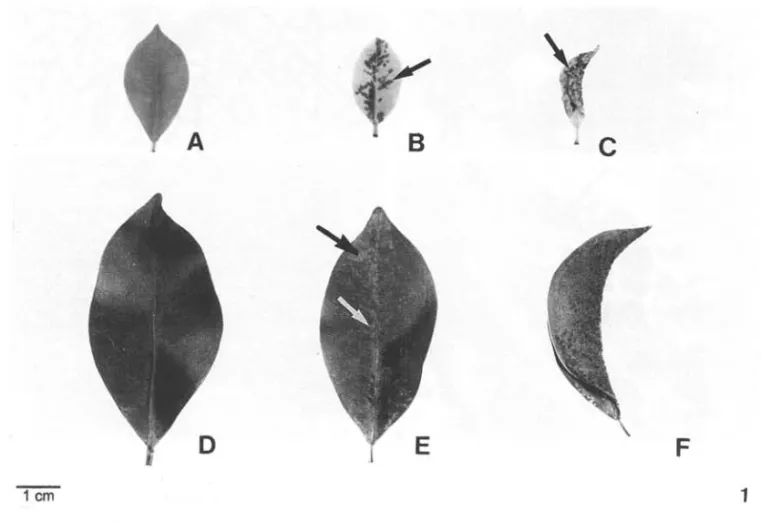
Hea1thy and galled young leaves - Young hea1thy leaves presented a light green color, membranous texture and were on an average 1.9 ± 0.5cm long and 1,1 ± 0.3cm wide (Fig. IA). Trichomes were present on both leaf surfaces. Fig. 2 shows a trichome on the adaxial surface. Stomata were sparse and only observed on the abaxial surface (Fig. 3).
In the rnidrib region, the 2-3layered adaxial epiderrnis was covered by a thin cuticle. The internaI epidermal layer presented larger cells. The parenchyma presented 4-5 layers of cells next to the adaxial portion of the vascular system, which had an elliptical shape and consisted of primary xylem and phloem. The pith was distinct and located inside the vascular
· C
F
1
Figure. I. Ficus microcarpa. A. You~g and healthy leaf; B. Young leaf showing the early stage of gall development. Note the leslOns (arrow); C. Young leaf wlth mature fold gall. Note the lesions (arrow); D. Mature and healthy leaf; E. Mature leaf showmg the early stage of gall development. Note the lesions concentrated next to the middle vein (arrows)' F
60 Souza, Kraus, Isaias & Neves: Anatomical and ultrastructural aspects of leaf galls in Ficlls microcarpa L.f.
Acta 001. bras. 14(1): 57-69. 2000
system. Several parenchyma cells, full of phenolic compounds, were interspersed among the conductive cells. The pericycle had 2-3 cell layers, while the endodermis was indistinct. Next to the abaxial side of the vascular system 4-6 parenchyma layers and 3-4 angular collenchyma layers were present. Phenolic compounds were detected in some cells of the pith and cortical parenchyma. The epidermis on the abaxial surface was uni-stratified, with partially differentiated cells, which were slightly smaller when compared to those occurring on the adaxial surface. The cuticle was relatively thin and similar to that on the adaxial surface (Fig. 8).
In the lateral vein region, the epidermis was 2-3 layered, covered by a thin cuticle. The cells were larger than those on the midrib. The inner epidermal layer presented larger cells. A large amount of cystoliths were observed, mainly on the adaxial side of the lamina. The partially differentiated mesophyll was made up of nine celllayers, the frrst with anticlinally elongated cells. Cells with phenolic compounds were frequent. Procambial cells constitute the vascular bundles, where few tracheary elements were differentiated. The endodermis was already differentiated at this stage of leaf development and involved the minor and the major bundles. The pericycle was distinct just involving the larger bundles. On the abaxial side of the lamina, the epidermis was 2-layered and possessed few stomata. Some stomata were not totally differentiated. The cuticle was similar to that on the adaxial surface (Fig. 9).
The early stages of gall development were characterized by the fully expanded leaflamina. Small reddish to brownish spots, due to G.
ficorum induction, were visible on the adaxial
and abaxial surfaces of the leaf lamina. These small spots were generally concentrated next to the midrib when the infestation was intense (Fig. 1B). Figures 4 and 5 show the lesions on both surfaces. The epidermal cells around the lesions accumulate phenolic compounds. Stomata on the abaxial surface were numerous and
61
differentiated. In some cases, however, stomata development is interrupted, ostiole and guard cell are not formed anel/or the subsidiary cells become deformed (Fig. 5).
In the midrib region, the cuticle was thin but somewhat thicker than on the healthy leaf. The epiderrnis on the abaxial surface showed a higher degree of differentiation, the cells possessing convex periclinal externai walls. There was some inhibition in the development of vascular tissues, and at the lateral parenchyma portions a few slightly hypertrophied cells were observed. As a consequence, the midrib region was not convex (Fig. 10). In the region of lateral veins, the tracheary elements of the major veins were differentiated. The mesophyll had more layers (14), with some hypertrophied cells, which generally had phenolic compounds. Because of the hypertrophy of the cells and hyperplasia of the tissues, swelled areas were observed in the lamina (Fig. 11).
The mature gall was characterized by the complete folding of the lamina (Fig. 1C). This folding always occurred next to and along the midrib, and, therefore, half of the adaxial surface was either totally or partially covering the other half. Thus, the larval chamber was formed having permanent communication with the externai environment. The majority of the stomata on the abaxial surface showed structural alterations similar to an early stage of gall development and abnormal distribution (Fig. 7). The epidermis had perforations caused by
Gynaikothrips mouthparts (Fig. 6). The midrib
62 Souza, Kraus, Isaias & Neves: AnatomicaI and ultrastructural aspects of leaf galls in ncus miCrocorpo L.f.
Acta boI. bras. 14(1): 57-69. 2000
surface, alterations were more conspicuous, especially cell hypertrophy (Fig. 13). Similar to the preceding stage, leaf lamina exhibited several swollen areas, probably due to the action of the galling insect.
In general, during gall development, leaf lamina became thicker and in mature galls there was a larger number of injured areas when compared to those in the initial stage of development. That half of the lamina which folded over the other lamina, presented a larger concentration of injured areas.
Healthy and galled mature leaves - Mature and healthy leaves showed a dark green color, coriaceous texture, and were on an average 5.8 ± 3.1cm long and 3.1 ± O,4cm wide (Fig. lD). There were conspicuous differences between the adaxial and the abaxial side of the lamina when compared to the young healthy leaf. Trichomes were scarce ar absent on both lamina surfaces (Fig. 14 and 15). Figure 15 shows a largerdensity of differentiated stomata and a trichome on the abaxial surface. The subsidiary cells were situated above the guard cells, and presented cuticular projections which covered the guard cells partially (Fig. 16).
In the midrib region of the mature healthy leaf, the epidermis on the adaxial surface was 2-3 layered, similar to the young leaf. Nevertheless, the epidermis of mature leaves was covered by a thicker cuticle, 114 the height of the cell. The inner layer had larger cells. Below the epidermis, there were 2-3 layers of small parenchyma cells, which were continuous up to the palisade parenchyma. About 3-4 layers of lignified pericyclic fibers involved the vascular system, which was composed of primary and secondary xylem and phloem, and cambium. The vascular system presented a round shape and was formed by two arcs, one abaxially placed, which was larger and continuous, and a smaller one, adaxially placed. Below the vascular system, there were three layers of parenchyma cells and 3-4 layers of angular
63
collenchyma cells. The endodermis was indistinct. On the abaxial surface, the cuticle was thicker than that on the adaxial surface. The epidermis was uni-stratified and the cells had convex periclinal walls (Fig. 21).
The epidermis covered the region of the lateral veins with 2-3 layers in the adaxial surface of the lamina. Epidermal cells were larger than those on the midrib region. The palisade parenchyma was 2-layered. The first layer had anticlinally elongated cells, with few intercellular spaces; the cells of the second layer were shorter and presented small intercellular spaces. Five to seven cell layers with larger intercellular· spaces formed the spongy parenchyma; the cells of the inner layer were more closely disposed. The epidermis on the abaxial side of the lamina had two layers and a higher density of stomata (Fig. 22). In the major veins, the cells of the endodermis were sclerified and larger than those on the minor veins. Bundle sheath extensions with sclerified cells were present.
The early stages of gall development were characterized by fully expanded lamin~, with small reddish to brownish spots on the abaxial (Fig. lE) and adaxial surfaces. Even though these spots were visible, they were less distinct when compared to the young leaves, because of the dark green color of the lamina. The spots tended to concentrate next to the midrib when the degree of infestation had increased, similar to what is found in young leaves. Figures 17 and 18 show the lesions which accumulated phenolic compounds on the adjacent cells. The cell walls of the adaxial surface are thicker (Fig. 17). Altered stomata are also present on the abaxial surface (Fig. 18).
64 Souza, Kraus, Isaias & Neves: Anatorrllcal and ultrastructural aspects of leaf galls in FtClIS II/Icrocorpo L.f.
Acta boI. bras. 14(1): 57-69. 2000 65
66 Souza, Kraus, Isaias & Neves: Anatomical and ultrastructural aspects of leaf galls in FiclIs microcarpa LJ.
the lateral vein region, the mesophyll showed hyperplasia and some of its cells were hypertrophied, leading to the reduction of intercellular spaces (Fig. 24). The swellings of the leaflamina on mature galled leaves were less evident when compared to those of young ones. Galls in the mature stage were characterized by the folded leaf along the midrib forming the larval chamber, similar to what was observed in the young leaf (Fig. lF). Galls were very hard but broke easily. On the abaxial surface the swollen areas were evident and the stomata presented morphological modifications (Fig. 19). An altered stoma is seen in Figure 20.
In the midrib region of the mature galled leaf, the vascular system did not show great alterations when compared to the preceding stage. Parenchyma cells showed some hypertrophy, and most of the pericyciic fibers were not lignified. Cells with phenolic compounds were present (Fig. 25). Because of hyperplasia and hypertrophy next to the midrib, the rib was not formed. In the lateral vein region, cell hypertrophy was concentrated on the abaxial side of the lamina while tissue hyperplasia was concentrated on the adaxial side. Both phenomena were intensified in this region. On the adaxial side, cells with phenolic compounds were more common (Fig. 26). The swollen areas on the leaf surface were conspicuous.
During gall development leaf thickening occurred, similar to what was observed on young leaves. Areas with all the morphological alterations already described were more numerous on the mature galls, and were more concentrated on the folded portions next to the midrib.
Discussion
Mound e/ ai (1980) referred to at least 6.000 known species of thrips in the world, and stated that the majority occurred in the tropics. Many species have received attention because they are pests on plants of economical interest (Ananthakrishnan 1992). According to Carrera
(1967), there are several species ofThysanoptera that produced galls in several plants, such as
Gynaiko/hrips jicorum, which infested Ficus
microcarpa. In Brazil, the first study describing
the attack by G jicorum on Ficus was that of Amante & Almeida (1962), which erroneously identified the plant species as F. re/usavar. nítida Thunb. Meyer (1987) also stated that Gjicorum is the galling agent in F. re/usa, and calls attention to the importance ofthis insect because of its relatively large geographic distribution in Marrocos, Argelia, Israel, India and Malaysia. All the information available on this species was that the small insects made the thickening and folding of the leaf lamina next to the midrib, and formed a larval chamber, where they were found at different developmental stages (Meyer 1987).
In this study, we have confirmed that G
jicorum attacks young and mature leaves of F.
microcarpa. Rohfritsch (1992) stated that young
tissues are more sensitive to the induction than mature tissues. Therefore, the author conc1uded that it was an error to generalize that only meristematic or young tissues react to gall inducer attacks. The studied galling species is an example of this processo
The early stages in the development of G
j'icorum galls on F. microcarpa were
Acta bot. bras. 14(1): 57-69. 2000
(Mani 1964; Meyer & Maresquelle 1983; Hori 1992).
In young leaves of F. microcarpa the vascular system was differentiated just in the midrib region, where the lignified pericyclic fibers were absent. Generally, lignification of the tissues was incipient, contributing to the flexibility of the leaf lamina. In contrast, the presence oflignified pericyclic fibers around the vascular tissues of the mature leaf might contribute to its rigidity.
An anatomical analyses of the mature healthy leaf of F. microcarpa showed many similarities to the previous observations made by Mello Filho et aI. (1983). These authors considered the leaves as glabrous, nevertheless, our SEM observations evidenced the presence of some trichomes on the adaxial and abaxial surfaces of mature leaves, and their conspicuous presence on young leaves, indicating that during organogenesis they were lost. Mello Filho et aI. (1983) refer to a hypodermis and a simple epidermis on the abaxial surface, but did not describe them. Here we observed that the abaxial epidermis on the region of the midrib is uni-stratified, but becomes bi-stratified in the region of the lateral veins. Thus, the layer considered hypodermis is, in reality, an epidermallayer.
Meyer (1987) described that very often the epidermis on galls caused by thysanopterans became necrotic and disintegrated. In F.
microcarpa galls, the disintegrated and necrotic
areas were observed which might be considered cell reactions to the feeding activity of G.
ficorum.
In galls, the swollen areas of the lamina observed in young and mature leaves coincided with the areas of cellular hypertrophy and tissue hyperplasia. These swellings were detected in a larger amount and more precociously in young leaves. In young leaves, the presence of less differentiated cells, possibly allowed a greater number of cell divisions and, thus, the swellings might in part be due to this characteristic. Moreover, the minor quantity of cells with
67
lignified walls in young leaves might also have favored cell hypertrophy.
In young healthy leaves, few differentiated stomata were observed in the epidermis, differing from the tissues of the gall, where they were already differentiated, more numerous and with structural alterations. Thus, cecidogenesis induced a quicker differentiation of stomata, and also altered the normal development of the guard and subsidiary cells. In the mature galled leaf, stomata predominantly presented less alteration, due to the stomata having already been differentiated before the insect attack. On young and adults galled leaf stomata distribution is also modified. Meyer & Maresquelle (1983) described that epidermal cells on the abaxial surface of the galled leaf as being hypertrophied and the stomata less numerous. These authors, therefore, did not define whether the leaves were young or mature. In F. microcarpa gall, the smaller number of stomata observed on some regions could be caused by the greater distance of the cells as an consequence of cell hyperplasia or hypertrophy of the non-specialized epidermal cells.
Leaf folding causing the formation of the larval chamber in mature galls, even in young or mature leaves, might be due to the more pronounced structural alterations in this stage. As the majority of insect attacks occur near the midrib, leaf folding in this region may be a consequence of this attack. In this gall there generally were no alterations in the leaf margin. In the affected region, cell hypertrophy was more conspicuous on the abaxial portion of the leaf, causing leaf folding. At this stage of gall development, the mesophyll was totally disorganized, being compact, without intercellular spaces, with numerous hypertrophied cells containing a great amount of phenolic compounds.
68 Souza, Kraus, Isaias & Neves: AnatomicaI and ultrastructuraI aspects of Ieaf gaIls in ncus microcorpo L.f.
etal. 1991; Arduin & Kraus 1995; Kraus etal.
1996). The presence of phenolic compounds is known as a defense against herbivory (Levin 1971; Feeny 1976; Harborne 1990) and it rnight be proposed that these substances should basically favor the inducer against parasitoids and predators or against fungi and microorganisms infestation (Cornell 1983; Rhoades 1985). Besides their defensive role, phenolic compounds are also involved in hormonal regulation. The presence of these compounds should inhibit indoleacetic acid oxidases (IAA-oxidases), increasing the auxin action responsible for cell hypertrophy (Bhansali
et aI. 1978; Hori 1992). The quantification of
phenolics, auxin and related enzymes in the galls
of F. microcarpa has not been studied yet.
The absence of lignified cells, as well as pericyclic fibers in mature galls formed either in young or mature leaves rnight involve different processes. In young leaves the results indicated that there was an inhibition of the differentiation of the pericyclic fibers, while in mature leaves there might be the degradation of the lignified wall. According to Herrns & Mattson (1992), phenilalanin is the limiting precursor for the synthesis of such phenilpropanoids as lignin, flavonoids, and condensed tannins, indicating that these were derived from the same metabolic route. In young leaves, we propose that there was a prioritization for the forrnation of soluble phenolic compounds in detriment of lignin production. Otherwise, in mature leaves, the degradation of lignin in the wall of pericycle derivatives is unexplained. Therefore, more detailed studies are required to explain our hypothesis. On the other hand, it is known that there are mechanical and chernical investments in defensive compounds in galls (Corne1l1983) and so, the presence of a great quantity of phenolics should corroborate the chemical investment of F. microcarpa gall.
The fact that the tissues of young and mature leaves responded to the stimuli of G. ficorum is an indication that these tissues, independent of
the stage of development, acquired competence for cecidogenesis. In young leaves of F.
microcarpa, cecidogenetic responses were faster
and led to more structural differences since the cells were not completely differentiated. Moreover, it should be taken into consideration that the development of the gall was related not only to the stage of development of the host organ, but also to the number of insects present, sites of feeding, etc., as was proposed by Ananthakrishnan (1980 apudRohfritsch 1992).
G. ficorum might use young as well as
mature tissues of F. microcarpa for its feeding and oviposition activities, indicating a high leveI of interaction between host and parasite. The structural modifications in leaf tissues resulted in galls that were very simple and where cell hypertrophy and tissue hyperplasia were involved in their morphogenesis. These two processes were generally observed, in the formation of some other galls caused by thrips (Rohfritsch 1992; Ananthakrishnan 1992). F.
microcarpa gall study was also relevant because
the cecidogenetic action by G. ficorum was extensive, and involved the entire leaf larnina. Nevertheless, in spite of the numerous sites of feeding and the wide area of attack, F.
microcarpa galls can be considered rudimentary
as no new tissue differentiation was observed.
Acknowledgments
This investigation was supported by CNPq (301776/83 and PIBIC NIl08362/95-7) and CAPES/ PICDT research grants.
References
Amante, E. & Almeida, E. 1962. Insetos que ocorrem em
FiClIS re/usa e F1cus benjamina. Arquivos do Instituto
Biológico 29: 93 -lO 1.
Ananthakrishnan, T. N. 1992. Uni que aspects in the biology of thrips-induced galls. Pp. 185-195. In: 1. D . Shorthouse & O. Rohfritsch (Eds.), Biology
ofinsect-induced galls. Oxford University Press, New York.
Arduin, M .; Kraus, J. E. & Venturelli , M . 1991. Estudo morfológico de galha achatada em folha de
S/ru/han/hus vulgaris Mart. (Loranthaceae). Revista
Acta bot. bras. 14(1): 57-69. 2000
Arduin, M. & Kraus, 1. E. 1995. Anatomia e ontogenia de
galhas foliares de Piptadenia gonoacantha (Fabales,
Mimosaceae). Boletim de Botânica, Universidade de
São Paulo 14: 109-130.
Bhansali , R. R.; Kumar, A. & Arya, H. C. 1978.
Polyphenols and related enzymes in normal and gall
tissues of Ficus tnysorensis Heyne. Indian Journal of
Experimental Biology 16: 850-851 .
Carrera, M. 1967. (Ed.) Ordem dos Tisanópteros. pp.
54-55. In: Entomologia para você. 3'. ed . EDART
Livraria Editora Ltda., São Paulo.
Condit, I. 1. 1969. Ficus: the exotic species. University of
California Press, Arcadia.
Cornell, H. V. 1983. The secondary chemistry and complex
morphology of galIs formed by Cynipidae
(Hymenoptera): why and how? The American
Midland Naturalist 110: 225-234.
Feeny, P. 1976. Plant apparency and chemical defense. Pp.
1-40. In: G. W WalIace & R. L. ManselI (Eds.),
Biochemical interaction between plaots aod insects.
Plenum Press, New York.
Fernandes, G. W & Price, P. W 1988. Biogeographical
gradients in gaJling species richness: tests of
hypotheses: Oecologia 76: 161-167.
Ghouse, A K. M. & Yunus, M. 1972. Preparation of
epidermal peels from leaves of gymnosperms by
treatment with hot, 60% HN03• Stain Technology 47:
322-324.
Harborne , 1.B . 1990. Constraints on the evolution of biochemical pathways. Biological Journal ofLinnean Society 39: 135-151.
Herms, D. A. & Mattson, W 1. 1992. The dilemma of
plants: to grow or defendo The Quarterly Review of Biology 67: 283-335.
Hori, K. 1992. Insect secretions and their effect on plant
growth, with special reference to hemipterans. pp.
157-170. In: 1. D. Shorthouse & O. Rohfritsch (Eds.),
Biology or insect-induced galls. Oxford University Press, New York.
10hansen, D. A. 1940. Plaot microtechnique. McGraw-Hill Book Co, New York.
Kraus, J. E.; Sigiura, H. C. & Cutrupi, S. 1996. Morfologia
e ontogenia em galhas entomógenas de Guarea
tnacrophy/la subsp. tuberculata (Meliaceae).
Fitopatologia Brasileira 21 : 349-356.
Kraus, 1. E. & Arduin, M. 1997. Manual básico de
métodos em morfologia vegetal. Editora Universidade Rural, Seropédica.
Krau s, J. E.; Sousa, H. c.; Rezende, M. H., Castro; N. M.,
Vecchi , C. & Luque, R. 1998. Astra blue and basic
fuchsin double staining ofplant materials . Biotechnic
& Histochemistry 73: 235-243.
69
Levin, D. A. 1971. Plant phenolics: an ecological
perspecti ve. The American Naturalist 105: 157-181.
Mani, M. S. 1964. Ecology or plaot galls. Df. W lunk Publishers, The Hague.
Mello Filho, L. E.; Neves, L. 1. & Caldas, R. L. S. 1983.
Anatomia foliar de Flcus InlCrocarpa L. f. (Moraceae).
Bradea 3: 387-398.
Meyer, 1. 1987. Plaot galls aod galls inducers. Gebrüder Borntraeger, Berlin.
Meyer, 1. & Maresquelle, H. J. 1983. Anatomiedesgalles.
Gerbrüder Borntraeger, Berlin.
Mound, L. A.; Heming, B. S. & Palmer, 1. M. 1980.
Phylogenetic relationships between the families of recent Thysanoptera. Zoological Journal or Linnean Society 69: 111-141.
Neves , L. 1. 1987. Morfologia, desenvolvimento e
anatomia de Ficus Iomentel/o Miq. (Moraceae). Tese
de Doutorado, Universidade de São Paulo, São Paulo.
Neves, L. J. & Isaias, R. M. S. 1987. Ocorrência de agente
galhador em flores de Fl"cus tnlcrocarpa L. f. Bradea
4: 327-330.
Price, P. W ; Waring, G . L. & Fernandes, G . W. 1986.
Hypotheses on the adaptive nature of gaJls. Proceedings of Entomological Society or Washington 88: 361-363.
Price, P. W ; Fernandes , G. W & Waring, G. L. 1987.
Adaptative nature of insect galls. Environmental
Entomology 16: 15-24.
Raman, A. & Gopinathan, K.. 1987. On the structure and
morphogenesis of leaf galls of Flcus religiosa Linn.
(Moraceae) induced by Pipaldiplosis pipaldiplosis
Mani (Cecidomyiidae: Diptera). Beitriige zur Biologie
der Pflanzen 62: 69-77.
Rhoades , D. F. 1985. Offensive-defensive interactions
between herbivores and plants: their relevance in herbivore population dynamics and ecological theory. The American Naturalist 125: 205-238.
Rohfritsch, O. 1992. Patterns in gall development. Pp.
60-86. In : J . D. Shorthouse & O. Rohfritsch (Eds.),
Biology or insect-induced galls. Oxford University Press, New York.
Silveira, M. 1989. Preparo de amostras biológicas para microscopia eletrônica de varredura. Pp. 71-90. In: W.
Souza (Ed.), Manual sobre técnicas básicas em