Lo ng-te rm o utco m e o f 2 5 childre n and
ado le sce nts with se ve re aplastic ane m ia
tre ate d with antithym o cyte glo bulin
1Departamento de Medicina Interna, Serviço de Transplante de Medula Ó ssea,
Hospital das Clínicas, Universidade Federal do Paraná, Curitiba, PR, Brasil
2Department of Hematology-O ncology, and International O utreach Program,
St. Jude Children’s Research Hospital, and Department of Pediatrics, College of Medicine, University of Tennessee, Memphis, TN, USA C.R. de-Medeiros1,
R.C. Ribeiro2,
M.A. Bittencourt1,
J. Zanis-Neto1
and R. Pasquini1
Abstract
Severe aplastic anemia (SAA) is probably an immune-mediated disor-der, and immunosuppressive therapy is recommended for patients with no available donor for bone marrow transplant. Between October 1984 and November 1987, 25 consecutive children and adolescents with SAA with no HLA-compatible marrow donor received equine antithymocyte globulin (ATG) (15 mg kg-1 day-1) for 10 days. The patients were evaluated 6 weeks, 6 months, and 12 months after starting ATG treatment. Thereafter, patients were evaluated yearly until July 1998. Median age was 10 years (range, 1.5-20 years), granulocyte counts on referral ranged from 0.032 to 1.4 x 10⁹/l (median 0.256 x 10⁹/l), and 12 patients had granulocyte counts <0.2 x 10⁹/l. At a median follow-up of 9.6 years (range, 8.6-11.8 years), 10 patients (40%) remained alive with good marrow function. No mor-phologic evidence of hematological clonal disorders has been ob-served, although two patients probably have acquired clonal chromo-somal abnormalities (trisomy 8 and del(6)q21, respectively). Re-sponses to ATG were observed between 6 weeks and 6 months from the start of treatment in 60% of evaluable patients. The response rate was not different in patients whose granulocyte count at diagnosis was <0.2 x 10⁹/l, or in those who were <10 years of age. This study supports the view that, when compared with supportive measures, ATG is an effective treatment for children or adolescents with SAA. Although these results are inferior to those reported for marrow transplantation or more intensive immunosuppressive regimens, these patients who responded to ATG are long-term survivors with stable peripheral blood counts and a low rate of relapse.
Co rre spo nde nce
C.R. de-Medeiros Serviço de Transplante de Medula Ó ssea, HC
Universidade Federal do Paraná Rua General Carneiro, 180 80060 Curitiba, PR Brasil
Fax: + 55-41-264-5472
E-mail: medeiros@ avalon.sul.com.br
Received May 10, 1999 Accepted February 14, 2000
Ke y wo rds
·Aplastic anemia
·Children ·Adolescents
·Antithymocyte globulin
Intro ductio n
Severe aplastic anemia (SAA) is a hetero-geneous group of disorders characterized by pancytopenia and bone marrow failure. SAA has several causes, but an immune-mediated
(ATG) or antilymphocyte globulin prepara-tive regimen. The cytotoxicity of these agents to T lymphocytes, and the consequent re-duced production of bone marrow inhibitory lymphokines are probably the chief mechan-isms of action (6). In large cohorts of pa-tients, approximately 50% of those with SAA have responded to ATG; in rare cases, there was complete normalization of blood counts (7-9). But evolution to other clonal hemato-logic disorders, such as paroxysmal noctur-nal hemoglobinuria, myelodysplasia, and leu-kemia was observed, and patients with se-vere neutropenia (<0.2 x 10⁹
/l) who were <20 years of age had the poorest prognosis (10-14). Although responses to ATG have been consistently documented in children and adolescents with SAA, the quality of response, the predictors of response, and the long-term consequences of treatment are not as well established.
This report is the first to describe a Bra-zilian sizable cohort of children treated with ATG at one institution in which we investi-gated the quality of long-term marrow func-tion (>10 years post-diagnosis) and exam-ined factors associated with the hematologic response.
Mate rial and Me tho ds
Patie nts
Twenty-five patients with SAA, less than 20 years of age, were treated with ATG at the University Hospital, Federal University of Parana, Brazil (Table 1). Patients, or their legal representatives, signed an informed con-sent to participate in the study, which was approved by the Ethics Commission of the hospital. All patients had severe aplastic ane-mia as defined by established criteria. Cases of constitutional aplastic anemia were not included. None of the patients had HLA-compatible bone marrow donors, nor had any patient received prior immunosuppres-sive treatment.
Tre atme nt
All patients received horse antihuman thymocyte globulin (ATGAM, Upjohn Co., Kalamazoo, MI, USA), 15 mg kg-1
day-1 by 6-h intravenous infusion, daily for 10 days. ATG was diluted in 500 ml of 0.9% NaCl solution and administered via a central venous access device after premedication with oral acetaminophen (500 mg), intravenous diphenhydramine hydrochloride (50 mg) and intravenous hydrocortisone (50 mg). Before the first ATG dose, 0.1 mg of ATG was administered intradermally to detect sensiti-zation. All treated patients tested negatively. Several lots of ATG were utilized during the study. Serum sickness was treated with pred-nisone (1 mg kg-1
day-1
) until clinical mani-festations improved. By protocol design, a second course of ATG was not administered within 12 months after the start of the first course, regardless of patients hematologic responses.
Evaluatio ns and re spo nse crite ria
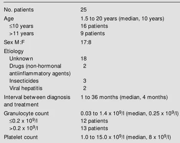
Clinical and laboratory evaluations were conducted 6 weeks, 6 months, and 12 months Table 1 - Clinical features of children at diagnosis of severe aplastic
anemia.
No. patients 25
Age 1.5 to 20 years (median, 10 years)
£10 years 16 patients
>11 years 9 patients
Sex M :F 17:8
Etiology
Unknow n 18
Drugs (non-hormonal 2
antiinflammatory agents)
Insecticides 3
Viral hepatitis 2
Interval betw een diagnosis 1 to 36 months (median, 4 months) and treatment
Granulocyte count 0.03 to 1.4 x 10⁹/l (median, 0.25 x 10⁹/l)
£0.2 x 10⁹/l 12 patients
>0.2 x 10⁹/l 13 patients
after the end of the 10-day ATG course. The last follow-up date was July 1998. The first three evaluations included physical exami-nation, complete blood count, platelet count, and bone marrow aspiration and biopsy. The Ham test and cytogenetic studies of the bone marrow cells were included in the subse-quent yearly evaluations.
Responses to treatment were defined pro-spectively as type I (patients became inde-pendent of red cell or platelet transfusions and were not hospitalized for fever), type II (patients required red cell or platelet support and/or were admitted due to fever and neu-tropenia, despite increased hemoglobin, platelet counts, or granulocyte counts), and type III (no increase in hemoglobin, platelet count, or granulocyte count within 12 months after the completion of ATG).
Suppo rtive care
All patients were treated in double-occu-pancy nonsterile rooms. Patients who had an absolute neutrophil count less than 0.5 x 10⁹
/ l and an axillary temperature exceeding 38.0o
C were treated empirically with broad-spectrum antibiotics. All blood products were irradiated. Red cells and platelets were transfused when the hemoglobin level fell below 7.0 g/dl and the platelet count below 20 x 109/l, respectively. All patients received care at the University Hospital for the first 12 months and were then discharged in the care of the referring physician. Return visits to the University Hospital for follow-up evaluation were scheduled on a yearly basis.
Statistical analysis
Survival time was measured from the first day of ATG infusion. Product limit estimates of the survival time distribution were calculated using the method of Kaplan and Meier. The log-rank test was used to compare survival distribution.
Re sults
Re spo nse
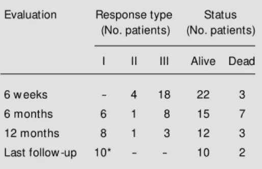
The clinical responses to ATG are listed in Table 2. At the 6-week evaluation, four patients (16%) had responded (type II re-sponses) and three had died from hemor-rhagic or infectious complications. The re-mainder had type III responses. At 6 months, six patients achieved a type I response and one patient had a type II response. Seven patients died during this period from compli-cations of bone marrow failure. At one year, two more patients had achieved type I re-sponses and three patients continued to show no response. Of these three patients, two later achieved a type I response (one after further immunosuppression with cyclo-sporine plus prednisone), and two died.
Of the eight patients who responded within one year of ATG treatment, one de-veloped severe thrombocytopenia 6 years later that resolved after cyclosporine and prednisone therapy. Currently, ten patients (40%) are alive with good bone marrow function (Figure 1). Response to ATG
ap-Table 2 - Outcome of children treated w ith anti-thymocyte globulin for severe aplastic anemia.
Type I: Patients became independent of red cell or platelet transfusions and w ere not hospitalized for fever; type II: patients required red cell or platelet support and/or w ere admitted due to fe-ver and neutropenia; type III: no increase in he-moglobin, platelet count, or granulocyte count by 12-month follow -up. * Includes tw o patients w ho received additional im m unosuppressive therapy.
Evaluation Response type Status
(No. patients) (No. patients)
I II III Alive Dead
6 w eeks - 4 18 22 3
6 months 6 1 8 15 7
12 months 8 1 3 12 3
pears to be independent of age or severity of neutropenia. Seven of the 10 responders were less than 10 years of age, and five had abso-lute neutrophil counts less than 0.2 x 10⁹
/l at diagnosis. However, the number of patients is too small for meaningful statistical analy-sis.
Co m plicatio ns o f the rapy
Fever was the most common clinical sign, occurring in 19 patients (76%). A maculo-papular skin rash occurred in 17 patients (68%), and 6 (24%) developed cutaneous vasculitis. Five patients (20%) developed hypertension, which was managed with sub-lingual nifedipine and diuretics. Serum sick-ness, characterized by fever, skin rash, ar-thralgia, and serpentinous lesions on palms and soles, was observed in two patients (8%) and resolved with administration of pred-nisone. Treatment was not discontinued in any patient due to ATG-associated compli-cations.
Although no hematologic disease has been noted in the long-term survivors (ex-cept for the patient with thrombocytopenia), one patient has abnormal mean corpuscular volume (>100 fl), and two patients have developed clonal karyotypic abnormalities (trisomy 8 and del(6)q21, respectively). Cen-tral nervous system bleeding was the cause of death for 10 patients, bacterial infection for four, and fulminant viral hepatitis for one.
D iscussio n
We set out to characterize the quality of bone marrow function in long-term survi-vors of SAA who were initially managed with ATG. The bone marrow function of our ten long-term survivors has been normal. Interestingly, two of these patients devel-oped clonal cytogenetic abnormalities (tri-somy 8 and del(6)q21), and a third patient has an elevated red cell mean corpuscular volume. Although these abnormalities can be found in patients with myelodysplastic syndromes (MDS) and lymphoid malignan-cies, our patients bone marrow studies have not been revealing evidence of them. Re-cently, Geary et al. (15) reported the follow-up of 13 SAA patients with an abnormal cytogenetic clone (including trisomy 8), de-tected at or sometime after diagnosis. They received immunosuppressive therapy and transformation to acute leukemia or MDS was not observed after 4.1 years (range 1.2 to 11.2 years). Surprisingly, in four patients the abnormal clone disappeared after treat-ment (15). So, the clinical significance of these findings is presently unknown.
Adults with SAA who are treated with ATG alone have a 9.6% and 6.6% cumula-tive risk at 10 years for myelodysplasia and leukemia, respectively (10). Those treated mainly with ATG have the same probability (9% at 4 years) (16). Approximately 15% of these patients develop evidence of paroxys-mal nocturnal hemoglobinuria (17). The in-cidence of MDS and acute leukemia as a late complication of immunosuppressive treat-ment in children may vary with the type of drugs used, being more frequent when cy-closporine and G-CSF are used together (18). Children with SAA may be less prone to developing clonal hematologic disorders af-ter treatment with ATG. Paroxysmal noctur-nal hemoglobinuria has not been detected in any of our patients, although we have used only the acidified Ham test for screening.
Early mortality, defined as death within
P
e
rc
e
n
t
a
liv
e
100
80
60
40
20
0
0 2 4 6 8 10 12
Years Figure 1 - Kaplan-M eier
100 days of the beginning of therapy, had an important negative impact on the overall survival of our patients. Seven patients (47%) died during this period, most from CNS hem-orrhage or infectious complications, in con-trast to recent reports of mortality below 10% (19). However, most patients in our series were admitted with serious medical complications, such as active infection and bleeding.
The intensity of the initial immunosup-pressive regimen can also influence early mortality. ATG is only mildly immunosup-pressive, prolonging the time for bone mar-row recovery. The mortality rates reported for patients treated with more intense regi-mens, such as combinations of cyclosporine and ATG with or without G-CSF, are 8 to 12% (20). Taken together, the data support the opinion that early intensive immunosup-pression is associated with decreased rates of early death and increased rates of long-term survival.
Relapse can be a significant problem for patients whose SAA initially responded to immunosuppression. Rates of relapse reach 35% at 10 years, as reported by Schrezen-meier et al. (21). In our series, only one patient had a relapse after achieving a type I response. Rescue with cyclosporine A plus prednisone was effective, and after 4 years the patient maintains normal hematologic parameters, demonstrating that this immu-nosuppressive combination should be con-sidered for patients who relapse or initially fail to respond to ATG (22).
The number of patients in our study was too small to allow meaningful analysis of prognostic factors. In general, patients with presenting granulocyte counts less than 0.2 x 10⁹/l tend to have a worse prognosis than others, mainly with respect to early mortality (23). In our series, 4 of the 12 patients who had granulocyte counts <0.2 x 10⁹
/l re-sponded to ATG, and another subsequently responded to cyclosporine plus prednisone. This rate of response did not differ from that of patients who presented with higher locyte counts, suggesting that initial granu-locyte count is probably not associated with outcome if patients survive the first few months after immunosuppressive therapy.
Young age at diagnosis has been associ-ated with poor outcome in some studies, but not in others (24-26). In our series, 6 of 16 patients <10 years of age (37.5%) are long-term disease free survivors, as compared with 4 of 9 patients (44.4%) older than age10 years. Thus, age <10 years does not appear to be associated with poorer outcome.
The overall response and survival rates achieved in our study are inferior to those reported for sibling HLA-matched stem cell transplantation (24,25) and for more inten-sive immunosuppresinten-sive regimens (20,26). However, our survivors have enjoyed long-term stable peripheral blood counts, Karnofsky scores of or near 100, and a low rate of relapse, demonstrating that normal long-term survival is possible for children treated with ATG.
Re fe re nce s
1. Young NS & Alter BP (1994). Definitive treatment of acquired aplastic anemia. In: Young NS & Alter BP (Editors), Aplastic Anemia: Acquired and Inherited.1st edn. W.B. Saunders Company, Philadelphia, PA.
2. M aciejew ski JP, Selleri C, Sato T, Ander-son S & Young NS (1996). A severe and consistent deficit in marrow and
circulat-ing primitive hematopoietic cells (long-term culture-initiating cells) in acquired aplastic anemia. Blood, 88: 1983-1991. 3. M arsh JC, Chang J, Testa NG, How s JM
& Dexter TM (1991). In vitro assessment of marrow ‘stem cell’ and stromal cell function in aplastic anaemia. British Jour-nal of Haematology, 78: 258-267. 4. Champlin RE, Feig SA, Sparkes RS & Gale
RP (1984). Bone marrow transplantation from identical tw ins in the treatment of aplast ic anaem ia: im plicat ion f or t he pathogenesis of the disease. British Jour-nal of Haematology, 56: 455-463. 5. Hinterberger W, Row lings PA,
tw ins into patients w ith aplastic anemia. Annals of Internal M edicine, 126: 116-122.
6. Young NS & M aciejew ski J (1997). M ech-anism of disease: the pathophysiology of acquired aplastic anemia. New England Journal of M edicine, 336: 1365-1372. 7. M athé G, Amiel JL & Schw arzenberg L
(1970). Bone marrow graft in man after conditioning by antilymphocytic serum. British M edical Journal, 2: 131-136. 8. Champlin RE, Ho WG & Gale RP (1983).
Antithymocyte globulin treatment in pa-tients w ith aplastic anemia: a prospective randomized trial. New England Journal of M edicine, 308: 113-118.
9. Young NS, Griffith P, Brittain E, Elfenbein G, Gardner F, Huang A, Harm on D, Hew llet J, Fay J, M angan K, M orrison F, Sensebrenner L, Shadduck R, Wang W, Zarouilis C & Zuckerman K (1998). A mul-ticentric trial of antithymocyte globulin therapy for aplastic anemia and related diseases. Blood, 72: 1861-1869. 10. Bacigalupo A, How s J, Gluckman E,
Nis-sen C, M arsh J, Van Lint M T, Congiu M , De Planque M M , Ernst P, M cCann S, Raghavachar A, Frickhofen N, Wursch A, M armont AM & Gordon-Smith EC (1988). Bone marrow transplantation (BM T) ver-sus immunosuppression for the treat-ment of severe aplastic anaemia (SAA): a report of the EBM T SAA Working Party. British Journal of Haematology, 70: 177-182.
11. Socie G, Henry-Amar M , Bacigalupo A, How s J, Tichelli A, Ljungman P, M cCann SR, Frickhofen N, Van Veer-Korthof E & Gluckman E for the European Bone M ar-row Transplantation Severe Aplastic Ane-mia Working Party (1993). M alignant tu-mors occurring after treatment of aplastic anemia. New England Journal of M edi-cine, 329: 1152-1157.
12. De Planque M M , Kluin-Nelemans HC, van Krieken HJ, Kluin PM , Brand A, Beverstock JC, Willenze R & Van Rood JJ (1988). Evolution of acquired severe aplas-tic anaemia to myelodysplasia and subse-quent leukaemia in adults. British Journal of Haematology, 70: 55-62.
13. Tichelli A, Gratw ohl A, Wursch A, Nissen C & Speck B (1988). Late haematological
complications in severe aplastic anaemia. British Journal of Haematology, 69: 413-420.
14. Paquette RL, Tebyani N, Frane M , Ireland P, Ho WG, Champlin RE & Nimer SD (1995). Long-term outcome of aplastic anemia in adults treated w ith antithymo-cyte globulin: comparison w ith bone mar-row transplantation. Blood, 85: 283-290. 15. Geary CG, Harrison CJ, Philpott NJ, How s
JM , Gordon-Smith EC & M arsh JCW (1999). Abnormal cytogenetic clones in patients w ith aplastic anemia: response to immunosuppressive therapy. British Journal of Haematology, 104: 271-274. 16. Doney K, Pepe M , Storb R, Bryant E,
Anasetti C, Appelbaum FR, Buckner CD, Sanders J, Singer J, Sullivan K, Weiden P & Hansen J (1992). Immunosuppressive therapy of aplastic anemia: results of a prospective, randomized trial of antithy-mocyte globulin (ATG), methylpredniso-lone, and oxymetholone to ATG, very high-dose m et hylprednisolone, and oxymetholone. Blood, 79: 2566-2571. 17. Speck B, Tichelli A, Gratw ohl A & Nissen
C (1990). Treatment of severe aplastic anemia: a 12 year follow -up of patients after bone marrow transplantation or af-ter therapy w ith antilymphocyte globulin. In: Sahid NT (Editor), Aplastic Anemia and Other Bone M arrow Failure Syndromes. Springer-Verlag, New York, NY. 18. Ohara A, Kojima S & Hamajima N (1997).
M yelodysplastic syndrome and acute my-elogenous leukemia as a late clonal com-plication in children w ith acquired aplastic anemia. Blood, 90: 1009-1012.
19. Bacigalupo A, Broccia G, Corda G, Arcese W, Carotenuto M , Gallamini A, Locatelli F, M ori PG, Saracco P, Todeschini G, Coser P, Iacopino P, Van Lint M T & Gluckman E for the European Group for Blood and M arrow Transplantation Working Party on SAA (1995). Antilym phocyte globulin, cyclosporin, and granulocyt e colony-stimulating factor in patients w ith ac-quired severe aplastic anemia (SAA): a pilot study of the EBM T SAA Working Party. Blood, 85: 1348-1353.
20. Frickhofen N, Kaltw asser JP, Schrezen-m eier H, Raghavachar A, Vogt HG, Herm ann F, Freund M , M eusers P,
Salama A & Heimpel H (1991). Treatment of aplastic anemia w ith antilymphocyte globulin and methylprednisolone w ith or w ithout cyclosporine. The German Aplas-tic Anemia Study Group. New England Journal of M edicine, 324: 1297-1304. 21. Schrezenmeier H, M arin P, Raghavachar
A, M cCann S, How s J, Gluckman E, Nis-sen C, Van Veer-Korthof ET, Ljungman P, Hinterberger W, Van Lint M T, Frickhofen N & Bacigalupo A (1992). Relapse of aplas-tic anaem ia after im m unosuppressive treatment: a report from the European Bone M arrow Transplantation Group SAA Working Party. British Journal of Haema-tology, 85: 371-379.
22. Gluckm an E, Esperou-Bourdeau H, Baruchel A, Boogaert s M , Briere J, Donadio D, Leverger G, Leporrier M , Reif f ers J, Janvier M , M ichallet M , Strickmans P & The Cooperative Group on the Treatment of Aplastic Anemia (1992). M ulticenter random ized study com paring cyclosporine-A alone and antithymocyte globulin w ith prednisone for treatment of severe aplastic anemia. Blood,79: 2540-2546.
23. Bayever E, Champlin R & Ho W (1982). Comparison betw een bone marrow trans-plantation and antithymocyte globulin in treatment of young patients w ith severe aplastic anemia. Journal of Pediatrics, 100: 307-316.
24. Halperin DS, Grisaru D, Freedman M H & Saunders EF (1988). Severe acquired aplastic anemia in children: 11-year expe-rience w ith bone marrow transplantation and immunosuppressive therapy. Ameri-can Journal of Pediatric Hematology and Oncology, 11: 304-311.
25. Gillo A, Boulad F, Small T, Kernan N, Reyes B, Childs B, Castro-M alaspina H & O’Reilly RJ (1993). Treatment of severe aplastic anemia in children: the M SKCC experience. Blood, 82: 693 (Abstract). 26. Bittencourt M A, M edeiros CR, Zanis-Neto