REVERSIBLE POSTERIOR LEUCOENCEPHALOPATHY
SYNDROME ASSOCIATED WITH BONE MARROW
TRANSPLANTATION
Hélio A.G. Teive
1, Ivar V. Brandi
2, Carlos H.F. Camargo
2,
Marco A. Bittencourt
3, Carmem M. Bonfim
3, Maria L. Friedrich
3,
Carlos R. de Medeiros
4, Lineu C. Werneck
5, Ricardo Pasquini
6ABSTRACT - Reversible posterior leucoencephalopathy syndrome (RPLS) has previously been described in patients who have renal insufficiency, eclampsia, hypertensive encephalopathy and patients receiving immunosuppressive therapy. The mechanism by which immunosuppressive agents can cause this syndrome is not clear, but it is probably related with cytotoxic effects of these agents on the vascular endothelium. We report eight patients who received cyclosporine A (CSA) after allogeneic bone marrow transplantation or as treatment for severe aplastic anemia (SSA) who developed posterior leucoencephalopathy. The most common signs and symptoms were seizures and headache. Neurological dysfunction occurred preceded by or concomitant with high blood pressure and some degree of acute renal failure in six patients. Computerized tomography studies showed low-density white matter lesions involving the posterior areas of cerebral hemispheres. Symptoms and neuroimaging abnormalities were reversible and improvement occurred in all patients when given lower doses of CSA or when the drug was withdrawn. RPLS may be considered an expression of CSA neurotoxicity. KEY WORDS: neurological complications, cyclosporine A, bone marrow transplantation.
Leucoencefalopatia posterior reversível associada a transplante de medula óssea
RESUMO - A síndrome de leucoencefalopatia posterior reversível (SLPR) tem sido descrita em pacientes com insuficiência renal, eclâmpsia, encefalopatia hipertensiva e em pacientes que recebem terapia imunossupressora. O mecanismo pelo qual os agentes imunossupressores podem causar a síndrome ainda não são conhecidos, porém estão provavelmente relacionados aos efeitos citotóxicos destes agentes no endotélio vascular. Relatamos oito pacientes que receberam ciclosporina A (CSA) após transplante de medula óssea alogênico ou para tratamento de anemia aplástica severa e que desenvolveram a SLPR. Os sinais e sintomas mais comuns foram convulsões e cefaléia. A disfunção neurológica ocorreu simultaneamente ou precedida por elevação da pressão arterial sistêmica e disfunção renal aguda em seis pacientes. O exame de tomografia computadorizada do crânio demonstrou a presença de áreas de baixos valores de atenuação na distribuição da substância branca, envolvendo áreas posteriores de ambos os hemisférios cerebrais. O quadro clínico e as anormalidades tomográficas foram reversíveis; a melhora ocorreu em todos os pacientes em que as doses de CSA foram reduzidas ou quando a droga foi retirada. A SLPR pode ser considerada uma expressão de neurotoxicidade da CSA.
PALAVRAS-CHAVE: complicações neurológicas, ciclosporina A, transplante de medula óssea.
Serviço de Neurologia e Unidade de Transplante de Medula Óssea do Hospital de Clínicas da Universidade Federal do Paraná, Curitiba PR, Brazil: 1Professor Assistente de Neurologia; 2Neurologista; 3Hematologista; 4Professor Adjunto de Hematologia; 5Professor Titular e
Chefe do Serviço de Neurologia; 6Professor Titular e Chefe da Unidade de Transplante de Medula Óssea. Presented in part at the 50th
American Academy of Neurology Meeting in Minneapolis, MN, April 1998.
Received 21 March 2001, received in final form 25 May 2001. Accepted 31 May 2001.
Dr. Hélio A.G. Teive - Serviço de Neurologia, Hospital de Clínicas, UFPR - Rua General Carneiro 181 / 12o andar - 80060-900 Curitiba PR Brasil. FAX 41 244 5060. E-mail: [email protected]
Allogeneic bone marrow transplantation (AlloBMT) is an important therapeutic option for hematologi-cal malignant and non-malignant diseases, some im-munodeficiency disorders and certain inborn errors of metabolism. It is associated with several unusual complications1. Neurological events are frequent in
AlloBMT recipients and they lead to high morbidity
and mortality2, although their frequency and type
vary among different series3. One of these
complica-tions is reversible posterior leucoencephalopathy syndrome (RPLS). The most common symptoms in-clude headache, seizures, visual abnormalities, de-creased alertness and altered mental status4.
areas on computerized tomography (CT) and bright T2-weight signal changes on magnetic resonance imaging (MRI) scan4,5.
We report eight patients who developed RPLS du-ring the treatment with Cyclosporine A (CSA), uti-lized for graft versus host disease (GVHD) prophy-laxis after AlloBMT.
METHOD
From March 1992 to August 2000, eight patients ad-mitted at our Service presented neurological events and lesions on CT compatible with RPLS after AlloBMT.
The patients received CSA as part of prophylaxes to graft versus host disease (Table 1). They underwent BMT due to severe aplastic anemia (4 patients), chronic my-eloid leukemia (2 patients), Fanconis anemia (1 patient) and myelodysplastic syndrome (1 patient).
CSA levels in plasma were performed routinely on a weekly basis until 8 weeks after the drug starting on all patients, by a fluorescence polarization immunoassay me-thod. In general, CSA levels between 200 400 ng/ml were acceptable. CSA levels, when possible, were also measured at the time of suspected drug toxicity. CSA was withdrawn only if neurological toxicity was considered Grade IV (in-tractable seizures or coma).
RESULTS
The most common present signs and symptoms were seizures and headache (Table 2). Neurological dysfunction occurred preceded by or concomitant with high blood pressure and abnormal renal func-tion in 6 patients undergoing BMT (Tables 2 and 3). One patient also presented hypomagnesemia. CT stu-dies showed low-density white matter lesions
involv-. s g n i d n i f d n a s c i t s i r e t c a r a h c s t n e i t a P . 2 e l b a T t n e i t a P
# Age/Sex Diagnosis/Procedure Clinicalfindings
1 27/F CML/BMT Hypertension,headache,blurredvision,seizures
2 16/F SAA/BMT Hypertension,headache,statusepilepticus
3 10/F FA/BMT Hypertension,headache,drowsiness,seizures
4 17/M SAA/BMT Hypertension,headache,seizures
5 36/F SAA/BMT Hypertension,seizures
6 21/F CML/BMT Seizures
7 15/F SAA/BMT Seizures
8 63/M MDS/NM-BMT Hypertension,blindness,seizures
; e m o r d n y s c i t s a l p s y d o l e y m , S D M ; a i m e n a i n o c n a F , A F ; a i m e k u e l d i o l e y m c i n o r h c , L M C ; a i m e n a c i t s a l p a e r e v e s , A A S w o r r a m e n o b e v i t a l b a o l e y m -n o n , T M B M N ; y p a r e h t e v i s s e r p p u s e n u m m i , T S I ; n o i t a t n a l p s n a r t w o r r a m e n o b , T M B . n o i t a t n a l p s n a r t . D H V G t s n i a g a s i x a l y h p o r P . 1 e l b a T e s a e s i
D ProphylaxisagainstGVHD
A A
S IntravenousCSA3mg/kgtwicedailyfromday-1untiltheycouldbetreatedwithoralmedications,at l i t n u d e n i a t n i a m s a w e s o d s i h T . o . p A S C f o g k / g m 2 1 h t i w d e t r a t s s a w y p a r e h t l a r o t n i o p h c i h w s a w g u r d e h t t a h t r e t f A . 0 8 1 + y a d n o y l i a d g k / g m 5 2 , 6 o t y l w o l s d e c u d e r n e h t d n a 0 5 + y a d . r a e y a n i h t i w d e u n i t n o c s i d d n a y l e v i s s e r g o r p d e c u d e r 1 1 d n a 6 , 3 s y a d n o g k / g m 0 1 h t i w d n a y a d t s r i f e h t n o y a d a e c n o g k / g m 5 1 X T M s u o n e v a r t n I L M
C IntravenousCSA5mg/kgtwicedailyfromday-1until+2,3mg/kgivtwicedailyfromday3untilday y a d m o r f n o i t c u d e r e v i s s e r g o r p h t i w s o r e p g k / g m 5 1 , 5 3 y a d l i t n u 5 1 y a d m o r f v i g k / g m 5 7 , 3 , 4 1 . 0 8 1 y a d l i t n u 6 3 . 1 1 d n a 6 , 3 s y a d n o g k / g m 0 1 h t i w d n a y a d t s r i f e h t n o y a d a e c n o g k / g m 5 1 X T M s u o n e v a r t n I . f f o d e r e p a t r e t a l d n a 8 2 y a d l i t n u 4 1 y a d m o r f d e r e t s i n i m d a s a w g k / g m 1 e n o l o s i n d e r p l y h t e M a i m e n a s 'i n o c n a
F IntravenousCSA3mg/kgtwicedailystartingonday-1.
. 1 1 d n a 6 , 3 s y a d n o g k / g m 0 1 h t i w d n a y a d t s r i f e h t n o y a d a e c n o g k / g m 5 1 X T M s u o n e v a r t n I e m o r d n y s c i s a l p s i d o l e y
. e m o c t u o d n a s e i d u t s T C , s e r u t a e f y r o t a r o b a L . 3 e l b a T t n e i t a
P SerumCSA ) l m / g n ( l e v e l m u r e S e n i n i t a e r C ) l d / g m ( l e v e l T C n o s n o i s e
L Improvement
g n i r e w o l n o e s o d A S C e m o c t u O
1 NR 1.5 Bilaterallowdensityareasinvolvingwhite s e b o l l a t i p i c c o -l a r o p m e t f o r e t t a m s y a d
4 Alive,normalneurologicalstatus
2 416 2.0 Bilaterallowdensitycorticaland -o t e i r a p g n i v l o v n i s n o i s e l l a c i t r o c b u s s e b o l l a t i p i c c o s y a d
3 Alive,normalneurologicalstatus
3 464.47 1.7 Bilateralwhitematterlowdensitylesions s e b o l l a t i p i c c o -o t e i r a p n i s y a d
3 Diedday+34,acuteGVHD
4 NR 2.4 Low-densitylesioninleftoccipital lobe 3days Diedday+66,acuteGVHD
5 NR 1.5 Rightlowdensityareasinvolvingwhite t f e l d n a s e b o l l a t i p i c c o -o t e i r a p n o r e t t a m e b o l l a t n o r f g n i v l o v n i n o i s e l y t i s n e d -w o l s y a d
2 Alive,normalneurologicalstatus
6 540.12 0.8 Bilaterallowdensitylesionsonparietal s e b o l y a d
1 Alive,normalneurologicalstatus
7 180.83 0.5 Rightlowdensitylesioninvolving e b o l l a t i p i c c o s y a d
3 Alive,normalneurologicalstatus
8 350 1.5 Bilaterallowdensitylesionsinvolving s e b o l l a t i p i c c o s y a d
4 Alive,normalneurologicalstatus
. e s a e s i d t s o h s u s r e v t f a r g , D H V G ; A e n i r o p s o l c y c , A S C ; y h p a r g o m o t d e z i r e t u p m o c , T C ; d e z i l a e r t o n , R N
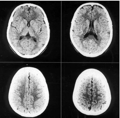
ing the posterior areas of cerebral hemispheres (Table 3). In one patient, follow-up CT scan five months later showed complete resolution of the previous abnormalities (Figs 1 and 2).
Five patients had plasma level of CSA measured at the time of neurological toxicity. It ranged from 180,83 ng/ml to 540,12 ng/ml. Symptoms and neu-roimaging abnormalities were reversible. Improve-ment occurred in all patients, when lowering the dose of CSA (Table 3).
DISCUSSION
BMT is associated with several neurological com-plications that may be related to underlying diseases, bone marrow transplant procedure and severe im-munossupression6,7. Allogeneic transplants usually
require prolonged immunosuppression and are mos-tly associated with neurological complications6.
Cere-brovascular events, central nervous system infections, metabolic encephalopathy and treatment with CSA are the major causes of neurological complications in different series2-9.
Hinchey et al.4 reported fifteen patients who
de-veloped neurological symptoms and signs associated with reversible white matter lesions in neuroimaging exams characterizing RPLS. This may occur associa-ted with hypertensive encephalopathy, eclampsia, use of recombinant human erythropoietin10 and use
of immunosuppressive agents, mainly CSA. Until the study of Lanzino et al.5 the morphological substrate
of these lesions was hypothesized on the basis of neuroradiological appearance. They reported one patient who underwent stereotaxic biopsy of the lesions. Microscopic examination demonstrated only mildly edematous white matter.
CSA proved to be effective in the prevention and treatment of GVHD11, the most life-threatening
com-plication after allogeneic BMT12. It is also considered
part of first line therapy to patients with moderate to severe aplastic anemia13,14. CSA is a cyclic
undeca-peptide of fungal origin, and acts by blocking the release of interleukin-2 and several other cytokines, thereby preventing the second stage of T cells acti-vation12. Its use is associated with multiple side
fects and the most common systemic complications are renal and hepatic toxicity and arterial hyperten-sion. Fifteen to 40% of patients receiving CSA also experience neurological side effects14, like tremor,
paresthesia, headache, seizures, confusion, visual hal-lucinations, cortical blindness, ocular flutter, parkin-sonism, cerebellar-like syndromes and leukoencepha-lopathy14-17. Imaging studies demonstrate reversible
cortical and white matter low density areas on CT and bright T2-weighted signal changes on MRI18,19.
The mechanism of these neurological effects is not completely understood. Reports from both in vivo animal models and in vitro endothelial cell cul-tures suggest that CSA induces endothelial cell inju-ries20-22.Its mediated endothelial cell damage is
as-sociated with alteration in the pattern of endothelin, a potent vasoconstrictor, prostacyclin and trombo-xane A2 release22,23.Elevated plasma levels of
endothelin have been found in patients treated with CSA after BMT24.
Patients with CSA-induced neurotoxicity may also have a disturbance in cerebral blood flow similar to that produced by hypertensive encephalopathy19.
Ho-wever, hypertension is not present in all patients with CSA neurotoxicity, such as Patients 6 and 7 of this series. The susceptibility of the posterior regions of the brain to the lesions is not well understood. It is probably related to altered vascular reaction to circu-lating pressor agents4, like endothelin, and the
rela-tively poorer sympathetic innervation of posterior circulation25-26. Foci of blood brain barrier damage
in the occipital and parietal lobes were also demon-strated in patients taking CSA after BMT27.
The neurological symptoms and signs are revers-ible when the drug is decreased or withdrawn, but may recur when it is reinstituted. One of our pa-tients (Patient 2) experienced seizure in two differ-ent occasions, and in both improvemdiffer-ent occurred with decrease of the CSA dose. Resolution of neuro-logical signs and symptoms occurred within four days in all patients of this series.
Associated factors may increase central nervous system toxicity induced by CSA, such as hypomag-nesemia, arterial hypertension, acute renal failure, hypocholesterolemia and corticosteroids thera-py4,14,28.Adverse neurologic events may occur in
pa-tients with therapeutic blood levels of the drug and without known risk factors4,29.All patients reported
in this series were receiving CSA when they devel-oped RPLS; six patients presented arterial hyperten-sion and some degree of renal dysfunction, one also
presented hypomagnesemia. The presence of hypo-magnesemia also predisposes the patients to neu-rotoxicity and its prevention seems to reduce the risk of associated neurological dysfunction29.One patient
had no risk factor other than therapy with corticos-teroids. The administration of methylprednisolone associated with CSA increases the risk of general-ized seizures, probably secondary to an increase in serum levels of CSA30-31. Blood pressure should be
closely monitored and elevation treated without delay. Seizures can be treated by magnesium replace-ment, anticonvulsivants, or preferably, by reduction or withdrawal of CSA. Continuous anticonvulsant therapy is not necessary unless a previous seizure disorder is present.
The reversible posterior leucoencephalopathy presented by these patients may be considered an expression of CSA neurotoxicity. Blood pressure and renal function monitoring and magnesium replace-ment may help to prevent neurological dysfunction but it is important to be aware that this condition may occur despite therapeutic blood levels of CSA.
REFERENCES
1. Armitage JO. Bone marrow transplantation. N Engl J Med 1994;330:827-838.
2. Gallardo D, Ferrà C, Berlanga JJ, et al. Neurologic complications after allogeneic bone marrow transplantation. Bone Marrow Transplant 1996;18:1135-1139.
3. Graus F, Saiz A, Sierra J, et al. Neurologic complications of autologous and allogeneic bone marrow transplantation in patients with leuke-mia: a comparative study. Neurology 1996;46:1004-1009.
4. Hinchey J, Chaves C, Appignani B, et al. A reversible posterior leuco-encephalopathy syndrome. N Engl J Med 1996;334:494-500. 5. Lanzino G, Cloft H, Hemstreet MK, et al. Reversible posterior
leuco-encephalopathy following organ transplantation. Description of two cases. Clin Neurol Neurosurg 1997;99:222-226.
6. Patchell RA. Neurological complications of organ transplantation. Ann Neurol 1994;36:688-703.
7. Patchell RA, White CL 3d, Clark AW, et al. Neurologic complications of bone marrow transplantation. Neurology 1985;35:300-306. 8. Teive H, Zétola V, Zanis J Neto, et al. Neurological complications in
bone marrow transplantation: analysis of 458 patients. Neurology 1996;46(Suppl 2):A350.
9. Atkinson K, Biggs J, Darveniza P, et al. Cyclosporine associated cen-tral nervous system toxicity after bone marrow transplantation. N Engl J Med 1984;310:527.
10. Delanty N, Vaughan C, Frucht S, Stubgen P. Erythropoietin-associated hypertensive posterior leukoencephalopathy. Neurology 1997;49:686-689.
11. Powles RL, Clink HM, Spence D, et al. Cyclosporin A to prevent graft-versus-host disease in man after allogeneic bone marrow transplanta-tion. Lancet 1980;327-329.
12. Ferrara JLM, Deeg HJ. Graft-verus-host disease. N Engl J Med 1991;324:667-674.
13. Faulds D, Goa KL, Benfield P. Cyclosporin: a review of its pharmaco-dynamic and pharmacokinetic properties, and therapeutic use in can-cer chemotherapy. Drugs 1993;45:953-1040.
14. Kahan BD. Ciclosporine. N Engl J Med 1989;321:1725-1738. 15. Wasserstein PH, Honig LS. Parkinsonism during cyclosporine
treat-ment. Bone Marrow Transplant 1996;18:649-650.
17. Aksamit AL, de Groen PC. Cyclosporine-related leucoencephalopathy and PML in a liver transplant recipient. Transplantation 1995;60:874-890.
18. Pace MT, Slovis TL, Kelly JK, Abella SD. Cyclosporin A toxicity: MRI appearance of the brain. Pediatr Radiol 1995;25:180-183.
19. Schwartz RB, Bravo SM, Klufas RA, et al. Cyclosporin neurotoxicity and its relationship to hypertensive encephalopathy: CT and MR find-ings in16 cases. AJR 1995;165:627-631
20. Fogo A, Hakim RC, Sugiura M, et al. Severe endothelial injury in a renal transplant patient receiving Cyclosporine. Transplantation 1990;49:1190-1192.
21. Zoja C, Furci L, Ghilardi F, et al. Cyclosporin-induced endothelial cell injury. Lab Invest 1986;55:455-462.
22. Bunchman TE, Brookshire. Cyclosporin-induced synthesis of endo-thelin by cultured human endothelial cells. J Clin Invest 1991;88:310-314.
23. Haug C, Duell T, Lenich A, et al. Elevated plasma endothelin concen-trations in cyclosporine-treated patients after bone marrow transplan-tation. Bone Marrow Transplant 1995;16:191-194
24. Tsakiris DA, Luescher TF, Loeffler BM, et al. Endothelin in the early phase of bone marrow transplantation. Transplantation 1994;57:300. 25. Edvinson L, Owmam C, Sjoberg NO. Autonomic nerves, mast cells,
and amina receptors in human brain vessels: a histochemical and phar-macological study. Brain Res 1976;115:377-393.
26. Beausang-Linder M, Bill A. Cerebral circulating in acute hypertension - protective effects of sympathetic nervous activity. Acta Physiol Scand 1981;111:193-199.
27. Reece DE, Frei Lahr DA, Shepard JD, et al. Neurologic complications in allogeneic bone marrow transplant patients receiving cyclosporin. Bone Marrow Transplant 1991;8:393-401
28. Thompson CB, June CH, Sullivan KM, Thoma ED. Association between cyclosporine neurotoxicity and hypomagnesaemia. Lancet 1984;116:1120 29. Lucey MR, Kolars JC, Merion RM, et al. Cyclosporin toxicity at thera-peutic blood lavel and cytochrome P-450 IIIA Lancet 1990;335:11-15. 30. Durrant S, Chipping PM, Palmer S, Gordon-Smith EC. Cyclosporin A,
methylprednisolone, and convulsions. Lancet 1982;2:829-830. 31. Boogaersts MA, Zachee P, Verwilghen RL. Cyclosporin,