Authors
Camila Crensiglova 1 Bárbara Benevides Rehme 1 Letícia Raysa Schiavon Kinasz 1
Domingos Candiota Chula 1
Marcelo Mazza do Nascimento 1
Maria Fernanda Sanches Soares 1
1 Universidade Federal do
Paraná.
Submitted on: 03/16/2015. Approved on: 10/16/2015.
Correspondence to:
Camila Crensiglova.
Hospital de Clínicas, Universidade Federal do Paraná.
Rua Padre Camargo, nº 280, 6º
Andar, Alto da Glória, Curitiba, PR, Brazil.
CEP: 80069-240.
E-mail:camila.crensiglova@gmail. com
Frequency and clinical histological analysis of glomerular
diseases in a tertiary hospital in southern Brazil
Frequência e avaliação clínico-histológica das doenças
glomerula-res em um hospital terciário da região Sul do Brasil
Introdução: As glomerulopatias são as doenças renais mais frequentemente diag-nosticáveis por biópsia. O levantamento epidemiológico das glomerulopatias per-mite identificar sua distribuição e principais etiologias e serve de subsídio para definição de estratégias de prevenção e tratamento.
Objetivo: O presente estudo pretende iden-tificar a frequência e a correlação clínico-patológica das glomerulopatias diagnos-ticadas por biópsia no HC-UFPR durante 5 anos. Métodos: Foram realizadas 131 biópsias no período de 1 de janeiro de 2008 a 31 de dezembro de 2012, submetidas à microscopia óptica e de imunofluorescên-cia. Todas as lâminas de microscopia óptica foram revistas por um patologista. Dados clínicos e laboratoriais e resultados da mi-croscopia de imunofluorescência foram ob-tidos por revisão dos prontuários. Resulta-dos: Foram reanalisados 128 de 131 casos; 46,5% foram obtidos em homens. A idade média de realização da biópsia foi 43 anos para os homens e 38 anos para as mulheres. Em 99 casos identificou-se a indicação da biópsia; 49,5% apresentaram síndrome ne-frótica; 17,17%, insuficiência renal aguda e 15,15% insuficiência renal crônica; 8,08%, síndrome nefrítica; 6,06%, proteinúria iso-lada e 4,04%, hematúria isoiso-lada. 61,21% tratavam-se de glomerulopatia secundária, 33,62% glomerulopatia primária e 5,17% não puderam ser classificados. Dentre as glomerulopatias secundárias, a mais fre-quente foi a nefrite lúpica (49,29%), e, dentre as primárias, glomeruloesclerose seg-mentar e focal (30,77%) e nefropatia mem-branosa (25,64%). Conclusão: O paciente com glomerulopatia neste serviço é adulto e portador de síndrome nefrótica. Ao con-trário de outros relatos, observamos pre-domínio das glomerulopatias secundárias, refletindo possivelmente o perfil terciário de atendimento do HC-UFPR.
R
ESUMOIntroduction: The glomerulopathies are the most common biopsy-proven kidney disea-ses. The epidemiological investigation of glomerulopathies allows the identification of their distribution and main causes and ena-bles the development of prevention and treat-ment strategies. Objective: This study aims to identify the frequency and clinical-pathologi-cal correlation of glomerular diseases diag-nosed at the HC-UFPR over the period of 5 years. Methods: 131 biopsies were perfor-med between January 1, 2008 and Decem-ber 31, 2012 and were analysed by light and immunofluorescence microscopy. Histopa-thological slides were reviewed by a patholo-gist. Clinical and laboratory data and the im-munofluorescence microscopy results were extracted from medical records. The findings were tabulated and analysed. Results: 128 of 131 cases were reanalysed. 46.5% were obtained from men. Patients´ age averaged 43 years for men and 38 for women. In 99 cases, the indication of biopsy was identified; 49.5% cases presented nephrotic syndrome, 17.17%, acute renal failure and 15.15%, chronic renal failure; 8.08%, nephritic syn-drome; 6.06%, isolated proteinuria and 4.04% isolated hematuria. In 61.21% an underlying disease related to the glomeru-lopathy could be identified; 33.62% corres-ponded to primary disease and in 5.17% of cases the nature of the glomerulopathy could not be determined. Among seconda-ry glomerulopathies, the most frequent was Lupus Nephritis (49.29%), and among the primary, Focal Segmental Glomerulosclerosis (30.77%) and Membranous Nephropathy (25.64%). Conclusion: The average patient with glomerulopathy in this service is an adult with nephrotic syndrome. Unlike other reports, secondary glomerulopathies were predominant. These findings may reflect the tertiary characteristic of the assistance at HC-UFPR.
A
BSTRACTI
NTRODUCTIONGlomerulopathies are the kidney diseases most often diagnosed through biopsy.1 Cellular and humoral im-mune mechanism of primary or secondary nature are very much involved in the pathogenesis of this disease category.2 Its progression often results in the need for renal replacement therapy, procedure which, besides being inconvenient for patients, generates high costs for the healthcare system - in 2012 alone, the Ministry of Health invested R $ 1.8 billion in hemodialysis.3
Urinalysis, the estimated glomerular filtration rate and individual patient characteristics may indicate the possible diagnosis of a glomerular disease, but only the pathological evaluation of material obtained from renal biopsy enables the physician to establish a precise diagnosis and possible prognostic indicators. The main primary glomerulopathies found in surveys carried out around the world are: focal segmental glomerulosclerosis (FSGS), IgA (IgAN) nephropathy, minimal change disease (MCD), membranous ne-phropathy (MN), rapidly progressive glomerulone-phritis (RPGN), post-infectious glomeruloneglomerulone-phritis, and undetermined chronic glomerulonephritis.4
Secondary glomerulopathies include: diabetic nephropathy (DN), lupus nephritis (LN), renal amyloidosis, and crescent glomerulopathy, related and unrelated to neutrophyl anticytoplasmic antibodies (ANCA).5 The epidemiological survey of glomerulopathies enables the physician to identify its frequency and main causative factors, and it serves as an input for defining their clinical, laboratory and histological features in a particular region, assisting in the development of prevention and treatment strategies.6 This study aims to identify the frequency and clinic-pathological correlation of the glomerular diseases diagnosed by renal biopsy at our institution over a five-year period.
M
ETHODSThis research project was approved by the Research Ethics Committee under number 329.124.
The survey of patients was carried out by retrospective search in the biopsy records of the Pathology Service (SAP), dated January 1st, 2008 to
Palavras-chave: epidemiologia; biópsia; síndrome ne-frótica; nefrite lúpica; glomerulonefrite membranosa; glomerulosclerose segmentar e focal.
Keywords: epidemiology; biopsy; nephrotic syn-drome; lupus nephritis; glomerulonephritis; membra-nous; glomerulosclerosis, focal segmental.
December 31st of 2012. Using the keywords: kidney, glomerulopathy, nephritis, glomerulonephritis and nephritis, we found 396 cases, broken down into an initial spreadsheet.
After initial screening we consulted the pathology report files. We excluded the cases pertaining to: neoplasia; nephrectomy; pyelonephritis and transplantation. We included only those reports which really dealt with glomerulopathies and renal biopsies with inconclusive diagnoses pending further analysis. This selection resulted in 131 cases, whose medical records were reviewed. We extracted the following information: age; gender; underlying disease and/or comorbidities (diabetes mellitus, hypertension, systemic lupus erythematosus, other); serology (HIV, hepatitis B, hepatitis C and syphilis); clinical presentation (nephrotic syndrome, nephritic syndrome, isolated hematuria, isolated proteinuria, acute renal failure, chronic renal failure); 24-hour proteinuria; partial data from urinalysis (proteinuria, hematuria; cylinders; leukocyturia); serum creatinine; creatinine clearance; serum albumin; lipid profile (total cholesterol and fractions and triglycerides); hemodialysis or peritoneal dialysis at the time of biopsy. The corresponding histological slides, stained by HE, PAS, PAMS and Masson’s were reviewed by a single pathologist. The results from the concomitant immunofluorescence microscopy examinations were consulted.
The findings were tabulated in a Microsoft Excel spreadsheet 12.0. Descriptive statistics were performed using the same software, and the statistical parameters were expressed as mean, median and standard deviation. We used the Fisher’s exact test to compare data at the GraphPad InStat application. We considered a p≤ 0.005 as significant.
R
ESULTSOf the 131 selected patients, 128 had material that could be reevaluated.
chronic renal failure; 8 (8.08%) nephritic syndrome; 6 (6.06%), proteinuria alone and 4 (4.04%), hematuria alone (Table 1).
TABLE 1 CLINICALSITUATIONATTHETIMEOFBIOPSY
Clinical Presentation Number of
cases %
Nephrotic syndrome 49 49.5
Acute renal failure 17 17.17
Chronic renal failure 15 15.15
Nephritic syndrome 8 8.08
Proteinuria alone 6 6.06
Hematuria alone 4 4.04
In all the 128 cases, the biopsy technique adopted was the incisional biopsy through a cutting needle. Biopsies obtained an average of 13 glomeruli; one case had no glomeruli. In this and 11 other cases (9.38%) there were no signs of glomerular disease within the methods employed, so that glomerulopathy was confirmed in 116 cases.
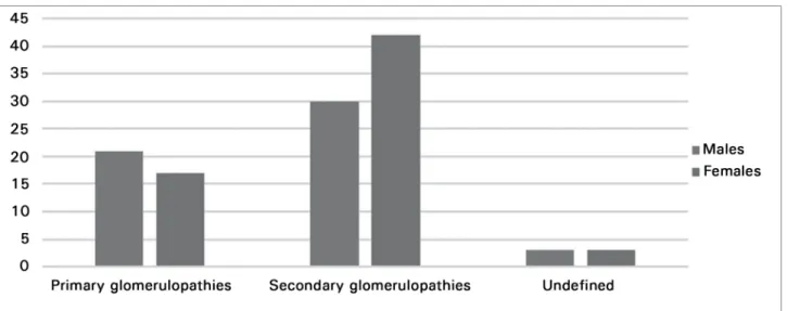
Of the 116 confirmed cases of glomerulopathy, 54 (46.5%) were men (p = 0.0739). The mean patient age was 40 years; among men, the mean was 43 years, and for women, 38. Six subjects were aged 70 years or higher. In 72 cases (62.06%), the glomerulopathy was secondary; in 38 cases (32.75%) it was considered primary; and in 6 cases (5.17%) we were unable to classify it as primary or secondary. Among men, there were 30 cases of secondary glomerulopathy (58.82%); and among women, 42 (71.19%). The distribution between men and women can be seen on Figure 1. Among the most frequently identified comorbidities we found: hypertension (70 cases, 60.34%); systemic lupus erythematosus (43 cases, 37.06%); diabetes (20 cases; 17.24%); HIV (11 cases; 9.48%); hepatitis C (11 cases, 9.48%); hepatitis B (4 cases; 3.45%) and hepatitis B associated with hepatitis C (1 case; 0.86%). This data and its distribution between the genders can be found on Table 2.
We found 35 cases of lupus nephritis (30.17%), 23 cases of FSGS (19.83%) - of which 12 (10.34%) were identified as primary FSGS, membranous nephropathy (10 cases, 8.62%) and diabetic nephropathy (9 cases; 7.76%) (Figure 1). The other results can be found on Table 3. Of the 35 cases identified as lupus nephritis, 26 (74.28%) were women (p = 0.0024). Chronic tubulointersticial changes were present in 50% or more of the kidney cortex from 20 patients.
These patients had FSGS, diabetic nephropathy, hypertensive arterial-arteriolar nephrosclerosis and lupus nephritis.
D
ISCUSSIONDue to the high cost3 and relevant social and economic factors,7 for which kidney diseases are responsible, countries like Japan8 and Italy9 regularly record the nationally diagnosed glomerulopathies. In Brazil, data on the prevalence and epidemiological characteristics of glomerulopathies are scarce. A few Brazilian hospitals have proposed the creation of a local database.10-15 The latest national survey on glomerular diseases until this study was published in 2009, and involved 9,617 renal biopsies in the country.6 To date, there are no studies in the literature on the distribution of glomerulopathies in patients treated at our institution or even in our state, which assigns relevance to this study.
In Brazil, the main indication for biopsy was nephrotic syndrome, which coincides with data from other studies carried out in our country6,10,11,13,14 and the world.16-24 Indeed, nephrotic syndrome is considered one of the Absolute indications for renal biopsy in individuals older than 6 years of age.
The average obtained from 13 glomeruli per sample meets the bulk of its adequacy regarding the guidelines, which impose the appropriate number of 8-10 glomeruli per sample.25-28 This finding, coupled with the presence of only one sample without glomeruli shows that obtaining a histopathological sample has been satisfactory in our institution, which partly ensures diagnostic accuracy. Of the 128 selected patients, 12 showed no signs of glomerular disease through light microscopy or immunofluorescence; that is, during this period, in almost 10% of the suspected glomerulopathies, we were facing other illnesses, normal histopathological kidney or diseases which diagnosis could have been better elucidated through an ultrastructural investigation, not available in our country during the study period. However, it is noteworthy that 6 (50%) of these cases were from patients who were infected with H1N1 during the epidemic of July and August 2009, with samples obtained due to a specific and scientific epidemiological interest, which revealed mainly tubular degenerative changes, as previously published.29
TABLE 2 DISTRIBUTIONOFCOMORBIDITIESPERGENDER
Comorbidities Total #
of cases Males Females
Arterial
hypertension 70 33 37
Systemic lupus
erythematosus 43 12 31
Diabetes mellitus 20 10 10
HIV 11 8 3
Hepatitis C 11 7 4
Hepatitis B 4 3 1
Hepatitis C +
Hepatitis B 1 1 0
TABLE 3 DISTRIBUTIONOFGLOMERULOPATHIESBY
ORDEROFFREQUENCY
Histopathology Diagnosis Number of Cases %
Lupus nephritis 35 30.17
Primary FSGS 12 10.34
Secondary FSGS 11 9.48
Membranous nephropathy 10 8.6
Diabetic nephropathy 9 7.7
Membranous proliferative
glomerulonephritis 6 5.17
Glomerulopathy secondary to
vascular disease 5 4.31
Minimal lesion disease 4 3.44
Proliferative and exudative
glomerulopathy 4 3.44
IgA Nephropathy 3 2.58
Necrotizing glomerulopathy 2 1.72
Terminal kidney 2 1.72
no significant difference between genders, a similar pattern to that of the 20096 national record and the one from the Federal District.10 The average age found reflects the pattern of the patients enrolled in the clinic where the study was carried out. When the study was designed, we sought to find a possible change in the pattern of glomerular disease after 70 years of age, as seen in the literature;14,30,31 however, we found only six cases in this age group, a very limited number to make any extrapolations.
In our study, secondary glomerulopathies were more common than the primary ones. This goes against the overwhelming majority of results presented in the literature,16,20,30,32,33 as well as national6 and regional results (Paulista Registration of Glomerulonephritis,14 Federal District,10 Belém13 and Amazonas12), in agreement only with studies carried out in Belgium34 and Paraguai.35 Possibly, this is due to the fact that the series came from a tertiary hospital.
The prevalence of secondary glomerulopathies was more significant among females, a reflection of the high prevalence of lupus nephritis, since systemic lupus erythematosus (SLE) is classically predominant in women.2 In fact, lupus nephritis was more frequent among women than men (26 women and 9 men) and glomerulonephritis was the most frequently diagnosed in our series. This result is in agreement with the Paulista Registration of Glomerulonefritis14 and most of the series studied, in which it is first among secondary glomerulopathies.6,16,22,24,36-38 Because it is an illness with high morbidity and mortality, especially when associated with common risk factors in developing countries, such as unfavorable
socioeconomic conditions, it is important to pay attention to the kidney health of patients with LES.39
The second most found diagnosis on histopathologic analysis was focal segmental glomerulosclerosis, also highly frequent in national results6,14 and in results from Hispanic populations.16 is important to consider that frequent diagnosis of FSGS in our series and in the series above, can be attributed to population characteristics, i.e., selection of adults and nephrotic patients. However, FSGS is a morphological diagnosis, a common consequence of various renal diseases. In our series, we can distinguish the primary or secondary source of FSGS with some reliability, according to the analysis of 24-hour proteinuria, albumin and their own histopathology features.
The cases that progressed to proteinuria in the nephrotic range, hypoalbuminemia, diffuse podocyte disease signals and global sclerosis pattern, predominantly solidified were classified as primary, while those who had subnephrotic proteinuria, normoalbuminemia, focal podocyte disease and global sclerosis pattern predominantly obsolescent, with possible hyalinizing-sclerotic vascular changes, were secondary. As a result, we obtained a very similar proportion of cases of membranous nephropathy and primary FSGS, which differs from the Brazilian results, which put FSGS ahead of membranous nephropathy.6,14 These studies, however, did not make the distinction between primary or secondary FSGS. The significant finding of membranous nephropathy, comparable to the prevalence of primary FSGS in our sample, coincides with findings from Europe,20 a fact that can be attributed to the European heritage in our population.
Chronic tubulointersticial changes (IFTA%) reflect the duration of kidney disease and the irreversibility of the alterations, and are directly related to serum creatinine levels. Twenty samples were found with IFTA below 50%, so that these cases were very likely diagnosed later. The glomerular diseases found in these cases were in similar proportion to each other - focal segmental glomerulosclerosis, diabetic nephropathy, lupus nephritis and hypertensive arteriolar nephrosclerosis. This information corroborates previous findings2 and highlights the fact that, in our country, such diseases are still diagnosed at a late stage.
Although this is a glomerulonephritis that had the highest increase in prevalence in the past 15 years
in Brazil,6 IgA nephropathy was found in only three samples. This contradicts what was expected, because according to the 2009 survey, the South Region had the highest proportion of IgA nephropathy, among all regions of Brazil.6 Taking into account that the number of patients who had hematuria alone was small (4 cases), the low frequency of this disease in our study may be related to non-routine indication of biopsy in patients with this clinical condition,11 to the low identification or the low frequency of hospital admissions for glomerular hematuria in our center.
The limitation of this study was the small number of biopsies carried out in this center and the retrospective review. Still, this was an unprecedented study in our state, which enabled the recognition of the population affected by glomerulopathies and lays the foundation for a future Glomerulopathies Register in the State. It is important to point out that all records and biopsies were extensively revised, so that the conclusions were based on consistent and clinically relevant findings.
C
ONCLUSIONThrough this study, we were able to trace the profi-le of patients seen because of glomerulonephritis in most tertiary institution of our state. We found equal distribution of men and women, in agreement with national studies,6,14 and average age of 40 years. Most cases were secondary glomerulopathies, which was concordant with only two discordant previous stu-dies34,35 and most published national6,10,13,14 and inter-national studies.16,20,30,32,33 The most often diagnosed glomerular disease in our country was lupus nephri-tis, which was the main cause of secondary kidney disease in several studies.6,14,16,22,24,36-38 It is important to stress that the incidence of IgA nephropathy in our series was low, which may reflect the strategy of glo-merular hematuria study in our country.
The profile of glomerulopathies found is a reflection not only of the tertiary profile of our hospital care, but also of our biopsy indications. The small number of samples in our series suggests that we should intensify some of the renal disease search strategies, to better identify them in our population.
R
EFERENCES2. Braunwald E, Fauci AS, Hauser SL, Longo DL, Kasper DL, Jameson JL, eds. Medicina interna de Harrison. 18a ed. Rio de Janeiro: McGraw-Hill; 2013.
3. Rabelo C, Pinheiro D. Recursos para hemodiálise aumen-tam em R$181,6 mi. Portal da saúde, 2012 [Acesso: 17 Fev 2013]. Disponível em: http://portalsaude.saude.gov.br/ portalsaude/noticia/4458/162/recursos-para-hemodialise--aumentam-em-r$-1816-mi.html
4. Kumar V, Perkins JA. Robbins e Cotran Patologia: Bases Patoló-gicas das Doenças. 8a ed. Rio de Janeiro: Elsevier; 2010. 5. Bogliolo L, Brasileiro Filho G, Rocha A, eds. Bogliolo
pato-logia. 7a. ed. Rio de Janeiro: Guanabara Koogan; 2010. 6. Polito MG, de Moura LA, Kirsztajn GM. An overview on
frequency of renal biopsy diagnosis in Brazil: clinical and pathological patterns based on 9,617 native kidney biop-sies. Nephrol Dial Transplant 2010;25:490-6.
7. Oliveira MB, Romão JE Jr, Zatz R. End-stage renal disease in Brazil: epidemiology, prevention, and treatment. Kidney Int Suppl 2005:S82-6. PMID: 16014106
8. Schena FP. Survey of the Italian Registry of Renal Biopsies. Frequency of the renal diseases for 7 consecutive years. The Italian Group of Renal Immunopathology. Nephrol Dial Transplant 1997;12:418-26.
9. Sugiyama H, Yokoyama H, Sato H, Saito T, Kohda Y, Nishi S, et al.; Committee for Standardization of Renal Patholo-gical Diagnosis and Working Group for Renal Biopsy Data-base, Japanese Society of Nephrology, Tokyo, Japan. Japan Renal Biopsy Registry: the first nationwide, web-based, and prospective registry system of renal biopsies in Japan. Clin Exp Nephrol 2011;15:493-503.
10. Ferraz FHRP, Martins CGB, Cavalcanti JC, Oliveira FL, Quirino RM, Chicon R, et al. Perfil das doenças glomerula-res em um hospital público do Distrito Federal. J Bras Ne-frol 2010;32:249-56.
11. Queiroz MMM. Silva Júnior GB, Lopes MSR, Nogueira JOL, Correia JW, Jerônimo ALC. Estudo das doenças glo-merulares em pacientes internados no hospital geral César Cals - Fortaleza, Ceará. J Bras Nefrol 2009;31:6-9.
12. Cardoso ACD, Kirstajn GM. Padrões histopatológicos das doenças glomerulares no Amazonas. J Bras Nefrol 2006;28:39-43.
13. Alves Júnior JM, Pantoja RKS, Barros CV, Braz MN. Estu-do clínico-patológico das Glomerulopatias no Hospital de Clínicas Gaspar Vianna. Rev Para Med 2008;22:39-47. 14. Malafronte P, Mastroianni-Kirsztajn G, Betônico GN,
Romão JE Jr, Alves MA, Carvalho MF, et al. Paulista Re-gistry of glomerulonephritis: 5-year data report. Nephrol Dial Transplant 2006;21:3098-105. DOI: http://dx.doi. org/10.1093/ndt/gfl237
15. Rocha LP, Carminati CR, Machado JR, Laterza VL, dos Reis MA, Corrêa RR. Prevalence of nephropathies in chil-dren and adolescents and alterations in renal biopsies in Mi-nas Gerais, Brazil, from 1996 to 2010. Ann Diagn Pathol 2013;17:22-7. DOI:http://dx.doi.org/10.1016/j.anndiagpa-th.2012.04.006
16. Arias LF, Henao J, Giraldo RD, Carvajal N, Rodelo J, Ar-beláez M. Glomerular diseases in a Hispanic population: review of a regional renal biopsy database. São Paulo Med J 2009;127:140-4. DOI: http://dx.doi.org/10.1590/S1516-31802009000300006
17. Covic A, Schiller A, Volovat C, Gluhovschi G, Gusbeth-Tato-mir P, Petrica L, et al. Epidemiology of renal disease in Roma-nia: a 10 year review of two regional renal biopsy databases. Nephrol Dial Transplant 2006;21:419-24. DOI: http://dx.doi. org/10.1093/ndt/gfi207
18. Ossareh S, Asgari M, Abdi E, Nejad-Gashti H, Ataipour Y, Aris S, et al. Renal biopsy findings in Iran: case series report from a referral kidney center. Int Urol Nephrol 2010;42:1031-40. PMID: 20052543 DOI: http://dx.doi. org/10.1007/s11255-009-9684-0
19. Batinić D, Sćukanec-Spoljar M, Milosević D, Subat-Dezulović M, Saraga M, Delmis J, et al. Clinical and histopathological characte-ristics of biopsy-proven renal diseases in Croatia. Acta Med Croa-tica 2007;61:361-4. PMID: 18044469
20. Naumovic R, Pavlovic S, Stojkovic D, Basta-Jovanovic G, Ne-sic V, et al. Renal biopsy registry from a single centre in Serbia: 20 years of experience. Nephrol Dial Transplant 2009;24:877-85. DOI: http://dx.doi.org/10.1093/ndt/gfn564
21. Rivera F, López-Gómez JM, Pérez-García R; Spanish Registry of Glomerulonephritis. Clinicopathologic correlations of renal pa-thology in Spain. Kidney Int 2004;66:898-904. PMID: 15327378 DOI: http://dx.doi.org/10.1111/j.1523-1755.2004.00833.x 22. Garyal, Kafle RK. Hisopathological spectrum of glomerular
disease in Nepal: a seven-year retrospective study. Nepal Med Coll J 2008;10:126-8.
23. Razukeviciene L, Bumblyte IA, Kuzminskis V, Laurinavi-cius A. Membranoproliferative glomerulonephritis is still the most frequent glomerulonephritis in Lithuania. Clin Nephrol 2006;65:87-90. DOI: http://dx.doi.org/10.5414/CNP65087 24. Werner T, Brodersen HP, Janssen U. Analysis of the spectrum
of nephropathies over 24 years in a West German center based on native kidney biopsies. Med Klin (Munich) 2009;104:753-9. DOI: http://dx.doi.org/10.1007/s00063-009-1160-1 25. Working Group of the International IgA Nephropathy
Ne-twork and the Renal Pathology Society; Cattran DC, Cop-po R, Cook HT, Feehally J, Roberts IS, Troyanov S, te la. The Oxford classification of IgA nephropathy: rationale, cli-nicopathological correlations, and classification. Kidney Int 2009;76:534-45.
26. Weening JJ, D'Agati VD, Schwartz MM, Seshan SV, Alpers CE, Appel GB, et al. The classification of glomerulonephritis in systemic lupus erythematosus revisited. J Am Soc Nephrol 2004;15:241-50. DOI: http://dx.doi.org/10.1097/01.ASN.0000108969.21691.5D 27. Tervaert TW, Mooyaart AL, Amann K, Cohen AH, Cook HT,
Drachenberg CB, et al. Pathologic classification of diabetic ne-phropathy. J Am Soc Nephrol 2010;21:556-63. DOI: http:// dx.doi.org/10.1681/ASN.2010010010
28. Solez K, Colvin RB, Racusen LC, Haas M, Sis B, Mengel M, et al. Banff 07 classification of renal allograft pathology: updates and future directions. Am J Transplant 2008;8:753-60. PMID: 18294345 DOI: http://dx.doi.org/10.1111/j.1600-6143.2008.02159.x
29. Sevignani G, Soares MF, Marques GL, Freitas AKE, Gentili A, Chula, DC, et al. Insuficiência renal aguda em pacientes infec-tados pelo H1N1 - correlação clínico-histológica em uma série de casos. J Bras Nefrol 2013;35:185-90. DOI: http://dx.doi. org/10.5935/0101-2800.20130030
30. Pinçon E, Rioux-Leclercq N, Frouget T, Le Pogamp P, Vigneau C. Renal biopsies after 70 years of age: a retrospective longitudinal study from 2000 to 2007 on 150 patients in Western France. Arch Gerontol Geriatr 2010;51:e120-4. PMID: 20447700
31. Mazzarolo Cruz HM, Cruz J, Silva AL Jr, Saldanha LB, de Oli-veira Penna D. Prevalence of adult primary glomerular diseases: retrospective analysis of 206 kidney biopsies (1990-1993). Rev Hosp Clin Fac Med Sao Paulo 1996;51:3-6. PMID: 8762646 32. Chang JH, Kim DK, Kim HW, Park SY, Yoo TH, Kim BS, et al.
Changing prevalence of glomerular diseases in Korean adults: a review of 20 years of experience. Nephrol Dial Transplant 2009;24:2406-10. DOI: http://dx.doi.org/10.1093/ndt/gfp091 33. Heaf J, Løkkegaard H, Larsen S. The epidemiology and
prog-nosis of glomerulonephritis in Denmark 1985-1997. Nephrol Dial Transplant 1999;14:1889-97. PMID: 10462267 DOI: http://dx.doi.org/10.1093/ndt/14.8.1889
34. Mesquita M, Fosso C, Bakoto Sol E, Libertalis M, Corazza F, Vanden Houte K, et al. Renal biopsy findings in Belgium: a re-trospective single center analysis. Acta Clin Belg 2011;66:104-9. PMID: 21630606
36. Rychlík I, Jancová E, Tesar V, Kolsky A, Lácha J, Stejskal J, et al. The Czech registry of renal biopsies. Occurrence of re-nal diseases in the years 1994-2000. Nephrol Dial Transplant 2004;19:3040-9. DOI: http://dx.doi.org/10.1093/ndt/gfh521 37. Naini AE, Harandi AA, Ossareh S, Ghods A, Bastani B.
Preva-lence and clinical findings of biopsy-proven glomerulonephriti-dis in Iran. Saudi J Kidney Dis Transpl 2007;18:556-64.
38. Parichatikanond P, Chawanasuntorapoj R, Shayakul C, Choen-suchon B, Vasuvattakul S, Vareesangthip K, et al. An analysis of 3,555 cases of renal biopsy in Thailand. J Med Assoc Thai 2006;89:S106-11. PMID: 17044461