The Effects of Extracorporeal Shock Wave
Therapy in Patients with Coccydynia: A
Randomized Controlled Trial
Shih-Feng Lin1☯, Yi-Jen Chen1,2,3☯, Hung-Pin Tu4, Chia-Ling Lee1,2,5, Ching-Lin Hsieh6, Wen-Lan Wu7, Chia-Hsin Chen1,2,8*
1Department of Physical Medicine and Rehabilitation, Kaohsiung Municipal Ta-Tung Hospital, Kaohsiung, Taiwan,2Department of Physical Medicine and Rehabilitation, Kaohsiung Medical University Hospital, Kaohsiung, Taiwan,3Graduate Institute of Clinical Medicine, College of Medicine, Kaohsiung Medical University, Kaohsiung, Taiwan,4Department of Public Health and Environmental Medicine, School of Medicine, College of Medicine, Kaohsiung Medical University, Kaohsiung, Taiwan,5Department of Physical Medicine and Rehabilitation, Kaohsiung Municipal Hsiao-Kang Hospital, Kaohsiung, Taiwan,6School of Occupational Therapy, College of Medicine, National Taiwan University, Taipei, Taiwan,7Department of Sports Medicine, College of Medicine, Kaohsiung Medical University, Kaohsiung, Taiwan,8Department of Physical Medicine and Rehabilitation, School of Medicine, College of Medicine, Kaohsiung Medical University, Kaohsiung, Taiwan
☯These authors contributed equally to this work. *[email protected]
Abstract
Coccydynia is pain in the coccygeal region, and usually treated conservatively. Extracorpo-real shock wave therapy (ESWT) was incorporated as non-invasive treatment of many mus-culoskeletal conditions. However, the effects of ESWT on coccydynia are less discussed. The purpose of this study is to evaluate the effects of ESWT on the outcomes of coccydynia. Patients were allocated to ESWT (n = 20) or physical modality (SIT) group (n = 21) ran-domly, and received total treatment duration of 4 weeks. The visual analog scale (VAS), Oswestry disability index (ODI), and self-reported satisfaction score were used to assess treatment effects. The VAS and ODI scores were significantly decreased after treatment in both groups, and the decrease in the VAS score was significantly greater in the ESWT group. The mean proportional changes in the ODI scores were greater in the ESWT group than in the SIT group, but the between-group difference was not statistically significant. The patients in the ESWT group had significantly higher subjective satisfaction scores than SIT group. We concluded that ESWT is more effective and satisfactory in reducing discomfort and disability caused by coccydynia than the use of physical modalities. Thus, ESWT is rec-ommended as an alternative treatment option for patients with coccydynia.
Trial Registration
ClinicalTrials.govNCT02313324 OPEN ACCESS
Citation:Lin S-F, Chen Y-J, Tu H-P, Lee C-L, Hsieh C-L, Wu W-L, et al. (2015) The Effects of
Extracorporeal Shock Wave Therapy in Patients with Coccydynia: A Randomized Controlled Trial. PLoS ONE 10(11): e0142475. doi:10.1371/journal. pone.0142475
Editor:Jan P. A. Baak, Stavanger University Hospital, NORWAY
Received:January 3, 2015
Accepted:October 21, 2015
Published:November 10, 2015
Copyright:© 2015 Lin et al. This is an open access article distributed under the terms of theCreative Commons Attribution License, which permits unrestricted use, distribution, and reproduction in any medium, provided the original author and source are credited.
Data Availability Statement:All relevant data are within the paper and its Supporting Information files.
Introduction
Coccydynia is pain around the coccygeal region that may be caused by sudden impact over the coccyx area from falls or traumatic injuries, resulting in pain and inflammatory changes of the surrounding ligaments and muscles [1]. Pain in these conditions is associated with coccygeal instability or subluxation, and patients develop subsequent coccydynia [2]. The radiographic orientations of the coccyx were introduced by Postacchini and Massobrio [3], who observed that patients with symptoms of coccydynia showed more mobility over the first intercoccygeal joint. The pain drawings were used to identify the correct symptoms and offer valuable meth-ods for relieving the symptoms of coccydynia.
Patients with coccydynia often complain of pain and local tenderness around the coccyx [4]. Surgical interventions with the excision of the mobile coccyx or a total coccygectomy relieve approximately 80% to 90% of symptoms [5]. The most common complication of coccy-gectomy is wound infection; therefore, the surgery is rarely performed, and nonsurgical strate-gies remain the major treatment for coccydynia. Management typically includes medications such as nonsteroidal anti-inflammatory agents (NSAIDs), gentle massage over the ligaments attached to the sacrococcygeal joint [6], manual manipulation for mal-alignment of the coccyx [7,8], local steroid injections combined with anesthesia [9], and physical therapy with interfer-ential current (IFC) [10] or shortwave diathermy (SWD) [11]. SWD is used to provide heat to deep tissues [12] and is useful in relieving pain and muscle spasms in inflammatory tissue [13]. SWD is regarded as an effective treatment modality for chronic osteoarthritis [14] and is used for somatic pain generated by the ligamentous and muscular elements inserted into the coccyx [15]. SWD can be effective for the treatment of patients with chronic low back pain [16] and was reported to reduce pain in patients with coccydynia [17]. IFC has been reported to reduce inflammation-induced central sensitization [18], and reduce low back pain [19,20]. Combined with other physical modalities, IFC showed better outcomes in reducing the pain intensity associated with musculoskeletal disorders [10].
In recent years, extracorporeal shock wave therapy (ESWT) has been suggested for non-invasive treatment of many musculoskeletal conditions, including plantar fasciitis [21], epicon-dylitis [22] and shoulder calcification [23]. The effectiveness of pain relief with ESWT might be due to stimulation analgesia [24] and increased tissue regeneration [25]. However, the effects of ESWT on low back pain and coccydynia are less discussed till now. Lee et al. reported exer-cise program combined with ESWT relieved chronic low back pain and improved dynamic bal-ance more than exercise program with conservative physical therapy [26]. There is only one recent report by Marwan et al., presenting 2 cases with coccydynia, and reported three sessions of ESWT effective in relieving pain, and the pain did not recur during one year follow-up period [27]. Most patients prefer conservative treatment with non-invasive methods for coccy-dynia, including physical modalities and ESWT. There are few studies comparing the effects between ESWT and SWD combined with IFC therapy (SIT) in patients with coccydynia. The purpose of this study is to evaluate the effects of non-invasive ESWT on the outcomes of coccydynia.
Materials and Methods
Study population
This clinical study was approved by the Institutional Review Board (IRB) of Kaohsiung Medi-cal University Hospital (IRB number: KMUH-IRB-20120218). The study was carried out from November, 2012 to November 2013 according to the IRB approval of the study period. This clinical study was conducted after the approval of IRB for its study design and ethical concerns,
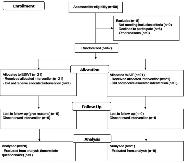
without initial registration as clinical trial, since researchers can start recruiting subjects and executing clinical studies once the clinical studies are approved by the IRB. The study was later registered on ClinicalTrials.gov Protocol and Results Registration System (registration number: NCT02313324) as suggested. The authors confirmed that all ongoing and related trials for this intervention are registered. The CONSORT checklist for randomized trial is included in Sup-porting Information (S1 Text). The patients with first-time diagnosis of coccydynia with pain score ranging from 1 to 10 were enrolled at the rehabilitation outpatient department (OPD). Patients with a history of direct traumatic events to the buttocks, such as falls or slipping, were also included. The physiatrists of rehabilitation OPD were responsible for patient enrollment. A total of 50 participants were screened. After providing informed consent in written form, the patients’histories were taken, and they underwent physical examination to identify the tender area and bony position. Roentgenography was used to assess the coccyx position and rule out major coccyx dislocations. Patients with cardiac pacemaker, tumors of the cauda equina, pelvic surgery, herniation of the lumbosacral disc, internal procidentia, genitourinary or gastrointesti-nal complaints or psychogenic factors were excluded. The basic demographic features and duration of coccygeal pain were recorded. Finally, 42 patients met the inclusion criteria, and were randomly allocated to 2 groups with allocation ratio of 1:1. The CONSORT diagram was shown inFig 1. The physiatrist in charge of randomization was not involved in the study assessment or data analysis.
Evaluation
Before treatment, all of the patients reported the pain intensity, using a visual analog scale (VAS), on a scale from 0 through 100 mm (0 for no pain and 100 mm for the worst pain) [28]. Disability and pain were measured using a spinal disorder-specific questionnaire, the Oswestry disability index (ODI, in Chinese) [29]. There are six statements to be ranked on a scale of 0 to 5 in each of the 10 sections of the questionnaire. The contents relate to impairments such as pain intensity and abilities in personal care, lifting, walking, sitting, standing, sleeping, sex life, social life and traveling. The patients were asked to identify the corresponding level of disability in each section. The total score ranges from 0 to 50; 0 represents the highest level of function, and 50 indicates completely disabled. In addition to the evaluations of the VAS and ODI prior to treatment, the scores were followed at the 5thand 8thweek post-enrollment follow-up visits. Self-reported satisfaction scales were used to assess the general condition at the 8thweek.
Interventions
The patients were randomly allocated to two parallel groups sequentially in a 1:1 ratio, the SIT group and the ESWT group, at the initial OPD visit. The enrolled patients were asked to dis-continue analgesics or other physical modalities for pain one week prior to and after the treat-ments. The patients in the SIT group received combined therapy with SWD and IFC
such that the two channels crossed each other with specific regions of interest concentrated on the gluteal area. The carrier frequency, typically 4000 Hz and 4100 Hz and designed to interfere with each other, resulted in a beat frequency of 100 Hz within the treated area. The treatment duration was 20 minutes. The protocol was set as 3 times a week for a period of 4 weeks.
The ESWT was performed by the same physiatrist, who was blinded to patients’enrollment, and was not involved in further follow-up assessment. A BTL-5000 radial ESWT with a median head applicator was used (BTL Industries Inc. Columbia, South Carolina, USA). The patients were maintained in the prone and knees separated position with a pillow under the abdominal area. The patients received 2000 shots of ESWT in the coccyx area per session for four sessions (one session a week for 4 consecutive weeks). The frequency used was 5 Hz and the pressure
Fig 1. Flow diagram of the enrollment.
was 3–4 bar. The detailed descriptions of the study protocol are included in the Supporting Information, both in original and translated forms (S2andS3Texts).
Statistical analysis
The results were assessed by another therapist who was blinded to the patient’s treatment method. All of the statistical analyses were performed by a statistician. We calculated the sam-ple size of 15 patients per group with the assumption of a mean pain intensity difference between the two groups of 20 mm (SD 16), which provided 90% power to detect such a differ-ence with a two-samplettest and a two-sided type I error of 0.05. Therefore we aimed to recruit 40 patients (20 per group) in our study. Power analysis and sample size determination were performed using the GPower version 3.1.92 [30]. Characteristics of the study
partici-pants for the continuous and categorical variables were analyzed byttest, the Wilcoxon rank-sum test, and the chi-squared test, as appropriate, for comparisons between the ESWT and SIT groups. The clinical evaluation was designed as a repeated measure, and werepeated measure, and we repeated measure, and we repeated measure, and we used random (PROC GLIMMIX) statements to account for a period of 8 weeks taken from the same individual between the ESWT and SIT groups. Therefore, the repeated measures data were analyzed with the quantita-tive data (VAS and Therefore, the repeated measures data were analyzed with the quantitaquantita-tive data (VAS and ODI) by a by a generalized linear mixed model ((PROC GLIMMIX statement), and conclusions were drawn according to the parameter (β, regression coefficient) and differ-ence means statement), and conclusions were drawn according to the parameter (â, regression coefficient) and difference means with Dunnett’s multiple-comparison post hoc test inin the ESWT and SIT groups. We next examined the interactions between ESWT and duration for VAS and. We next examined the interactions between ESWT and duration for VAS and ODI using repeated-measure using repeated-measure generalized linear mixed models. Differences between the ESWT and SIT groups in the subjective satisfaction score at the 8thweek follow-up were analyzed byt-test / Wilcoxon rank-sum test. The significance level was set at 0.05 by two-tailed tests. The analyses were performed using SAS version 9.3 statistical software (SAS Insti-tute Inc, Cary, North Carolina, USA).
Results
Patient characteristics
A total of 42 patients were recruited. The enrolled patients were equally allocated to ESWT and SIT groups, with 21 patients in each allocation. One patient in the ESWT group was excluded from analysis due to incomplete information from the questionnaire. There were eventually 20 patients in the ESWT group, and 21 patients in the SIT group. The basic characteristics of the included patients were shown inTable 1. There were no between-group differences in the age, gender, and onset duration prior to treatment.
Table 1. Baseline characteristics of the included patients.
ESWT (N = 20) SIT (N = 21)
Age, yrs 44.75(14.85) 44.46(18.88)
Gender, female, n (%) 15(75.0) 15 (71.43)
BMI, kg/m2 24.22(5.57) 22.45(3.12)
Time since onset, months 10.51(13.05) 12.57(16.48)
BMI: body mass index; ESWT: extracorporeal shock wave therapy; SIT: physical modality.
Clinical outcomes
The VAS score decreased by 30.5 mm and 41.0 mm in the ESWT group at the 5thand 8thweek post-treatment evaluations, respectively (p<0.001). In the SIT group, the VAS scores decreased
by 16.2 mm and 21.0 mm at the 5thand 8thweek post-treatment evaluations, respectively (p<0.001). The decrease in the VAS score was significantly greater in the ESWT group
com-pared with the SIT group at the 5thweek evaluation (p = 0.004,Table 2); the between-group difference was more significant at the 8thweek assessment. Differences between 5thweek assessment and 8thweek assessment in ESWT group were -10.5 mm (p<0.05) and differences
in SIT group were -4.8 mm (p>0.05) with Dunnett’s multiple-comparison post hoc test.
Calcu-lating the proportional change in the VAS score following the formula below, we found that the mean changes at the 5thweek assessment and 8thweek assessment were greater in the ESWT group (-49.19% to -66.13%) than in the SIT group (-26.77% to -34.66%). The mean pro-portional change in the VAS score in the ESWT group was 31.47% greater than in the SIT group (66.13%−34.66% = 31.47%).
Proportional change in VAS score (%) = 100 × (5thor 8thweek assessment—Initial assess-ment) / Initial assessment
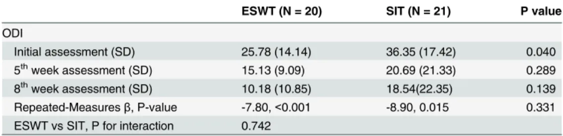
There were significant decreases in the ODI scores at the 8thweek post-treatment evalua-tions in the ESWT and SIT groups (p<0.001 and p = 0.015, respectively,Table 3). The changes
in the ODI score were not significantly different between the groups (p>0.05). Calculating the
proportional change in the ODI score following the formula below, we found that the mean changes at the 5thweek assessment and 8thweek assessment were greater in the ESWT group (-41.31% to -60.51%) than in the SIT group (-43.08% to -49.00%). The mean proportional
Table 2. The visual analog scale (VAS) at baseline, 5thweek, and 8thweek.
ESWT (N = 20) SIT (N = 21) P value
VAS (mm)
Initial assessment (SD) 62.00(18.24) 60.48(16.87) 0.783
5thweek assessment (SD) 31.50(18.99) 44.29(19.89) 0.042
8thweek assessment (SD) 21.00(18.32) 39.52(24.39) 0.009
Repeated measuresa,β, P-value -20.50,<0.001 -10.48, 0.005
ESWT vs SIT, P for interaction 0.004
SD: standard deviation; ESWT: extracorporeal shock wave therapy; SIT: physical modality.
aβ, P-value and interaction by generalized linear mixed model; PROC GLIMMIX, repeated-measure.
repeated-measure.
doi:10.1371/journal.pone.0142475.t002
Table 3. The Oswestry disability index (ODI) at baseline, 5thweek, and 8thweek.
ESWT (N = 20) SIT (N = 21) P value
ODI
Initial assessment (SD) 25.78 (14.14) 36.35 (17.42) 0.040
5thweek assessment (SD) 15.13 (9.09) 20.69 (21.33) 0.289
8thweek assessment (SD) 10.18 (10.85) 18.54(22.35) 0.139
Repeated-Measuresβ, P-value -7.80,<0.001 -8.90, 0.015 0.331
ESWT vs SIT, P for interaction 0.742
SD: standard deviation; ESWT: extracorporeal shock wave therapy; SIT: physical modality.
change in the ODI score in the ESWT group was 11.51% greater than in the SIT group (60.51%
−49.00% = 11.51%).
Proportional change in ODI score (%) = 100 × (5thor 8thweek assessment—Initial assess-ment) / Initial assessment
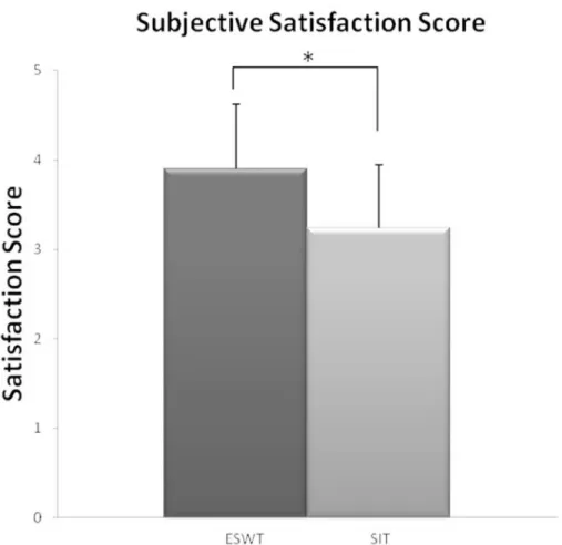
Subjective satisfaction of the patients
To evaluate the subjective satisfaction after treatment, a 5-level scale was used for evaluation at the 8thweek after treatment. The excellent and good scores were categorized into one group, and the acceptable and poor scores were categorized into another group for comparison. The patients in the ESWT group had better subjective satisfaction scores, with 70% reporting good to excellent satisfaction. The scores were significantly higher in the ESWT group than in the SIT group (p = 0.003,Fig 2).
Discussion
This study showed that non-invasive treatment options with ESWT and physical modalities with SWD and IFC were effective in reducing pain in coccydynia, and the effectiveness of
Fig 2. Patient satisfaction score.The subjective satisfaction score was higher in the ESWT group (*p<0.01). Differences between the ESWT and SIT groups in the subjective satisfaction score at the 8th
week follow-up were analyzed byt-test / Wilcoxon rank-sum test. ESWT: mean±SD = 3.95±0.76 versus SIT group: mean±SD = 3.24±0.76,t-test p = 0.003/Wilcoxon rank-sum test p = 0.007.
ESWT was superior to the physical modalities, with regard to pain reduction, grade of improvement in disability, and subjective satisfaction.
ESWT was first used to disintegrate renal stones in 1980 [31], and it has also been used in the treatment of various musculoskeletal disorders. Studies have shown that ESWT could enhance tendon repair and decrease pain. The success rate for tendinopathy ranges from 60% to 80% [32]. However, the mechanism of ESWT in pain relief is not completely understood, and many theories have been proposed to explain the effects of ESWT. One proposed mecha-nism for the effectiveness of ESWT in tendinopathy is increased tissue regeneration through the induction of mechanotransduction on the cytoskeleton and the stimulation of protein syn-thesis [33]. ESWT facilitates tendon repair through up-regulating extracellular matrix biosyn-thesis and increasing the expression of TGFβ1 and IGF-I [34]. ESWT also decreases the levels of inflammatory mediators (interleukins and matrix metalloproteinases) [35] and promotes vascularization of the injured tendon junction [25] to promote tissue healing [36]. ESWT has an effect on pain transmission by acting on substance P [37]. ESWT can promote neovasculari-zation by increasing the expression of factors including vascular endothelial growth factor (VEGF), endothelial nitric oxide synthase (eNOS), and proliferating cell nuclear antigen (PCNA) [25].
Patients with coccydynia show inflammatory changes in the area of the coccyx [8] and pain syndromes [9]. In a report evaluating the treatment and outcome analyses of patients with coc-cydynia, 66% of patients experienced significant improvement with conservative management using medication and/or local steroid injections [38]. Conservative treatments are generally recommended before surgical intervention with coccygectomy in the treatment of patients with coccydynia. Considering the effect of ESWT on reducing the inflammatory response, this non-invasive treatment modality might be beneficial for treating patients with coccydynia. There is only one recent report evaluating the effects of ESWT on pain relief in patients with coccydynia. Marwan et al. reported that three sessions of ESWT were effective in relieving pain, and the pain did not recur during a one-year follow-up period [27]. Our study random-ized patients to ESWT or physical modality treatment to compare the treatment outcomes. In addition to pain evaluation, we included a disability rating scale for a more comprehensive evaluation of the treatment outcomes. The results suggested favorable outcomes in patients treated with ESWT regarding the degree of improvement in pain, disability and subjective sat-isfaction. We concluded that ESWT could be an alternative treatment option for patients with coccydynia and have a satisfactory treatment response. Patients receiving ESWT visited the hospital once each week for 4 weeks. Conversely, patients receiving physical modalities visited the hospital 3 times per week. The patients’subjective satisfaction was better in the ESWT group, most likely because they had better treatment outcomes with fewer hospital visits and less time spent.
threshold improvements reported were mainly based on chronic low back pain populations; whether these thresholds are valid for the assessment of patients with coccydynia awaits further clinical studies.
There are some limitations to this study. The diverse etiologies of coccydynia should be studied with different treatments. The initial disability index differed between groups before therapy; however initial VAS scores were comparable between groups. The number of enrolled cases was not sufficiently large for etiological subgrouping, and further study with a larger number of cases is needed to compare ESWT with other treatment options, such as local ste-roid injections. Moreover, longitudinal follow-up over a longer period is indicated to evaluate the long-term effects of ESWT.
Conclusions
In this study, ESWT appeared to be useful in relieving the pain of coccydynia and more effec-tive in reducing pain syndromes than the use of physical modalities. Therefore, ESWT is rec-ommended as an alternative method for treating patients with coccydynia.
Supporting Information
S1 Text. CONSORT 2010 checklist of information to include when reporting a randomised trial.
(DOC)
S2 Text. Original protocol.
(PDF)
S3 Text. Translated protocol.
(PDF)
Author Contributions
Conceived and designed the experiments: SFL YJC CLH CHC. Performed the experiments: SFL YJC WLW CHC. Analyzed the data: YJC HPT CLL CLH WLW. Contributed reagents/ materials/analysis tools: HPT CLH CLL WLW CHC. Wrote the paper: SFL YJC CLH CHC.
References
1. Patel R, Appannagari A, Whang PG. (2008) Coccydynia. Curr Rev Musculoskelet Med 1: 223–226.
doi:10.1007/s12178-008-9028-1PMID:19468909
2. Maigne JY, Doursounian L, Chatellier G. (2000) Causes and mechanisms of common coccydynia: role of body mass index and coccygeal trauma. Spine 25: 3072–3079. PMID:11145819
3. Postacchini F, Massobrio M. (1983) Idiopathic coccygodynia. Analysis of fifty-one operative cases and a radiographic study of the normal coccyx. J Bone Joint Surg Am 65: 1116–1124. PMID:6226668
4. Fogel GR, Cunningham PY 3rd, Esses SI. (2004) Coccygodynia: evaluation and management. J Am Acad Orthop Surg 12: 49–54. PMID:14753797
5. Balain B, Eisenstein SM, Alo GO, Darby AJ, Cassar-Pullicino VN, Roberts SE, et al. (2006) Coccygect-omy for coccydynia: case series and review of literature. Spine 31: E414–420. PMID:16741442
6. Maigne JY, Chatellier G. (2001) Comparison of three manual coccydynia treatments: a pilot study. Spine 26: E479–483; discussion E484. PMID:11598528
7. Maigne JY, Chatellier G, Faou ML, Archambeau M. (2006) The treatment of chronic coccydynia with intrarectal manipulation: a randomized controlled study. Spine 31: E621–627. PMID:16915077
8. Wu CL, Yu KL, Chuang HY, Huang MH, Chen TW, Chen CH. (2009) The application of infrared ther-mography in the assessment of patients with coccygodynia before and after manual therapy combined with diathermy. J Manipulative Physiol Ther 32: 287–293. doi:10.1016/j.jmpt.2009.03.002PMID:
9. Nathan ST, Fisher BE, Roberts CS. (2010) Coccydynia: a review of pathoanatomy, aetiology, treatment and outcome. J Bone Joint Surg Br 92: 1622–1627. doi:10.1302/0301-620X.92B12.25486PMID:
21119164
10. Fuentes JP, Armijo Olivo S, Magee DJ, Gross DP. (2010) Effectiveness of interferential current therapy in the management of musculoskeletal pain: a systematic review and meta-analysis. Phys Ther 90: 1219–1238. doi:10.2522/ptj.20090335PMID:20651012
11. Rush J. (1996) Coccydynia. Current Orthopaedics 10: 128–131.
12. Goats GC. (1989) Continuous short-wave (radio-frequency) diathermy. Br J Sports Med 23: 123–127.
PMID:2691003
13. Mazza L, Formento E, Fonda G. (2004) Anorectal and perineal pain: new pathophysiological hypothe-sis. Tech Coloproctol 8: 77–83. PMID:15309642
14. Shields N, Gormley J, O'Hare N. (2002) Short-wave diathermy: current clinical and safety practices. Physiother Res Int 7: 191–202. PMID:12528575
15. De Andres J, Chaves S. (2003) Coccygodynia: a proposal for an algorithm for treatment. J Pain 4: 257–266. PMID:14622695
16. Rush PJ, Shore A. (1994) Physician perceptions of the value of physical modalities in the treatment of musculoskeletal disease. Br J Rheumatol 33: 566–568. PMID:8205406
17. Wray CC, Easom S, Hoskinson J. (1991) Coccydynia. Aetiology and treatment. J Bone Joint Surg Br 73: 335–338. PMID:2005168
18. Nijs J, Meeus M, Van Oosterwijck J, Roussel N, De Kooning M, Ickmans K, et al. (2011) Treatment of central sensitization in patients with 'unexplained' chronic pain: what options do we have? Expert Opin Pharmacother 12: 1087–1098. doi:10.1517/14656566.2011.547475PMID:21254866
19. Hurley DA, Minder PM, McDonough SM, Walsh DM, Moore AP, Baxter DG. (2001) Interferential ther-apy electrode placement technique in acute low back pain: a preliminary investigation. Arch Phys Med Rehabil 82: 485–493. PMID:11295009
20. Lara-Palomo IC, Aguilar-Ferrándiz ME, Matarán-Peñarrocha GA, Saavedra-Hernández M,
Granero-Molina J, Fernández-Sola C, et al. (2013) Short-term effects of interferential current electro-massage in adults with chronic non-specific low back pain: a randomized controlled trial. Clin Rehabil 27: 439–449.
doi:10.1177/0269215512460780PMID:23035006
21. Dizon JN, Gonzalez-Suarez C, Zamora MT, Gambito ED. (2013) Effectiveness of extracorporeal shock wave therapy in chronic plantar fasciitis: a meta-analysis. Am J Phys Med Rehabil 92: 606–620. doi:
10.1097/PHM.0b013e31828cd42bPMID:23552334
22. Childress MA, Beutler A. (2013) Management of chronic tendon injuries. Am Fam Physician 87: 486–
490. PMID:23547590
23. Peters J, Luboldt W, Schwarz W, Jacobi V, Herzog C, Vogl TJ. (2004) Extracorporeal shock wave ther-apy in calcific tendinitis of the shoulder. Skeletal Radiol 33: 712–718. PMID:15480643
24. Melzack R. (1979) Sensory modulation of pain. Int Rehabil Med 1: 111–115. PMID:233309
25. Wang CJ, Wang FS, Yang KD, Weng LH, Hsu CC, Huang CS, et al. (2003) Shock wave therapy induces neovascularization at the tendon-bone junction. A study in rabbits. J Orthop Res 21: 984–989.
PMID:14554209
26. Lee S, Lee D, Park J. (2014) Effects of extracorporeal shockwave therapy on patients with chronic low back pain and their dynamic balance ability. J Phys Ther Sci 26: 7–10. doi:10.1589/jpts.26.7PMID:
24567665
27. Marwan Y, Husain W, Alhajii W, Mogawer M. (2014) Extracorporeal shock wave therapy relieved pain in patients with coccydynia: a report of two cases. Spine J 14: e1–4.
28. Wewers ME, Lowe NK. (1990) A critical review of visual analogue scales in the measurement of clinical phenomena. Res Nurs Health 13: 227–236. PMID:2197679
29. Fairbank JC, Pynsent PB. (2000) The Oswestry Disability Index. Spine 25: 2940–2952; discussion
2952. PMID:11074683
30. Faul F, Erdfelder E, Lang AG, Buchner A. (2007) G*Power 3: a flexible statistical power analysis pro-gram for the social, behavioral, and biomedical sciences. Behav Res Methods 39: 175–191. PMID:
17695343
31. Chaussy C, Brendel W, Schmiedt E. (1980) Extracorporeally induced destruction of kidney stones by shock waves. Lancet 2: 1265–1268. PMID:6108446
32. Seil R, Wilmes P, Nuhrenborger C. (2006) Extracorporeal shock wave therapy for tendinopathies. Expert Rev Med Devices 3: 463–470. PMID:16866643
34. Abrahamsson SO. (1997) Similar effects of recombinant human insulin-like growth factor-I and II on cel-lular activities in flexor tendons of young rabbits: experimental studies in vitro. J Orthop Res 15: 256–
262. PMID:9167629
35. Notarnicola A, Moretti B. (2012) The biological effects of extracorporeal shock wave therapy (eswt) on tendon tissue. Muscles Ligaments Tendons J 2: 33–37. PMID:23738271
36. Kuo YR, Wang CT, Wang FS, Chiang YC, Wang CJ. (2009) Extracorporeal shock-wave therapy enhanced wound healing via increasing topical blood perfusion and tissue regeneration in a rat model of STZ-induced diabetes. Wound Repair Regen 17: 522–530. doi:10.1111/j.1524-475X.2009.00504.x
PMID:19614917
37. Hausdorf J, Lemmens MA, Kaplan S, Marangoz C, Milz S, Odaci E, et al. (2008) Extracorporeal shock-wave application to the distal femur of rabbits diminishes the number of neurons immunoreactive for substance P in dorsal root ganglia L5. Brain Res 1207: 96–101. doi:10.1016/j.brainres.2008.02.013
PMID:18371941
38. Hodges SD, Eck JC, Humphreys SC. (2004) A treatment and outcomes analysis of patients with coccy-dynia. Spine J 4: 138–140. PMID:15016390
39. Wright A, Hannon J, Hegedus EJ, Kavchak AE. (2012) Clinimetrics corner: a closer look at the minimal clinically important difference (MCID). J Man Manip Ther 20: 160–166. doi:10.1179/2042618612Y.
0000000001PMID:23904756
40. Schwind J, Learman K, O'Halloran B, Showalter C, Cook C. (2013) Different minimally important clinical difference (MCID) scores lead to different clinical prediction rules for the Oswestry disability index for the same sample of patients. J Man Manip Ther 21: 71–78. doi:10.1179/2042618613Y.0000000028
PMID:24421616
41. Ostelo RW, Deyo RA, Stratford P, Waddell G, Croft P, Von Korff M, et al. (2008) Interpreting change scores for pain and functional status in low back pain: towards international consensus regarding mini-mal important change. Spine 33: 90–94. doi:10.1097/BRS.0b013e31815e3a10PMID:18165753
42. Fritz JM, Hebert J, Koppenhaver S, Parent E. (2009) Beyond minimally important change: defining a successful outcome of physical therapy for patients with low back pain. Spine 34: 2803–2809. doi:10.