In Vivo
Deletion of the
Cebpa
+37 kb
Enhancer Markedly Reduces
Cebpa
mRNA in
Myeloid Progenitors but Not in
Non-Hematopoietic Tissues to Impair
Granulopoiesis
Hong Guo, Stacy Cooper, Alan D. Friedman*
Division of Pediatric Oncology, Johns Hopkins University School of Medicine, Baltimore, Maryland, United States of America
*afriedm2@jhmi.edu
Abstract
The murineCebpagene contains a +37 kb, evolutionarily conserved 440 bp enhancer that directs high-level expression to myeloid progenitors in transgenic mice. The enhancer is bound and activated by Runx1, Scl, GATA2, C/EBPα, c-Myb, Pu.1, and additional Ets fac-tors in myeloid cells. CRISPR/Cas9-mediated replacement of the wild-type enhancer with a variant mutant in its seven Ets sites leads to 20-fold reduction ofCebpamRNA in the 32Dcl3 myeloid cell line. To determine the effect of deleting the enhancerin vivo, we now characterize C57BL/6 mice in whichloxP sites flank a 688 bp DNA segment containing the enhancer. CMV-Cre mediated germline deletion resulted in diminution of the expected num-ber of viable Enh(f/f);CMV-Cre offspring, with 28-fold reduction in marrowCebpamRNA but normal levels in liver, lung, adipose, intestine, muscle, and kidney. Cre-transduction of line-age-negative marrow cellsin vitroreducedCebpamRNA 12-fold, with impairment of granu-locytic maturation, morphologic blast accumulation, and IL-3 dependent myeloid colony replating for>12 generations. Exposure of Enh(f/f);Mx1-Cre mice to pIpC led to 14-fold reduction ofCebpamRNA in GMP or CMP, 30-fold reduction in LSK, and<2-fold reduction in the LSK/SLAM subset. FACS analysis of marrow from these mice revealed 10-fold reduced neutrophils, 3-fold decreased GMP, and 3-fold increased LSK cells. Progenitor cell cycle progression was mildly impaired. Granulocyte and B lymphoid colony forming units were reduced while monocytic and erythroid colonies were increased, with reducedPu.1 andGfi1and increasedEgr1andKlf4in GMP. Finally, competitive transplantation indicated preservation of functional long-term hematopoietic stem cells upon enhancer deletion and confirmed marrow-intrinsic impairment of granulopoiesis and B cell generation with LSK and monocyte lineage expansion. These findings demonstrate a critical role for the +37 kb Cebpaenhancer for hematopoietic-specificCebpaexpression, with enhancer deletion lead-ing to impaired myelopoiesis and potentially preleukemic progenitor expansion.
OPEN ACCESS
Citation:Guo H, Cooper S, Friedman AD (2016)In VivoDeletion of theCebpa+37 kb Enhancer Markedly ReducesCebpamRNA in Myeloid Progenitors but Not in Non-Hematopoietic Tissues to Impair Granulopoiesis. PLoS ONE 11(3): e0150809. doi:10.1371/journal.pone.0150809
Editor:Tadayuki Akagi, Kanazawa University, JAPAN
Received:December 18, 2015
Accepted:February 19, 2016
Published:March 3, 2016
Copyright:© 2016 Guo et al. This is an open access article distributed under the terms of theCreative Commons Attribution License, which permits unrestricted use, distribution, and reproduction in any medium, provided the original author and source are credited.
Data Availability Statement:All relevant data are within the paper.
Introduction
CCAAT/enhancer binding proteinα(C/EBPα) is a basic region-leucine zipper transcription factor expressed preferentially within granulocytic and monocytic myeloid cells during hema-topoiesis [1]. C/EBPαlevels increase as long-term hematopoietic stem cells (LT-HSC) progress to the common myeloid progenitor (CMP) and subsequently to the granulocyte-monocyte progenitor (GMP), withCebpaopen reading frame (ORF) deletion preventing GMP formation associated with accumulation of upstream CMP and the Lin-Sca-1+c-kit+(LSK) stem/progeni-tor subsets [2,3]. As GMP mature, high-level C/EBPαexpression is required for granulopoiesis while reduced levels allow monopoiesis [4].
C/EBPαexpression or activity is commonly diminished in acute myeloid leukemia (AML) cases, includingCEBPApoint mutations impacting trans-activation or DNA-binding, RUN-X1-ETO expression reducingCEBPAtranscription, and C/EBPα(S21) phosphorylation also impairing trans-activation [5].
TheCebpapromoter is directly activated by C/EBPαand RUNX1 [6,7]. In addition, we identified a 440 bp DNA segment centered at +37.5 kb in the murineCebpagene, with 85% homology to the +42 kb region of the humanCEBPAlocus, harboring enhancer specific H3K4me1 histone marks and together with the promoter capable of directing high-level hCD4 transgene expression to GMP, CMP, and LSK cells but not to multiple non-hematopoietic tis-sues [7,8]. Runx1, C/EBPα, Pu.1, Erg, Fli-1, GATA2, Scl, Meis1, and Gfi-1b bind chromatin in the region of this enhancer in hematopoietic cells as determined by ChIP-Seq [9,10], Runx1, C/EBPα, Pu.1, Fli-1, Erg, Ets1, c-Myb, GATA2, and Scl bind conserved enhancerciselements in gel shift assays, and mutation of the Runx1, C/EBP, Ets, Myb, GATA, or E-box sites each reduce enhancer activity in 32Dcl3 myeloid cells in reporter assays [7,11]. Mutation of its seven Ets sites led to the greatest reduction in enhancer activity, and CRISPR/Cas9-mediated replacement of the endogenous enhancer alleles with a variant harboring point mutations in these Ets sites led to 20-fold reducedCebpamRNA expression in 32Dcl3 myeloid cells [11].
To determine whether the +37 kbCebpaenhancer is also critical for regulatingCebpa expressionin vivo, we have now generated and characterized mice in whichloxP sites flank the enhancer, designated as Enh(f/f) mice. Germline deletion using CMV-Cre revealed marked reduction ofCebpaexpression in marrow but not in other tissues, including liver, adipose, and lung, that normally express C/EBPα. As germline deletion or use of Vav-Cre to induce hemato-poietic-specific deletion led to significant early post-natal lethality, we focused on analysis of adult Enh(f/f);Mx1-Cre mice subjected to pIpC injections to induce enhancer deletion, fol-lowed by recovery for four weeks to reestablish homeostasis and to avoid transient pIpC effects. In this model,CebpamRNA was reduced 14-fold in GMP or CMP and 30-fold in the LSK mar-row population associated with a 3-fold reduction in GMP, LSK expansion, LSK/SLAM cell depletion, and impaired granulopoiesis relative to monopoiesis. Erythroid progenitor and platelet expansion and reduced numbers of B lymphoid colony forming units was also
observed, with preservation of functional LT-HSC. These findings demonstrate that the +37 kb Cebpaenhancer is central to regulation ofCebpatranscription and granulopoiesisin vivo.
Methods
Ethics Statement
This study was carried out in strict accordance with the recommendations in the Guide for the Care and Use of Laboratory Animals of the National Institutes of Health. The protocol (M013M116) was approved by the Johns Hopkins University Animal Care and Use Commit-tee. All efforts were made to minimize suffering.
supply support. The Giant Food Children's Cancer Research Fund, no grant number,www.giantfood. com. Giant foods makes an annual charitable donation to our Pediatric Oncology division and the monies are used in part for maintaining laboratory equipment. National Institutes of Health grant R01 HL130034 to Alan D. Friedman. The funders had no role in study design, data collection and analysis, decision to publish, or preparation of the manuscript.
Generation of Enhancer-Floxed Mice
The C57BL/6 (B6)-derived 123 kb BAC RP23-375B6 was obtained from CHORI. Recombi-neering methodology [12] was then utilized to transfer a 6,950 bp segment containing the 439 bp enhancer, a 940 bp 3’homology arm, and a 5,520 bp 5’homology arm to pBluescript II, fol-lowed by insertion of aloxP site 214 bp upstream and afrt-PGK-Neo-frt-loxP cassette derived from plasmid PL451 35 bp downstream of the enhancer, thus floxing a 688 bp genomic DNA segment. After removal of vector sequences, the plasmid insert was provided to the Johns Hop-kins Transgenic Core facility, which generated multiple G418-resistant B6 BL-1 embryonic stem cell (ESC) lines after electroporation. These were screened by 3’PCR using a forward primer near the 3’end of the Neo cassette and a reverse primer distal to the 3’homology arm. Homologous recombination was confirmed by Southern blotting afterSpeI digestion of geno-mic DNA, as described [13]. The 5’probe was a 1.3 kbKpnI/HindIII fragment centered 4.1 kb 5’to the enhancer, and the 3’probe was a 0.9 kbBamHI/XhoI fragment encompassing the 3’ homology arm. Targeted B6 ESC lines were then utilized to generate chimeric mice after injec-tion into B6-albino blastocysts. Briefly, ~12–15 targeted ESC were injected into blastocyst stage embryos obtained from superovulated B6(Cg)-Tyrc-2J/J females (Jackson Laboratories, #58). Following injection, surviving embryos were surgically transferred to oviducts of psuedopreg-nant ICR females (~15 embryos/female). Chimeric offspring were then bred to B6-albino mice and offspring with fully black fur were screened by tail clip DNA PCR for presence of the knockin (KI) DNA using primers that flank the 5’loxP site:
loxP5-F:5’-ACCTTCCGTGCTCAAGTCTGand
loxP5-R:5’-AAGTCCCCTTTGCCAGACAC, followed by 1.5% agarose gel electrophoresis.
Successfully targeted mice were then bred to homozygosity. Mx1-Cre (#3556), female CMV-Cre (#6054), female ROSA26-FLPo (#12930), and Vav-Cre (#8610) mice were obtained from Jackson Laboratories. Cre DNA was detected using:
Cre-F:3’-GTCCAATTTACTGACCGTACACand
Cre-R:5’-CTGTGACTTGGTCGTGGCAGC.
Deletion of both floxed alleles by CMV-Cre was determined by absence of the 5’loxP site and by detection of a band of appropriate size using primers upstream of the 5’loxP site and downstream of the 3’loxP site:
EnhΔ-F:5’-CCCAAGACAGCCAGGTTAGGAGTTCCand
EnhΔ-R:5’-ACATGATGTCCCGGAGAACAGAGCC.
Bialleleic deletion of the Neo cassette after FLPo expression was assessed using primers: Frt5-F:5’-GGTCTGAAGAGGAGTTTACGTCClocated just downstream of the 5’frtsite and
PGK-R:5’-AGAGGAGAACAGCGCGGCAGlocated in the PGK promoter and primers:
Enh-F:5’-CCACATCACACGGGGCCTGCand
3Arm-R:5’-ACACCAAGAGCTAAGAGGACACCCCflanking the entirefrt-PGK-Neo-frt
cas-sette. 8–12 wk old Enh(f/f);Mx1-Cre mice were injected intraperitoneally with 500μg of pIpC (Sigma) every other day for 6 doses. Blood or marrow was isolated 4 wks later for analysis.
Retroviral Transduction and Progenitor Assays
293T cells were cultured in Dulbecco’s modified Eagle medium (DMEM) with 10% heat-inacti-vated fetal bovine serum (HI-FBS). Forin vitrostudies, marrow isolated from Enh(f/f) or wild-type (WT) mice injected intraperitoneally with 150 mg/kg 5-fluorouracil (5-FU) 6 days earlier was subjected to red cell lysis with NH4Cl and cultured for 1 day in 10 ng/mL murine IL-3, 10
per 100 mm dish as described [7]. Three days later, 2μg/mL puromycin was added, and after 2 additional days viable cells isolated with Lympholyte M (Cedarlane Labs) were either plated in methylcellulose or subjected to lineage-depletion using biotin-conjugated B220, Gr-1, CD11b, Ter119, and CD3 mouse Lineage Cocktail (BD Pharmingen), anti-biotin microbeads, and MACS columns (Miltenyi Biotec) and placed in liquid culture with IMDM, 10% HI-FBS and IL-3, IL-6, and SCF. Myeloid colonies were enumerated 7–8 days later based on colony mor-phology. Myeloid colonies were also obtained using 30 ng/mL human G-CSF (Amgen), 30 ng/mL murine M-CSF, or 30 ng/mL murine GM-CSF (Peprotech). BFU-E were enumerated on day 10 after culture in Methocult M3120 (1% final concentration) with IMDM, 2 mM gluta-mine, 55 nMβ-mercaptoethanol, 10% plasma-derived serum (Animal Technologies), 20% BIT (Stem Cell Technologies), 5% PFHM-II (Invitrogen), and 10 U/mL (100 ng/mL) murine eryth-ropoietin (EPO). B lymphoid CFU were enumerated 7 days after culture in Methocult 3630, which contains IMDM, HI-FBS, and human IL-7. For myeloid colony replating, CFUs were pooled, washed with PBS, and replated at 1E3 cells/mL every 7 days. Cell morphology was assessed by Wright-Giemsa staining of cytospun cells. Photomicrographs were taken using a Zeiss Axiophot microscope (Carl Zeiss), a Kontron Electronik Progress 3012 camera (Kon-tron), and a 63X/1.40 NA oil objective.
Quantitative RNA Analysis and Western Blotting
RNA from hematopoietic cells was prepared using NucleoSpin RNA II, with use of RNase-free DNase (Machery-Nagel). Tissues were homogenized in Trizol using Tissue-Tearor (United Laboratory Plastics); RNA was extracted using chloroform, isopropanol precipitated, and fur-ther purified using NucleoSpin RNA II. First strand cDNA was prepared using AMV reverse transcriptase (Promega) and oligodT primer at 42°C for 1 hr. Quantitative PCR was carried out using 5–25 ng of each cDNA using iQ SYBR Green supermix (Bio-Rad).Cebpa,Cebpg, GMCSFRa, and ribosomal subunitmS16internal control primers were:
Cebpa-F:5’-TGGATAAGAACAGCAACGAG,
Cebpa-R:5’-TCACTGGTCAACTCCAGCAC,
Cebpg-F:5’-GCGCAGAGAGCGGAACAA,
Cebpg-R:5’-GTATCTTGAGCTTTCTGCTTGCT,
GMCSFRa-F:5’-CCAGGGATCAGGGACAAGG,
GMCSFRa-R:5’-CCTGTCAGTCACGTTGGGG,
mS16-F:5’-CTTGGAGGCTTCATCCACAT, and
mS16-R:5’-ATATTCGGGTCCGTGTGAAG.
Additional primer pairs were as described [4,8]. Western blotting for C/EBPαandβ-actin was carried out as described [7].
FACS Analysis and Flow Cytometry
A2F10.1) for MPP, ST-HSC, and LT-HSC. Alternatively, LSK cells were stained using PE— anti-CD150 (Q38-480) and Brilliant Violet 421–anti-CD48 (HM48-1) for LSK/SLAM LT-HSC. Marrow subsets for RNA analysis were obtained after lineage-depletion and antibody staining via a FACSAria II cell sorter (BD Biosciences).
Cell Proliferation and Apoptosis Assays
For cell cycle analysis using the BrdU Flow kit (BD Pharrmingen), mice were given a single intraperitoneal injection of 5-bromodeoxyuridine (BrdU; 100μg/g). 3 hrs later, bone marrow was isolated from femurs, tibias, iliac crest and spine. After red cell lysis, the cells were lineage depleted as above. Lineage negative cells were stained with LIVE/DEAD Fixable Aqua (Life Technologies) and for LSK, CMP, GMP, and MEP as above. The fixation, DNase treatment, and staining with FITC anti-BrdU and 7AAD were per the BrdU Flow kit protocol. For quies-cence analysis, lineage depleted marrow was stained with LIVE/DEAD Fixable Aqua and sur-face makers for LSK, CMP, GMP, and MEP, fixed, treated with DNase, and stained with FITC-anti-Ki67 (eBioscience, SolA15) and 7AAD. For analysis of apoptosis and cell death, marrow without red cell lysis was lineage depleted using biotin-conjugated mouse Lineage Cocktail (BD Pharmingen), MojoSort Streptavidin Nanobeads (Biolegend), and EasySep Magnet (Stem-cell Technology), followed by staining for progenitor subsets and with Alexa Fluor 488-anti-Annexin V (Life Technologies) and 7AAD.
Transplantation Studies
Enh(f/f);Mx1-Cre CD45.2+marrow cells, obtained 4 wks after pIpC exposure, were trans-planted by tail vein injection at a 1:1 ratio with 2E5 CD45.1+WT competitor cells into synge-neic WT CD45.1+recipient mice irradiated to 950 cGy. Mice were euthanized at 19 wks, and marrow and peripheral blood cells were analyzed using PE- or FITC-anti-CD45.1 (A20), and APC-anti-CD45.2 (104, Biolegend) and additional antibodies as above. 1E6 marrow cells from primary recipients were transplanted into irradiated secondary recipients, one secondary mouse per primary mouse, followed by similar blood analysis 16 wks later.
Statistics
Means and standard deviations (SD) are shown. The Studentttest was used for statistical comparisons.
Results
Generation of Mice with Floxed +37 kb
Cebpa
Enhancer Alleles
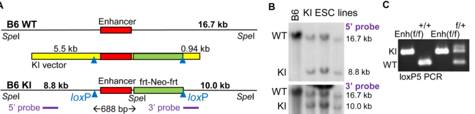
A targeting construct for homologous knockin (KI vector) was assembled in which the 439 bp +37 kbCebpaenhancer, 214 bp of genomic DNA upstream of the conserved enhancer seg-ment, 35 bp of downstream DNA, and a downstreamfrt-PGK-Neo-frtcassette are flanked by loxP sites (Fig 1A). This construct also includes a 5.5 kb 5’homology arm and a 940 bp 3’ homology arm. The 3’homology arm length was limited by a microsatellite repeat. The geno-mic DNA segments were derived from a B6 BAC. Of note, 129 strain BAC clones spanning the enhancer were not available. We positioned the 5’loxP site 214 bp upstream of the enhancer, rather than closer, due to a B6:129 homology gap at this location, to potentially facilitate 129 ESC cell targeting. The 3’arm was identical between B6 and 129 DNA, but the 5’arm contains 10 single nucleotide differences and a 4 bp gap`.
electroporation into a B6 ESC line yielded six subclones with HR among 176 lines screened. Genomic DNA isolated from the parental B6 ESC line and from three targeted B6 lines was digested withSpeI and subjected to Southern blotting using a 1.3 kb probe located in the 5’ homology arm or a 0.9 kb probe encompassing the 3’homology arm (Fig 1B). A 16.7 kb band was detected with either probe in all four lines, representing unmodified genomic DNA. In addition, 8.8 kb or 10.0 kb bands were detected in the three KI ESC lines with the 5’or 3’ probe, respectively, indicating presence of a properly targeted allele. Two of these lines were microinjected into albino blastocysts, yielding chimeric offspring with black and white coat col-ors. These were bred to albino mice, and tail snip DNAs from offspring with all black coat color were screened by PCR with a primer pair surrounding the 5’loxP site. Heterozygous Enh (f/+) mice were then bred to generate homozygous Enh(f/f) mice, as assessed also by genomic DNA PCR using the loxP5 primer pair (Fig 1C).
Effect of
In Vitro
Enhancer Deletion on
Cebpa
RNA Expression and
Myelopoiesis
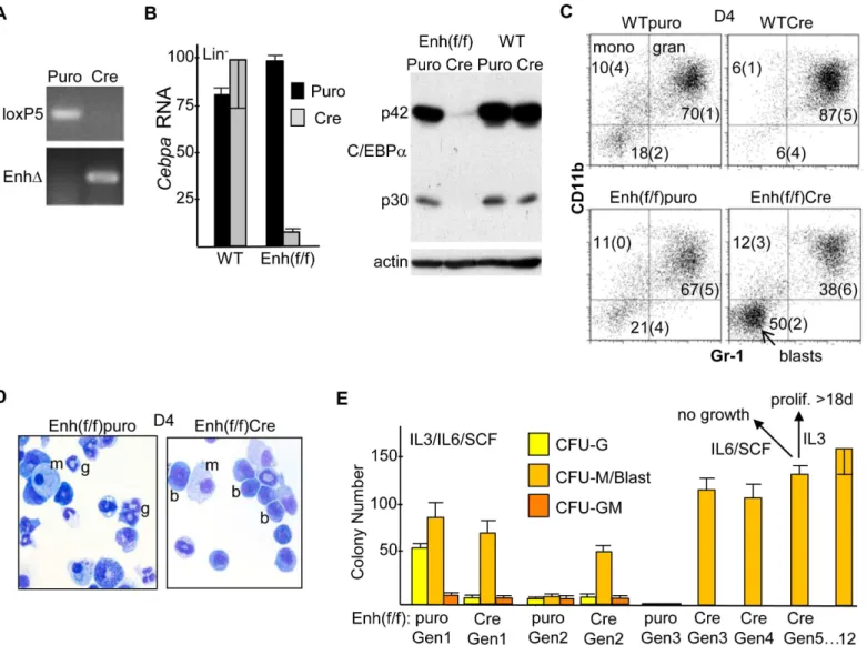
Mononuclear marrow cells from 8–12 wk old WT or Enh(f/f) mice exposed six days earlier to 5-FU were cultured in IMDM/FBS with the myeloid cytokines IL-3, IL-6, and SCF, transduced with pBabePuro (Puro) or pBabePuro-Cre (Cre), subjected to puromycin selection followed by removal of dead cells, and finally lineage-depleted. PCR analysis of DNA from Cre-transduced cells demonstrates highly efficient enhancer deletion, as indicated by complete loss of the floxed, KI 5’loxP site PCR product using the loxP5 primer pair and gain of a PCR product resulting from enhancer deletion (EnhΔ,Fig 2A). Quantitative RT-PCR analysis demonstrated equivalentCebpamRNA expression in Puro-transduced WT versus Enh(f/f) cells, indicating lack of effect of the PGK-Neo cassette onCebpaexpression, equivalent expression in Puro-compared with Cre-transduced WT cells, indicating lack of effect of Cre onCebpaexpression, and 12-fold average reduction inCebpaRNA in Enh(f/f) cells transduced with Cre versus Puro, indicating a key role for the +37 kb enhancer in regulating myeloid cell autonomous Cebpaexpressionin vitro(Fig 2B, left). The same Puro- or Cre-transduced, puromycin-selected, lineage-depleted samples were also subjected to Western blotting for C/EBPαandβ
-Fig 1. Generation of B6 Mice with FloxedCebpa+37 kb Enhancer Alleles. A) Diagram of a wild-type genomic allele in the vicinity of the +37 kbCebpa enhancer (B6 WT), the knockin (KI) vector, and a targeted genomic allele (B6 KI). The conserved 439 bp enhancer is flanked by afrt-PGK-Neo-frtcassette and twoloxP sites. Cre-mediated deletion of the DNA between theloxP sites will remove the enhancer, 214 bp of DNA 5’to the enhancer, and 35 bp of DNA 3’to the enhancer, 688 bp in total. Positions ofSpeI sites, fragment sizes expected afterSpeI digestion, and locations of the 5’and 3’probes used for Southern blotting are also shown.B) Southern blots obtained afterSpeI digestion of genomic DNA isolated from parental B6 BL-1 ESC (B6) or from three targeted lines (KI ESC lines) using the 5’or 3’probes. Locations and sizes of fragments derived from the WT and KI alleles are indicated.C) PCR of tail snip DNA, using loxP5 primers spanning the 5’loxP site, from homozygous Enh(f/f), WT (+/+), and heterozygous (f/+) mice obtained after breeding. Locations of bands obtained from the KI and WT alleles are indicated.
actin (Fig 2B, right). Cre-transduction of Enh(f/f) marrow led to marked reduction in C/EBPα protein, both its full-length p42 and shorter p30 isoforms.
Transduced, puromycin-selected lineage-negative (Lin-) cells were also placed in liquid cul-ture with IL-3, IL-6, and SCF. Four days later (D4), the cells were analyzed using FACS for CD11b and Gr-1 expression (Fig 2C). Under these culture conditions, CD11b+Gr-1-cells rep-resent monocytes and CD11b+Gr-1+cells represent granulocytes, as we previously confirmed using additional FACS antibodies [7]. Cre-transduction of Enh(f/f) cells led to ~2-fold
Fig 2. Effect ofin vitroEnhancer Deletion onCebpaExpression and Myelopoiesis. A) Mononuclear marrow cells from WT or Enh(f/f) mice were placed in IMDM/FBS with IL-3, IL-6 and SCF for 24 hr, transduced with pBabePuro (Puro) or pBabePuro-Cre (Cre) for 48 hr, puromycin selected for an additional 48 hr, and finally lineage-depleted. Genomic DNA was then subjected to PCR using the loxP5 or EnhΔprimer pairs followed by agarose gel electrophoresis and visualization by ethidium bromide staining.B) Total cellular RNAs were analyzed forCebpaand large ribosomal subunitmS16mRNA expression.Cebpa RNA expression, normalized usingmS16expression and set to 100 for WT marrow transduced with Cre, is shown (left, mean and SD from 3 determinations). Total cellular proteins isolated from the same groups of Lin-cells were subjected to Western blotting for C/EBPαandβ-actin; locations of the p42 and p30 C/ EBPαalternative translation variants are indicated (right).C) Lin-cells were placed in liquid culture with IMDM/FBS, IL-3, IL-6, and SCF and analyzed for surface CD11b and Gr-1 expression on day 4 (D4; mean and SD from three determinations).D) The morphology of Puro- or Cre-transduced Enh(f/f) cells from these cultures was assessed on D4 by Wright’s Giemsa staining; g—granulocyte; m—monocyte; b—blast.E) Lin-cells were cultured similarly in methylcellulose at 1E3 cell/mL, and myeloid CFUs were enumerated 7–8 days later (Gen1). CFU cells were then collected, washed with PBS, replated at 1E3 cells/mL, and analyzed similarly each 7 days (Gen 2 to Gen 12). In addition, a proportion of Gen5 cells were evaluated for their ability to proliferate in liquid culture in IMDM/FBS with IL-6/SCF or IL-3.
reduction in the proportion of granulocytes and a 2.5-fold increase in an immature CD11b- Gr-1-“blast”population, compared with Puro-transduced Enh(f/f) cells. Morphologic evaluation of these populations confirmed reduction of mature granulocytes and an increase in immature blastic cells in response to enhancer deletion (Fig 2D). Finally, transduced Lin-cells were placed in methylcellulose culture with IL-3, IL-6, and SCF. Enumeration of first generation (Gen1) colony-forming units (CFU) demonstrated marked reduction in CFU-G in response to enhancer deletion, with little effect on CFU-M or CFU-GM (Fig 2E). The ability of these colo-nies to replate for successive generations in the same cytokines was then evaluated. Puro-trans-duced myeloid CFU did not replate past Gen2. In striking contrast, Cre-transPuro-trans-duced CFU replated for at least 12 generations, with the morphology of the large majority of CFU cells past the 5thgeneration having a blastic appearance. In addition to replating in methylcellulose, 5th generation CFU cells were placed in liquid culture with IMDM/FBS and either 6/SCF or IL-3. The cells did not proliferate in IL-6/SCF and rapidly died, whereas the cells proliferated con-tinuously for at least 18 days in IL-3, increasing ~2-fold each day. Together, these data indicate that reducedCebpaexpression consequent to +37 kb enhancer deletion impairs hematopoietic cell autonomous granulopoiesisin vitro, leading to preservation of immature myeloid progeni-tors capable of long-term, IL-3-dependent proliferation without complete terminal maturation, a preleukemic phenotype.
Effect of
In Vivo
Enhancer Deletion on
Cebpa
mRNA Expression
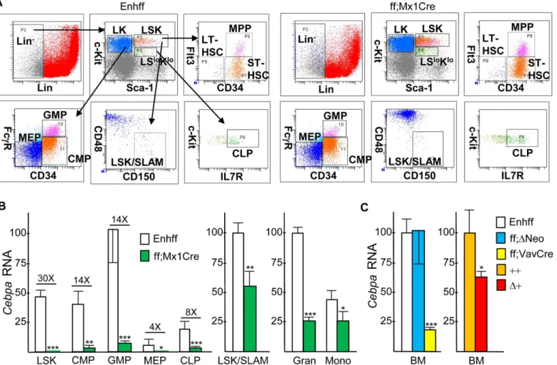
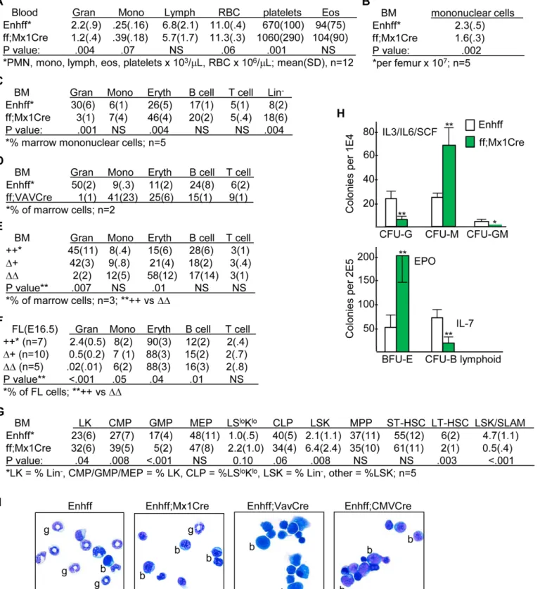
To evaluate thein vivoeffect ofCebpa+37 kb enhancer deletion onCebpaexpression in adult hematopoietic cells, we followed the example of investigators studying thein vivoconsequences of floxedCebpaORF deletion [3,14,15] and generated Enh(f/f);Mx1-Cre mice. The activity of the interferon-responsive Mx1 promoter can be induced by double-stranded pIpC RNA injec-tions, leading to efficient Cre induction in marrow cells and variable induction in other tissues. Enh(f/f);Mx1-Cre mice or Enh(f/f) littermates received six pIpC injections over a 12 day period. Four weeks after the last pIpC injection, to ensure maximal deletion efficiency, com-plete recovery from acute effects of pIpC, and reestablishment of hematopoietic homeostasis, mononuclear marrow cells were subjected to flow cytometry to allow isolation of the LSK, CMP, GMP, MEP, and LSK/SLAM populations. Representative FACS analyses of these and additional marrow hematopoietic stem/progenitor subsets, obtained from an Enh(f/f) and an Enh(f/f);Mx1-Cre mouse, is shown (Fig 3A). After total cellular RNA isolation,Cebpa expres-sion was evaluated via RT-PCR (Fig 3B). Due to their limited cell numbers, LSK/SLAM cells were obtained from a separate set of mice; in addition, marrow granulocytes and monocytes were isolated from a third set of mice. As expected, in marrow subsets isolated from Enh(f/f) mice,CebpamRNA increased as LSK or CMP progressed to GMP and was minimal in MEP. Reduced but evidentCebpain CLP may in part represent expression in a B/myeloid CLP sub-set.CebpamRNA was reduced upon Cre-mediated enhancer deletion by 30-fold, on average, in the LSK population, 14-fold in CMP or GMP, 4-fold in MEP, 8-fold in CLP, 1.5-fold in LSK/SLAM cells, 4.6-fold in granulocytes, and 1.6-fold in monocytes. These data indicate a critical dependence upon the presence of the +37 kbCebpaenhancer forCebpamRNA expres-sion in LSK, CMP, or GMP, intermediate dependence in CLP, and only mild dependence in the LSK/SLAM subset. Greater reduction ofCebpaRNA in GMP compared to granulocytes or monocytes may reflect maturation from a small number of GMP lacking complete enhancer deletion.
These data are consistent with the finding that Puro-transduced WT and Enh(f/f) Lin-marrow cells express equivalent levels ofCebpamRNA.
We also generated Enh(f/+);Vav-Cre mice and mated these together in an effort to obtain Enh(f/f);Vav-Cre offspring. The Vav promoter is expressed throughout hematopoiesis, begin-ning during the fetal liver stage of development, but not in non-hematopoietic tissues. How-ever, Enh(f/f);Vav-Cre offspring were obtained at 25% of the expected Mendelian ratio, and the three such mice identified were runted, potentially reflecting increased susceptibility of neonates to the infectious consequences of marked neutropenia. Analysis ofCebpamRNA expression in marrow cells from these Enh(f/f);Vav-Cre mice demonstrated 6-fold reduced expression (Fig 3C), further confirming that the +37 kbCebpaenhancer acts in a hematopoi-etic autonomous manner to play a key role in regulatingCebpatranscription.
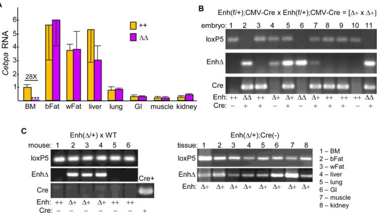
Finally, to assess the effect of enhancer deletion in non-hematopoietic tissues, we utilized CMV-Cre, which provides germline deletion [16]. In addition to marrow myeloid cells,Cebpa is expressed prominently in adipocytes, hepatocytes, and type II pneumocytes [17,18]. As with
Fig 3. Effect ofin vivoEnhancer Deletion onCebpaHematopoietic Expression. A) Representative FACS analyses of marrow stem/progenitor subsets. GMP, CMP, and MEP were analyzed within the Lin-Sca-1-c-kit+(LK) subset, CLP were analyzed within the Lin-Sca-1loc-kitlo(LSloKlo) subset, and MPP, ST-HSC, LT-HSC, or LSK/SLAM cells were enumerated within the Lin-Sca-1+c-kit+(LSK) subset.B) Total cellular RNAs from the LSK, CMP, GMP, MEP, CLP, LSK/SLAM, granulocyte, or monocyte marrow subsets isolated from Enh(f/f) or Enh(f/f);Mx1-Cre mice that had been subjected to pIpC injections and allowed to recover for 4 wks were analyzed for relativeCebpamRNA expression, normalized tomS16mRNA expression (mean and SD from three
determinations).C) RelativeCebpamRNA expression was analyzed similarly from marrow mononuclear cells isolated from Enh(f/f) mice versus mice lacking both PGK-Neo cassettes (ff;ΔNeo) or Enh(f/f);Vav-Cre mice and from wild-type (++) versus Enh(f+);CMV-Cre (Δ+) mice (n = 3).
Vav-Cre, upon mating Enh(f/+);CMV-Cre, designatedΔ+, mice few offspring with homozy-gous enhancer deletion (ΔΔ) were obtained, 40% of the predicted number, and those obtained were again smaller than their littermates.Cebpamarrow RNA was reduced 1.5-fold, on aver-age, inΔ+ vs Enh(f/f) control (++) littermates (Fig 3C). RNAs isolated from marrow or from seven non-hematopoietic tissues from Enh(f/f);CMV-Cre (ΔΔ) or age-matched wild-type (++) mice were subjected to analysis ofCebpaexpression (Fig 4A).CebpamRNA was reduced 28-fold in total marrow mononuclear cells, on average. In contrast, no significant reduction was seen in brown fat, white fat, liver, lung, small intestine, skeletal muscle, or kidney.
To confirm that CMV-Cre mediates germline enhancer deletion of the floxedCebpa enhancer, we first isolated DNA from a litter of E16.5 embryos derived from a cross between Enh(f/+);CMV-Cre (Δ+) parents, followed by PCR analysis for the 5’loxP site, non-floxed WT product (loxP5), enhancer deletion (EnhΔ), and Cre (Fig 4B). Embryos were used to increase the likelihood of obtaining homozygous enhancer deletion. Presence of the loxP5 and not the EnhΔband indicates enhancer genotype ++; presence of both bands indicates heterozygous deletion orΔ+, and presence of only the EnhΔband indicates homozygous deletion orΔΔ. Embryo 4 lacks Cre but has heterozygousΔ+ enhancer deletion, and embryo 6 lacks Cre but has homozygousΔΔenhancer deletion. Presence of enhancer deletion on one or both alleles in the absence of Cre indicates that in a prior generation CMV-Cre mediated germline enhancer deletion, which would then be passed on to subsequent offspring in all tissues. Second, we simi-larly evaluated tail DNA from 4 wk old offspring obtained from a cross between a wild-type B6 mother (Jackson Laboratories) and an Enh(Δ+) father who similar to Embryo 4 lacked CMV-Cre (Fig 4C, left). Three pups had an Enh(Δ+) genotype, despite absence of Cre in their or their parent’s genomes, indicating continued inheritance of enhancer deletion via the germ-line. Moreover, further analysis of one of these mice indicated that each of eight tissues ana-lyzed lacked an enhancer allele (Fig 4C, right). Taken together, these RNA and DNA data indicate that the +37 kbCebpaenhancer acts specifically in hematopoietic cells compared to those non-hematopoietic tissues analyzed.
Effect of
Cebpa
Enhancer Deletion on Hematopoietic Lineage
Development
Regarding marrow stem/progenitor subsets, Mx1-Cre mediated enhancer deletion increased CMP 1.4-fold, reduced GMP 3.4-fold, increased LSK 3-fold, reduced LSK;CD34-Flt3-LT-HSC 3-fold, and reduced the LSK/SLAM population 9.4-fold, on average (Fig 5G). In methylcellu-lose culture with IL-3/IL-6/SCF, enhancer deletion reduced CFU-G 5-fold while increasing CFU-M 3-fold; BFU-E obtained by culture with EPO were increased 4-fold, and B lymphoid CFU obtained in IL-7 were reduced 4-fold (Fig 5H). Morphologic analysis of marrow from Enh(f/f) compared with Enh(f/f);Mx1-Cre, Enh(f/f);Vav-Cre, or Enh(f/f);CMV-Cre mice dem-onstrates marked reduction in neutrophils and increased cells with blast morphology in the absence of theCebpaenhancer, the latter consistent with the expanded Lin-, CMP, and LSK populations (Fig 5I). In summary, these data demonstrate reduced GMP and marked inhibi-tion of GMP maturainhibi-tion along the granulocytic lineage, with preservainhibi-tion and even increased monopoiesis upon deletion of the +37 kbCebpaenhancer. Increased marrow erythropoiesis and blood platelets may represent redirection of CMP to MEP, and increased LSK and deple-tion of the LT-HSC and LSK/SLAM subsets may occur in response to GMP depledeple-tion.
Effect of
Cebpa
Enhancer Deletion on Transcription Factor and Myeloid
Cytokine Receptor Expression
The RNA samples used to evaluateCebpamRNA expression in GMP, CMP, or LSK marrow subsets from Enh(f/f) or Enh(f/f);Mx1-Cre mice exposed 4 wks earlier to pIpC were also
Fig 4. Effect of Germline Enhancer Deletion onCebpaExpression in Non-Hematopoietic Tissues. A) Total RNAs from marrow mononuclear cells, brown fat (bFat), white fat (wFat), liver, lung, small intestine (GI), skeletal muscle, or kidney from wild-type (++) and Enh(f/f);CMV-Cre (ΔΔ) mice were analyzed similarly for relativeCebpaexpression (n = 3).B) Total embryo DNA from a litter obtained at E16.5 from a cross between Enh(f/+);CMV-Cre (Δ+) mice was subjected to PCR using the loxP5, EnhΔ, and Cre primer pairs, followed by agarose gel electrophoresis and ethidium bromide staining. Enhancer and Cre genotypes are indicated.C) Tail DNA from a litter of 4 wk pups obtained from a cross between a WT andΔ+ mouse, both lacking CMV-Cre, was analyzed similarly (left). DNA from a mouse harboring Mx1-Cre served as a positive control for the Cre PCR (Cre+). DNAs from adult tissues of one of theΔ
+ pups were also analyzed using loxP5 and EnhΔ5 PCR (right).*- p<0.05,**- p<0.01,***- p<0.001.
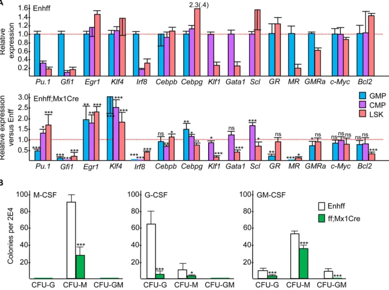
evaluated for expression of several myeloid and erythroid transcription factors. Relative expres-sion in GMP versus CMP versus LSK from Enh(f/f) mice and the ratio of expresexpres-sion in Enh(f/ f);Mx1-Cre versus Enh(f/f) mice for each subset is shown (Fig 6A).Pu.1andGfi1levels were increased in GMP compared with CMP or LSK. Enhancer deletion reducedPu.12-fold in GMP, potentially reflecting direct regulation ofPu.1transcription by C/EBPα[20,21], but increasedPu.11.3-fold in CMP and 3-fold in LSK.Gfi1was markedly reduced in GMP, CMP, and LSK, likely reflecting the role of Gfi-1 in mediating granulopoiesis [22], whileEgr1and Klf4levels were increased in all three subsets, potentially reflecting their role in monopoiesis [23,24].Irf8levels were markedly reduced in GMP, CMP, and LSK cells in response to enhancer deletion despite the positive role Irf8 plays during monopoiesis [25] but consistent with 5-fold reduction inIrf8evident in the expanded monocyte progenitor population present in the marrow ofCebpaORF(f/f);Mx1-Cre mice exposed to pIpC [26].Cebpblevels were mini-mally affected byCebpaenhancer deletion, of relevance given the ability of C/EBPβto compen-sate for absence of C/EBPαduring granulopoiesis [27,28].Cebpglevels were increased only 1.4-fold in GMP and 1.2-fold in CMP and were mildly reduced in LSK cells, of relevance given the 5-fold increase inCebpgevident in LSK cells from adult mice lacking both copies of the CebpaORF following Mx1-Cre-mediated deletion [29] and given the adjacency of theCebpa andCebpggenes. Finally, RNAs encoding the erythroid factorsGata1,Klf1, andSclwere only minimally changed in CMP and reduced in LSK despite the increased erythropoiesis observed, perhaps reflecting a post-transcriptional effect of reduced C/EBPαon the activity of one or more of their cognate transcription factors.
In conducting myeloid CFU assays, we utilized IL-3, IL-6, and SCF due to the ability of this cytokine combination to support growth of CFU-G, CFU-M, and CFU-GM and due to markedly reducedGcsfr,Mcsfr, andGmcsfrαmRNA expression in the absence of C/EBPα[30,
31]. We evaluated the levels of these myeloid cytokine receptor mRNAs in GMP and LSK from Enh(f/f) versus Enh(f/f);Mx1-Cre mice 4 wks after pIpC exposure (Fig 6A).Mcsfrwas reduced >12-fold by enhancer deletion in both subsets,Gcsfrwas reduced 5-fold in GMP but was not
affected in LSK, andGmcsfrαwas reduced only minimally in GMP or LSK. ReducedMcsfrin
GMP is evident despite increased CFU-M in IL-3/IL-6/SCF, whereas reducedGcsfrin GMP could reflect markedly diminished CFU-G numbers. To evaluate the functional consequences of these changes in receptor expression, we conducted CFU assays in M-CSF, G-CSF, or GM-CSF (Fig 6B). CFU-M were reduced 3-fold in M-CSF, and CFU-G were reduced 11-fold in G-CSF, whereas CFU-G were only reduced 4-fold and CFU-M 1.5-fold in GM-CSF. Relative sparing of CFU-G and CFU-M in GM-CSF may reflect the ability of this cytokine to mediate emergency granulopoiesis in the absence of C/EBPαvia induction of C/EBPβ[28,32].
Effect of
Cebpa
Enhancer Deletion on Marrow Progenitor Proliferation
and Survival
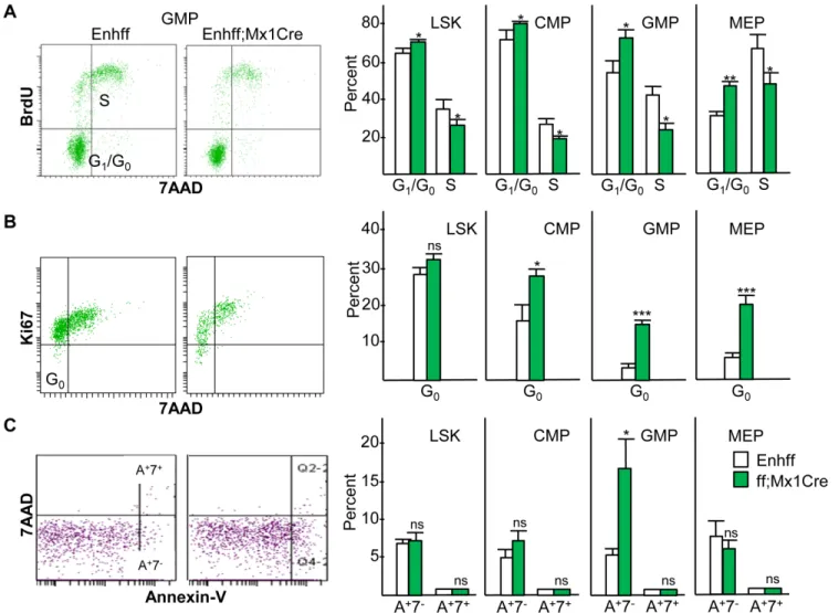
As C/EBPαcan directly inhibit cell cycle progression and apoptosis in myeloid cells [33–36], we evaluated the effect ofCebpaenhancer deletion on relevant parameters in the LSK, CMP, GMP, and MEP marrow subsets from Enh(f/f) versus Enh(f/f);Mx1-Cre mice 4 wks after com-pletion of pIpC injections. BrdU/7AAD staining 3 hr after BrdU injection demonstrated
embryos of indicated genotypes derived from Enh(f/+);CMV-Cre (Δ+) parents were subjected to FACS analysis for these same hematopoietic subsets.G) Marrow stem/progenitor subsets, as determined by FACS analysis, from the same mice evaluated in B and C.H) Myeloid, BFU-E, and B lymphoid CFUs were in enumerated after culture at 1E4 cells/mL in methylcellulose in the presence of IL-3/IL-6/SCF or at 2E5 cells/mL in EPO or IL-7, respectively (mean and SD from three determinations).I) Morphology of marrow obtained from Enh(f/f) or Enh(f/f);Mx1-Cre mice 4 wks after pIpC injections and from Enh(f/f); Vav-Cre or Enh(f/f);CMV-Cre mice.
increased G0/G1and reduced S cell cycle phase cells in each subset in the absence of the +37 kb
Cebpaenhancer, with the most evident effect in the GMP and MEP populations (Fig 7A). Of note, as expected the latter subsets had reduced G0/G1and increased S phase cells compared
with the earlier LSK or CMP subsets in Enh(f/f) mice. Similarly, Ki67/7AAD staining revealed reduced quiescent, G0phase cells as Enh(f/f) LSK cells progress to CMP and then to GMP or
MEP, and enhancer deletion increased the proportion of G0cells in the CMP, GMP, and MEP
subsets (Fig 7B). Thus, reduced GMP evident in the absence of theCebpaenhancer may in part reflect reduced proliferation, whereas LSK expansion occurs despite their mildly reduced pro-liferative status. Of note, although C/EBPαinhibits E2F activation of thec-Mycpromoter [37], c-MycRNA expression in GMP, CMP, or LSK was unaffected byCebpaenhancer deletion (Fig 6A). ExogenousCebpamarkedly reduces proliferation of marrow myeloid progenitors and
Fig 6. Effect ofin vivoEnhancer Deletion on Selected Transcription Factor, Myeloid Cytokine Receptor, or Bcl2 Expression. A) Total cellular RNAs from the GMP, CMP, or LSK marrow subsets isolated from Enh(f/f) or Enh(f/f);Mx1-Cre mice that had been subjected to pIpC injections and allowed to recover for 4 wks were analyzed for relative expression of the indicated mRNAs (mean and SD from three determinations). Relative expression of each RNA is compared in GMP versus CMP versus LSK cells in Enh(f/f) marrow, with the average value in GMP or CMP set to 1.0 (top panel). Expression in Enh(f/f); Mx1-Cre divided by expression in Enh(f/f) marrow is shown for each RNA in each subset analyzed (bottom panel). Dashed red line is the 1.0 level for both graphs. GR—Gcsfr, MR—Mcsfr, GMRa—Gmcsfrα.B) Myeloid CFUs obtained from Enh(f/f) or Enh(f/f);Mx1-Cre mice exposed 4 wks earlier to pIpC were in enumerated after culture at 2E4 cells/mL in methylcellulose in the presence of M-CSF, G-CSF, or GM-CSF (mean and SD from six determinations).
additional cell types [33,38]; therefore, reduced LSK, CMP, GMP, and MEP proliferation con-sequent toCebpaenhancer deletion apparently reflects direct or indirect pathways connecting C/EBPαto cell cycle progression not previously evaluated.
Annexin-V/7AAD staining demonstrated only minimal numbers of Annexin-V+7AAD+ non-viable cells in the LSK, CMP, GMP, or MEP populations, with an increased number of Annexin-V+7AAD-early apoptotic cells, from 5% to 16%, in response to enhancer deletion only in the GMP subset (Fig 7C). Increased apoptosis in the absence of theCebpaenhancer may thus also contribute to reduced GMP numbers. Of note, despite the ability of C/EBPαto activate theBcl2promoter in cooperation with NF-κB p50 [36], enhancer deletion did not affectBcl2mRNA levels in GMP or CMP, thoughBcl2was reduced 3-fold in the LSK subset (Fig 6A).
Fig 7. Effect ofin vivoEnhancer Deletion on Progenitor Cell Proliferation and Survival. A) Enh(f/f) or Enh(f/f);Mx1-Cre mice exposed 4 wks earlier to pIpC received a BrdU injection 3 hr prior to marrow harvest, followed by staining for surface markers to allow gating on the LSK, CMP, GMP, and MEP subsets and for intracellular incorporation of BrdU and 7AAD into DNA after fixation, permeabilization, and DNase exposure. G0/G1(BrdU-7AAD-) and S phase (BrdU+) cells were then enumerated (mean and SD from three determinations).B) Marrow from Enh(f/f) or Enh(f/f);Mx1-Cre mice exposed 4 wks earlier to pIpC were stained for LSK, CMP, GMP, and MEP and for intracellular Ki67 and 7AAD. G0(Ki67-7AAD-) cells were enumerated (mean and SD from three determinations).C) Marrow from a similar group of mice was stained for the same progenitors, for surface Annexin-V, and for permeability to 7AAD in the absence of fixation. Non-viable Annexin-V+7AAD+(A+7+) and early apoptotic Annexin-V+7AAD-(A+7-) cells were then enumerated (mean and SD from three determinations).
Effect of
Cebpa
Enhancer Deletion on Functional Long-Term
Hematopoietic Stem Cells
Functional LT-HSCs capable of long-term, multi-lineage hematopoietic reconstitution repre-sent a small subset of the FACS-defined LSK/SLAM or LT-HSC subsets. To evaluate the conse-quence ofCebpaenhancer deletion for their frequency in adult marrow, equal numbers of CD45.2+nucleated marrow cells isolated from Enh(f/f);Mx1-Cre mice exposed 4 wks earlier to pIpC and CD45.1+cells from WT mice were transplanted into lethally irradiated CD45.1+WT recipients. 19 wks later, at which point hematopoietic cells reflect output from functional LT-HSC, marrow and blood cells were analyzed for CD45.1 and CD45.2 expression, for lineage markers, and for Sca-1 and c-Kit expression (Fig 8A–8C). After primary transplantation, the proportion of CD45.1+and CD45.2+total nucleated cells in marrow or blood were equivalent, on average, indicating the presence of similar numbers of functional LT-HSC in the enhancer-deleted and WT marrow cells at the time of transplantation.
Granulocytes represented a much smaller fraction of the CD45.2+subset in primary trans-plant recipients compared with the CD45.1+subset, in marrow or blood, providing further evi-dence for a hematopoietic-intrinsic impairment in granulopoiesis consequent toCebpa enhancer deletion. Increased monocytes and reduced B cells in both marrow and blood and markedly expanded Lin-, LK, and LSK marrow populations in the CD45.2+compared with the CD45.1+population also confirmed the marrow-intrinsic nature of these changes when assessed in WT recipients. Increased marrow and blood T cells were also evident in the CD45.2+versus CD45.1+subsets.
Marrow from primary transplant recipients was transplanted into lethally irradiated CD45.1+WT secondary recipients. Among those mice that survived until 16 wks post-second-ary transplantation, the proportion of total CD45.2+and CD45.1+cells were not statistically different, with retention of multi-lineage CD45.2+cell engraftment, consistent with absence of a deficiency in functional LT-HSC in the initial Enh(f/f);Mx1-Cre graft (Fig 8D). There was again a trend towards reduced granulocytes in the CD45.2+subset, but this did not reach statis-tical significance. Early death of several secondary recipients may reflect their increased average CD45.2+proportion and so reduced total granulopoiesis compared to primary transplant recipients, predisposing to septic death in the setting of marrow transplantation where donor radiation weakens intestinal mucosa integrity facilitating bacterial entry into the bloodstream.
Discussion
The main conclusion of this study is that the +37 kbCebpaenhancer is a critical, hematopoi-etic-specific regulator ofCebpatranscription. In addition, availability of adult mice lacking the Cebpaenhancer provided a hypomorphic model that could be evaluated to gain new insight into regulation of hematopoiesis by C/EBPα. Consistent with results obtained with mice lack-ing both copies of the C/EBPαORF, deletion of theCebpaenhancer led to marked neutrope-nia, reduced GMP, expanded LSK, and increased erythroid progenitors. Enh(f/f);Mx1-Cre mice exposed to pIpC also manifested monocytosis, impaired B lymphopoiesis, and functional LT-HSC retention.
Mx1-Cre. In contrast, germline enhancer deletion by CMV-Cre did not alterCebpaexpression in adipocytes, liver, lung, small intestine, skeletal muscle, or kidney. Together, the transgenic and enhancer deletion data indicate that the +37 kbCebpaenhancer is necessary and sufficient for directing hematopoieticCebpagene transcription, with high-levelCebpaexpression in other lineages, e.g. adipocytes, hepatocytes, or type II pneumocytes, dependent on other regula-tory elements within theCebpalocus.
Fig 8. Effect ofin vivoEnhancer Deletion on Functional Long-Term Hematopoietic Stem Cells. A) Diagram of competitive transplantation assay. 2E5 CD45.2+nucleated marrow cells from Enh(f/f);Mx1-Cre mice exposed 4 wks earlier to pIpC were mixed with equal numbers of CD45.1+WT marrow cells and transplanted into lethally irradiated WT recipients. At 19 wks, peripheral blood (pB) and bone marrow (BM) cells were analyzed, and 1E6 marrow cells were transplanted into secondary transplant (2°TP) recipients, 1 recipient/donor. Those surviving were then analyzed 16 wks later.B) Percent of CD45.1+or CD45.2+cells amongst total marrow nucleated cells, and the percent of CD11b+Gr-1+granulocytes, CD11b+Gr-1-monocytes, Ter119+erythroblasts, B220+ B lymphoid cells, CD3+T lymphoid cells, Lin-, Lin-Sca-1-c-kit+(LK), or Lin-Sca-1+c-kit+(LSK) cells within the CD45.1+or CD45.2+subsets.C) Percent of CD45.1+or CD45.2+cells amongst nucleated peripheral blood cells in primary transplant recipients (mean and SD; n = 7).D) Percent of CD45.1+or CD45.2+ cells amongst nucleated peripheral blood cells in secondary transplant recipients (mean and SD; n = 3).
TheCebpagene is flanked by theCebpggene 64 kb upstream and by theSlc7a10gene, encoding an amino acid transport protein expressed only in neurons, 68 kb downstream. Cebpaenhancer deletion did not reduceCebpgexpression in marrow GMP, CMP, or LSK, sug-gesting a barrier to communication between the +37 kbCebpaenhancer and the -64 kbCebpg promoter.
CebpaORF(-/-), germline-deleted mice manifest neonatal lethality due to hepatic defects, with marked neutropenia and monocytopenia [19,30], andCebpaORF(-/-) fetal liver cells are also deficient in generating neutrophils and monocytes and manifest increased erythropoiesis [31,39].CebpaORF(f/f);Mx1-Cre mice exposed to pIpC have markedly reduced blood neutro-phils, monocytes, and eosinoneutro-phils, with increased platelets, 18-fold reduced marrow GMP, 5-fold increased CMP, 4-fold increased MEP, and 32-fold increased LSK cells [3].CebpaEnh (f/f);Mx1-Cre mice develop related changes, with neutropenia, reduced GMP, LSK expansion, and increased BFU-E. However, the degree of GMP and neutrophil reduction and LSK expan-sion was less, eosinophils were retained, and marrow CFU-M are increased rather than absent. These differences in myelopoiesis likely reflect the effect of residual, albeit low-levelCebpain enhancer-deleted GMP and supports our prior observations withCebpashRNA-transduced myeloid progenitors [4]. In the latter study, 3-foldCebpaknockdown impaired granulopoiesis while increasing monopoiesis, whereas 6-foldCebpaknockdown prevented commitment to either lineage, increased BFU-E formation even in the absence of EPO, and enabled morpho-logic blast accumulation with indefinite, cytokine-dependent myeloid CFU replating, the latter also seen in the current study uponCebpaenhancer deletion. High level C/EBPα, as seen in CFU-G, may homodimerize to direct granulopoiesis, whereas reduced C/EBPα, as seen in CFU-M, may heterodimerize with AP-1 proteins via their leucine zipper domains to mediate monopoiesis [4,40,41]. Homozygous enhancer-deleted (ΔΔ) fetal liver cells also had markedly reduced granulocytes, though monocytes were retained. Interestingly, heterozygous enhancer-deleted (Δ+) fetal liver had 5-fold reduced granulocytes whereas adultΔ+ marrow neutrophils were not reduced, suggesting greater sensitivity of fetal liver granulopoiesis to reduced C/ EBPα. Earlier work similarly revealed a>2-fold reduction in fetal liver granulocytes inCebpa ORF (+/-) embryos [39].
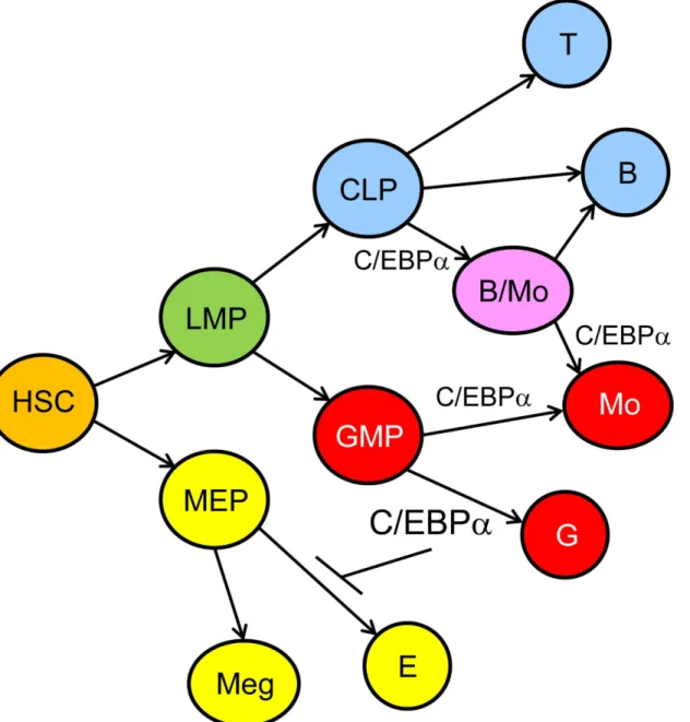
A model of hematopoietic lineage determination focused on the role of C/EBPαbased on our findings and those of others, is shown (Fig 9). In this model high-level C/EBPαis required for granulopoiesis and lower level for monopoiesis, high-level C/EBPαinhibits erythropoiesis, and C/EBPαcontributes to formation of a B/Mo progenitor arising from CLP.
Several prior studies addressed the effect of bialleleicCebpaORF deletion on functional LT-HSC in adult marrow, two finding increased LT-HSC after primary transplantation and one demonstrating impairment after both primary and secondary transplantation [3,14,15]. In the current study, functional LT-HSC were preserved afterCebpaenhancer deletion, per-haps reflecting the<2-fold reduction ofCebpamRNA observed in the LSK/SLAM population,
Fig 9. Model for the Role of C/EBPαDuring Hematopoiesis.In this model, HSC give rise to lymphoid-myeloid progenitors (LMP) and to MEP, MEP
generate megakaryocyte (Meg) and erythroid (E) progenitors, and GMP give rise to granulocytic progenitors (G) in the presence of high-level C/EBPαand to monocytic progenitors (Mo) in the presence of low-level C/EBPα. CLP give rise to T and B cell progenitors in the absence and to bipotent B/Mo progenitors in the presence of C/EBPα. The latter diverge to B cell or monocytic progenitors, again correlated with and potentially guided by loss or retention of C/EBPα.
which might mirror effects onCebpaexpression in functional LT-HSC. The ability of the Cebpaenhancer and promoter to nevertheless direct hCD4 transgene expression to long-term repopulating LT-HSC might either indicate that in this population anotherCebparegulatory element besides the +37 kb enhancer is also sufficient in this regard or that another element suppressesCebpatranscription in functional LT-HSC. Of note, FACS-defined LSK/SLAM cells are markedly depleted in the marrow of Enh(f/f);Mx1-Cre mice exposed to pIpC. This may represent depletion of the majority of this population, other than the more quiescent, func-tional LT-HSC, to enable LSK expansion in response to reduced myeloid and B lineage progenitors.
Exogenous C/EBPαinhibits G1 to S cell cycle progression in multiple cell types via several mechanisms, including via direct interaction with E2F1 in 32Dcl3 myeloid cells [35]. However, CebpaORF deletion did not alter BrdU incorporation into marrow LSK cells, andCebpa shRNA knockdown did not alter myeloid progenitor cell cycle parametersin vitro[3,4]. Simi-larly, we now find that reducedCebpaconsequent to enhancer deletion has minimal effect on proliferation of the LSK or CMP marrow subsets orc-Myclevels while reducing G1 to S cell cycle progression in GMP. Thus, the role of C/EBPαin regulation of cell cycle progression may depend on its level of expression, with high levels markedly slowing and low levels mildly slow-ing proliferation.
Exogenous C/EBPαinhibits apoptosis consequent to cytokine withdrawal from hematopoi-etic cells, in part via Bcl-2 induction in cooperation with NF-κB p50 [36,46]. Consistent with these findings,Cebpaenhancer deletion increases early apoptosis in GMP, perhaps together with slowed proliferation contributing to their reduced numbers.
Multiple mechanisms lead to diminished but generally not absent C/EBPαexpression or activity during the pathogenesis of acute myeloid leukemia, including alterations leading to reduced transcription, reduced translation, or reduced protein activity or stability [5]. Impaired but not absent myelopoiesis due to +37 kbCebpaenhancer deletion, with long-term, cytokine-dependent myeloid CFU replating, may represent preleukemic phenotypes absent in mice completely lacking C/EBPα. Of note, Bcr-abl expression inCebpaORF(-/-) hematopoietic cells generates erythroleukemia rather than myeloid leukemia [47], likely reflecting the need for a minimal number of GMP to act as substrates for myeloid transformation [48,49]. As we have not observed signs of leukemic transformation in a cohort of enhancer-deleted mice over a 55 wk period, expression of additional proliferative oncoproteins may be required, as we will pursue in future studies. Finally, as we discussed previously [11], sequencing of 110 human AML cases did not reveal point mutations or small insertions/deletions within the homologous +42 kb CEBPAenhancer [50]. This may reflect the fact that heterozygous absence of the +37 kb enhancer in adult mice does not significantly alter myelopoiesis and the greater efficiency of altering pathways that regulate the enhancer. For example, Runx1 binds and activates the +37 kb Cebpaenhancer [7], and ChIP-Seq demonstrated that the RUNX1-ETO AML oncoprotein binds specifically at the +42 kbCEBPAenhancer, but not theCEBPApromoter, in two patient samples and in the Kasumi-1 cell line [51], likely leading toCebpatrans-repression. In addition, our find-ing that Pu.1 and C/EBPαalso bind and activate the enhancer via conservedciselements [11] suggests that alterations that reduce the expression or activity of either of these transcription fac-tors might reduceCEBPAtranscription to further contribute to myeloid transformation.
Acknowledgments
Author Contributions
Conceived and designed the experiments: HG ADF. Performed the experiments: HG SC ADF. Analyzed the data: HG ADF. Wrote the paper: HG SC ADF.
References
1. Scott LM, Civin CI, Rorth P, Friedman AD. A novel temporal expression pattern of three C/EBP family members in differentiating myelomonocytic cells. Blood 1992; 80: 1725–1735. PMID:1391942
2. Iwasaki H, Mizuno S, Arinobu Y, Ozawa H, Mori Y, Shigematsu H, et al. The order of expression of tran-scription factors directs hierarchical specification of hematopoietic lineages. Genes Dev. 2006; 20: 3010–3021. PMID:17079688
3. Zhang P, Iwasaki-Arai J, Iwasaki H, Fenyus ML, Dayaram T, Owens BM, et al. Enhancement of hematopoietic stem cell repopulating capacity and self-renewal in the absence of the transcription fac-tor C/EBPα. Immunity 2004; 21: 853–863. PMID:15589173
4. Ma O, Hong S, Guo H, Ghiaur G, Friedman AD. Granulopoiesis requires increased C/EBPαcompared to monopoiesis, correlated with elevatedCebpain immature G-CSF receptor versus M-CSF receptor expressing cells. PLoS One 2014; 9: e95784. doi:10.1371/journal.pone.0095784PMID:24752325
5. Friedman AD. C/EBPαin normal and malignant myelopoiesis. Int J Hematol. 2015; 101: 330–341. doi:
10.1007/s12185-015-1764-6PMID:25753223
6. Christy RJ, Kaestner KH, Geiman DE, Lane MD. CCAAT/enhancer binding protein gene promoter: binding of nuclear factors during differentiation of 3T3-L1 preadipocytes. Proc Natl Acad Sci USA 1991; 88: 2593–2597. PMID:2006196
7. Guo H, Ma O, Speck NA, Friedman AD.Runx1deletion or dominant inhibition reducesCebpa transcrip-tion via conserved promoter and distal enhancer sites to favor monopoiesis over granulopoiesis. Blood 2012; 119: 4408–4418. doi:10.1182/blood-2011-12-397091PMID:22451420
8. Guo H, Ma O, Friedman AD. TheCebpa+37 kb enhancer directs transgene expression to myeloid pro-genitors and to long-term hematopoietic stem cells. J Leuk Biol. 2014; 96: 419–426.
9. Wilson NK, Foster SD, Wang X, Knezevic K, Schütte J, Kaimakis P, et al. Combinatorial transcriptional control in blood stem/progenitor cells: genome-wide analysis of ten major transcriptional regulators. Cell Stem Cell 2010; 7: 532–544. doi:10.1016/j.stem.2010.07.016PMID:20887958
10. Collins C, Wang J, Miao H, Bronstein J, Nawer H, Xu T, et al. C/EBPαis an essential collaborator in Hoxa9/Meis1-mediated leukemogenesis. Proc Natl Acad Sci USA 2014; 111: 9899–9904. doi:10. 1073/pnas.1402238111PMID:24958854
11. Cooper S, Guo H, Friedman AD. The +37 kb Cebpa enhancer is critical for Cebpa myeloid gene expres-sion and contains functional sites that bind SCL, GATA2, C/EBPα, PU.1, and additional Ets factors. PLoS One 2015; 10: e0126385. doi:10.1371/journal.pone.0126385PMID:25938608
12. Sharan SK, Thomason LC, Kuznetsov SG, Court DL. Recombineering: a homologous recombination-based method of genetic engineering. Nat Prot. 2009; 4: 206–223.
13. Suzow JG, Friedman AD. The murine myeloperoxidase promoter contains multiple functional elements —one element binds a cell type-restricted transcription factor, myeloid nuclear factor 1(MyNF1). Mol Cell Biol. 1993; 13: 2141–2151. PMID:8384306
14. Ye M, Zhang H, Amabile G, Yang H, Staber PB, Zhang P, et al. C/EBPαcontrols acquisition and main-tenance of adult haematopoietic stem cell quiescence. Nat Cell Biol. 2013; 15: 385–394. doi:10.1038/ ncb2698PMID:23502316
15. Hasemann MS, Lauridsen FK, Waage J, Jakobsen JS, Frank AK, Schuster MB, et al. C/EBPαis required for long-term self-renewal and lineage priming of hematopoietic stem cells and for the mainte-nance of epigenetic configurations in multipotent progenitors. PLoS Genet. 2014; 10: e1004079. doi:
10.1371/journal.pgen.1004079PMID:24415956
16. Schwenk F, Baron U, Rajewsky K. A cre-transgenic mouse strain for ubiquitous deletion ofloxP-flanked segments including deletion in germ cells. Nucl. Acids Res. 1995; 23: 5080–5081. PMID:8559668
17. Birkenmeier EH, Gwynn B, Howard S, Jerry J, Gordon JI, Landschulz WH, et al. Tissue-specific expres-sion, developmental regulation, and genetic mapping of the gene encoding CCAAT/enhancer binding protein. Genes Dev. 1989; 3: 1146–1156. PMID:2792758
18. Flodby P, Barlow C, Kylefjord H, Ahrlund-Richter L, Xanthopoulos KG. Increased hepatic cell prolifera-tion and lung abnormalities in mice deficient in CCAAT/enhancer binding proteinα. J Biol Chem. 1996; 271: 24753–24760. PMID:8798745
20. Kummalue T, Friedman AD. Cross-talk between regulators of myeloid development: C/EBPαbinds and activates the promoter of the PU.1 gene. J Leukoc Biol. 2003; 74: 464–470. PMID:12949251
21. Yeamans C, Wang D, Paz-Priel I, Torbett BE, Tenen DG, Friedman AD. C/EBPαbinds and activates the PU.1 distal enhancer to induce monocyte lineage commitment. Blood 2007; 110: 3136–3142. PMID:17671233
22. Hock H, Hamblen MJ, Rooke HM, Traver D, Bronson RT, Cameron S, et al. Intrinsic requirement for zinc finger transcription factor Gfi-1 in neutrophil differentiation. Immunity 2003; 18: 109–120. PMID:
12530980
23. Krishnaraju K, Hoffman B, Liebermann DA. Early growth response gene 1 stimulates development of hematopoietic progenitor cells along the macrophage lineage at the expense of the granulocyte and erythroid lineages. Blood 2001; 97: 1298–1305. PMID:11222373
24. Kurotaki D, Osato N, Nishiyama A, Yamamoto M, Ban T, Sato H, et al. Essential role of the IRF8-KLF4 transcription factor cascade in murine monocyte differentiation. Blood 2013; 121: 1839–1849. doi:10. 1182/blood-2012-06-437863PMID:23319570
25. Tamura T, Kurotaki D, Koizumi S. Regulation of myelopoiesis by the transcription factor IRF8. Int J Hematol. 2015; 101: 342–351. doi:10.1007/s12185-015-1761-9PMID:25749660
26. Paul F, Arkin Y, Giladi A, Jaitin DA, Kenigsberg E, Keren-Shaul H, et al. Transcriptional heterogeneity and lineage commitment in myeloid progenitors. Cell 2015; 163: 1663–1677. doi:10.1016/j.cell.2015. 11.013PMID:26627738
27. Jones LC, Lin ML, Chen SS, Krug U, Hofmann WK, Lee S, et al. Expression of C/EBPβfrom the C/ ebpa gene locus is sufficient for normal hematopoiesis in vivo. Blood 2002; 99: 2032–2036. PMID:
11877276
28. Hirai H, Zhang P, Dayaram T, Hetherington CJ, Mizuno S, Imanishi J, et al. C/EBPβis required for 'emergency' granulopoiesis. Nat Immunol. 2006; 7: 732–739. PMID:16751774
29. Alberich-JordàM, Wouters B, Balastik M, Shapiro-Koss C, Zhang H, Di Ruscio A, et al. C/EBPγ deregu-lation results in differentiation arrest in acute myeloid leukemia. J Clin Invest. 2012; 122: 4490–4504. doi:10.1172/JCI65102PMID:23160200
30. Zhang DE, Zhang P, Wang ND, Hetherington CJ, Darlington GJ, Tenen DG. Absence of granulocyte colony-stimulating factor signaling and neutrophil development in CCAAT enhancer binding proteinα -deficient mice. Proc Natl Acad Sci USA 1997; 94: 569–574. PMID:9012825
31. Heath V, Suh HC, Holman M, Renn K, Gooya JM, Parkin S, et al. C/EBPαdeficiency results in hyper-proliferation of hematopoietic progenitor cells and disrupts macrophage development in vitro and in vivo. Blood 2004; 104: 1639–1647. PMID:15073037
32. Zhang P, Nelson E, Radomska HS, Iwasaki-Arai J, Akashi K, Friedman AD et al. Induction of granulo-cytic differentiation by 2 pathways. Blood 2002; 99: 4406–4412. PMID:12036869
33. Wang X, Scott E, Sawyers CL, and Friedman AD. C/EBPαbypasses G-CSF signals to rapidly induce PU.1 gene expression, stimulate granulocytic differentiation, and limit proliferation in 32D cl3 myelo-blasts. Blood 1999; 94: 560–571. PMID:10397723
34. Porse BT, Pedersen TA, Xu X, Lindberg B, Wewer UM, Friis-Hansen L, et al. E2F repression by C/ EBPαis required for adipogenesis and granulopoiesis in vivo. Cell 2001; 107: 247–258. PMID:
11672531
35. Wang QF, Cleaves R, Kummalue T, Nerlov C, Friedman AD. Cell cycle inhibition mediated by the outer surface of the C/EBPαbasic region is required but not sufficient for granulopoiesis. Oncogene 2003; 22: 2548–2557. PMID:12730669
36. Paz-Priel I, Cai DH, Wang D, Kowalski J, Blackford A, Liu H, et al. C/EBPαand C/EBPαmyeloid onco-proteins induce Bcl-2 via interaction of their basic regions with NF-κB p50. Mol. Cancer Res. 2005; 3: 585–596.
37. Johansen LM, Iwama A, Lodie TA, Sasaki K, Felsher DW, Golub TR, et al. c-Myc is a critical target for C/EBPαin granulopoiesis. Mol Cell Biol. 2001; 21: 3789–3806. PMID:11340171
38. Umek RM, Friedman AD, McKnight SL. CCAAT-enhancer binding protein: a component of a differentia-tion switch. Science 1991; 251: 288–292. PMID:1987644
39. Suh HC, Gooya J, Renn K, Friedman AD, Johnson PF, Keller JR. C/EBPαdetermines hematopoietic cell fate in multipotential progenitor cells by inhibiting erythroid differentiation and inducing myeloid dif-ferentiation. Blood 2006; 107: 4308–4316. PMID:16469877
40. Cai DH, Wang D, Keefer J, Yeamans C, Hensley K, Friedman AD. C/EBPα:AP-1 leucine zipper hetero-dimers bind novel DNA elements, activate the PU.1 promoter, and direct monocyte lineage commit-ment more potently than C/EBPαhomodimers or AP-1. Oncogene 2008; 27: 2772–2779. PMID:
41. Hong SH, Skaist AM, Wheelan SJ, Friedman AD. AP-1 protein induction during monopoiesis favors C/ EBP:AP-1 heterodimers over C/EBP homodimerization and stimulates FosB transcription. J Leuk Biol. 2011; 90: 643–651.
42. Montecino-Rodriguez E, Leathers H, Dorshkind K. Bipotential B-macrophage progenitors are present in adult bone marrow. Nat Immunol. 2001; 2: 83–88. PMID:11135583
43. Balciunaite G, Ceredig R, Massa S, Rolink AG. A B220+ CD117+ CD19- hematopoietic progenitor with potent lymphoid and myeloid developmental potential. Eur J Immunol. 2005; 35: 2019–2030. PMID:
15971276
44. Richie Ehrlich LI, Serwold T, Weissman IL. In vitro assays misrepresent in vivo lineage potentials of murine lymphoid progenitors. Blood 2011; 117: 2618–2624. doi:10.1182/blood-2010-05-287102
PMID:21163922
45. Xie H, Ye M, Feng R, Graf T. Stepwise reprogramming of B cells into macrophages. Cell 2004; 117: 663–676. PMID:15163413
46. Paz-Priel I, Ghosal AK, Kowalski J, and Friedman AD. C/EBPαor C/EBPαoncoproteins regulate the intrinsic and extrinsic apoptotic pathways by direct interaction with NF-B p50 bound to the bcl-2 and FLIP gene promoters. Leukemia 2009; 23: 365–374. doi:10.1038/leu.2008.297PMID:18987666
47. Wagner K, Zhang P, Rosenbauer F, Drescher B, Kobayashi S, Radomska HS, et al. Absence of the transcription factor CCAAT enhancer binding proteinαresults in loss of myeloid identity in bcr/abl-induced malignancy. Proc Natl Acad Sci USA 2006; 103: 6338–6343. PMID:16606850
48. Kirstetter P, Schuster MB, Bereshchenko O, Moore S, Dvinge H, Kurz E, et al. Modeling of C/EBPα
mutant acute myeloid leukemia reveals a common expression signature of committed myeloid leuke-mia-initiating cells. Cancer Cell 2008; 13: 299–310. doi:10.1016/j.ccr.2008.02.008PMID:18394553
49. Ye M, Zhang H, Yang H, Koche R, Staber PB, Cusan M, et al. Hematopoietic differentiation is required for initiation of acute myeloid leukemia. Cell Stem Cell 2015; 17: 611–623. doi:10.1016/j.stem.2015. 08.011PMID:26412561
50. Cancer Genome Atlas Research Network. Genomic and epigenomic landscapes of adult de novo acute myeloid leukemia. New Engl J Med. 2013; 368: 2059–2074. doi:10.1056/NEJMoa1301689
PMID:23634996