Acquisition and manipulation of computed tomography
images of the maxillofacial region for biomedical
prototyping*
Aquisição e manipulação de imagens por tomografia computadorizada da região maxilofacial visando à obtenção de protótipos biomédicos
Maria Inês Meurer1, Eduardo Meurer2, Jorge Vicente Lopes da Silva3, Ailton Santa Bárbara4, Luiz Felipe Nobre5, Marília Gerhardt de Oliveira6, Daniela Nascimento Silva7
Biomedical prototyping has resulted from a merger of rapid prototyping and imaging diagnosis technologies. However, this process is complex, considering the necessity of interaction between biomedical sciences and engineering. Good results are highly dependent on the acquisition of computed tomography images and their subsequent manipulation by means of specific softwares. The present study describes the experience of a multidisciplinary group of researchers in the acquisition and manipulation of computed tomography images of the maxillofacial region aiming at biomedical prototyping for surgical purposes.
Keywords: Rapid prototyping; Biomedical prototyping; Computed tomography.
O processo de construção de protótipos biomédicos surgiu da união das tecnologias de prototipagem rápida e do diagnóstico por imagens. No entanto, este processo é complexo, em função da necessária interação entre as ciências biomédicas e a engenharia. Para que bons resultados sejam obtidos, especial atenção deve ser dispensada à aquisição das imagens por tomografia computadorizada e à manipulação dessas imagens em softwares específicos. Este artigo apresenta a experiência multidisciplinar de um grupo de pesquisadores
com a aquisição e a manipulação de imagens por tomografia computadorizada do complexo maxilofacial, visando à construção de protótipos biomédicos com finalidade cirúrgica.
Unitermos: Prototipagem rápida; Prototipagem biomédica; Tomografia computadorizada. Abstract
Resumo
* Study developed at Universidade Federal de Santa Catarina (UFSC), Florianópolis, SC, Pontifícia Universidade Católica do Rio Grande do Sul (PUCRS), Porto Alegre, RS, Universidade do Sul de Santa Catarina (Unisul), Tubarão, SC, and Centro de Pes-quisa Renato Archer (CenPRA), Campinas, SP, Brazil.
1. PhD of Clinical Stomatology, Associate Professor, Depart-ment of Pathology, Universidade Federal de Santa Catarina (UFSC), Florianópolis, SC, Brazil.
2. PhD of Oralmaxillofacial Surgery and Traumatology, Profes-sor at Universidade do Sul de Santa Catarina (Unisul), Tubarão, SC, Brazil.
3. Master, Electrical Engineering and Computation, Researcher at Centro de Pesquisa Renato Archer (CenPRA), Campinas, SP, Brazil.
4. PhD of Mechanical Engineering, Researcher at Centro de Pesquisa Renato Archer (CenPRA), Campinas, SP, Brazil.
violence is alarming(4). Additionally, pa-tients with severely affected by facial de-formities present with lower level of so-cial integration than patients with alteration in limbs or other parts of their body.
Computed tomography (CT) is the method of choice among the diagnostic imaging techniques utilized for preopera-tive evaluation of these patients. Besides the essential analysis of 2D images, 3D vi-sualization allows a privileged access to the structures of interest, particularly for the surgeon, highlighting some aspects which otherwise only could be accessed by means of a “mental reconstruction” of tomographic images. Despite the unques-tionably elucidative character of virtual 3D images, some distance still persists be-tween the virtual model and the actual han-dling of anatomic structures during a sur-gical procedure.
Biomedical prototyping from CT im-ages has shown to be an innovative
solu-Meurer MI, solu-Meurer E, Silva JVL, Bárbara AS, Nobre LF, Oliveira MG, Silva DN. Aquisição e manipulação de imagens por tomografia computadorizada da região maxilofacial visando à obtenção de protótipos biomédicos. Radiol Bras. 2008;41(1):49–54.
deformities(1,2), considering the necessity of long hospitalization periods, social se-curity costs during the recovery period, number of reconstructive surgical proce-dures, and costs of interdisciplinary surgi-cal teams as well as clinisurgi-cal ancillary per-sonnel(2,3). Besides congenital causes, la-bor-, traffic- and violence-related accidents show up as acquired causes of facial de-formities; in Brazil, the number of hospi-tal admissions as a result of accidents and
5. PhD of Medicine/Radiology, Associate Professor, Department of Clinical Practice, Universidade Federal de Santa Catarina (UFSC), Florianópolis. SC, Brazil.
6. PhD of Clinical Stomatology, Professor, Department of Sur-gery, Pontifícia Universidade Católica do Rio Grande do Sul (PU-CRS), Porto Alegre, RS, Brazil.
7. PhD of Oralmaxillofacial Surgery and Traumatology, Profes-sor, Department of Surgery, Faculdade de Odontologia da Ponti-fícia Universidade Católica do Rio Grande do Sul (PUCRS), Porto Alegre, RS, Brazil.
Mailing address: Dra. Maria Inês Meurer. Universidade Fede-ral de Santa Catarina, Departamento de Patologia. Campus Universitário – Trindade. Florianópolis, SC, Brazil, 88040-370. E-mail: [email protected]
Received May 21, 2007. Accepted after revision August 20, 2007.
INTRODUCTION
tion to minimize this distance. Similarly to products manufacturing, where proto-types are utilized to improve the final qual-ity, allowing the detection of possible mis-takes at early stages of the product devel-opment(5), biomedical prototyping allows the construction of physical objects repro-ducing anatomic structures and only is possible by means of an integration of medi-cal images acquisition and manipulation with computer-aided design (CAD) and rapid prototyping (RP) techniques, there-fore involving multidisciplinary teams.
With the direct visualization and ma-nipulation of an anatomic replica of bone structures, biomedical prototyping allows, for example, an accurate measurement of such structures as well as simulation of os-teotomies and resection techniques. As a result, a decrease in the surgical time, an-esthesia period and infection risk can be observed, with a decrease in costs of the global treatment and better clinical re-sults(1,6,7). Additionally, the utilization of these prototypes improves the communi-cation between practitioners and patients, aiding in the explanation about the surgi-cal procedure and in the obtention of the term of free and informed consent, besides being useful in the production of custom-ized prosthetic implants(8–11). Biomedical prototypes also can be utilized for didactic purposes(9).
The biomedical prototyping process re-quires an intense interaction of biomedi-cal sciences, information technology and engineering. For the understanding of the process, and also the communication among the professionals involved it is nec-essary for engineers to learn concepts of diagnostic imaging and surgery, and for radiologists and surgeons to explore the world of information technology and manufacturing processes.
This study is aimed at presenting a re-view of the key issues involved in the de-velopment of biomedical prototypes. The considerations presented in this paper re-sult from the integration of a team includ-ing surgeons involved in the Program of Post-Graduation in Oralmaxillofacial Sur-gery and Traumatology of Pontifícia Uni-versidade Católica do Rio Grande do Sul (CTBMF-PUCRS), engineers of the Divi-sion for Products Development at Centro
de Pesquisa Renato Archer (CenPRA) and medical and dental radiologists of Univer-sidade Federal de Santa Catarina (UFSC).
STEPS FOR THE CONSTRUCTION OF BIOMEDICAL PROTOTYPES
1. Patient selection
The option for biomedical prototyping instead of less costly techniques, should be reserved for the cases where there is a real benefit to the patient, disregarding short-lived techniques and market ap-peals(6,7,12). This technique will be mostly useful in surgical procedures for which re-liable techniques are not available or in cases where these techniques require fur-ther refinements and/or improvements. With the aid of a prototype during the sur-gical planning phase, a surgeon can elabo-rate the technique, evaluate details, opti-mize the procedure, anticipate difficulties and, mainly, the solution for these diffi-culties. Experiments involving the utiliza-tion of biomedical prototyping at CTBMF-PUCRS have demonstrated that the diffi-culties observed during the simulation of the surgical procedure with the biomodel are very similar to those found during the actual surgery, which corroborates the va-lidity of these prototypes during the surgi-cal planning.
2. Images acquisition
It is desirable that a single volume of the whole segment to be studied is imaged utilizing thin sections. It is essential to take into consideration that the patient exposure to radiation is a limiting factor, and the ra-diologist is responsible for choosing the best images acquisition protocol, in order to achieve a balance between the prototype quality and radiation dose.
Some steps are necessary to optimize the acquisition of the images for subse-quent computer processing:
– Theoretically, the slices thickness should be as low as possible to allow an appropriate image 3D reconstruction (ex-cellent results can be achieved with 1 mm-thick slices). However, some times this is not feasible in cases of extensive areas to be scanned.
– In cases where the area of interest in-cludes only the face, the change in the
ac-quisition plane from axial to coronal may reduce substantially the number of sec-tions.
– In the helical mode, an increase in the pitch may allow the obtention of more ex-tensive volumes with thick slices, espe-cially in multidetector CT equipment. This is a better solution than increasing the slice thickness. If the increase in the slices thick-ness is inevitable, the reconstruction inter-val for volume formation should be as small as possible.
– The field of view (FOV) should cover the whole region of interest. For the face and skull, a FOV of approximately 250 mm is sufficient; even smaller FOVs can be utilized, depending on the area to be re-produced in the prototype. The smaller the FOV, the higher the image quality, because the available matrix is applied to a smaller area, reducing the pixel size and, conse-quently, the partial volume effect(13).
– In principle, the gantry should not be inclined during the images acquisition, considering that some softwares for images processing do not allow the compensation of this inclination yet, producing proto-types with dimensional alterations(14).
– The utilization of filters during im-ages acquisition is controversial. Some studies have reported a higher rate of arti-facts when bone filters are utilized during images acquisition(15,16).
– Artifacts related to metal dental res-torations should be subsequently removed by means of graphic computing tools, al-though this is a time-consuming and bor-ing process which many times affects nega-tively the final outcomes. Aiming at mini-mizing the productions of such artifacts, the patient should be positioned in the oc-clusal plane (ococ-clusal surfaces of the teeth) parallel to the axial plane. With this strat-egy, artifacts are restricted to the region of the dental cusps, reducing the number of sections to be manually edited.
3. Image files storage and transference
all CT equipment (particularly the oldest ones) is able export DICOM files. So, it is important to previously confirm with the prototyping center the compatibility be-tween the images format and the editing application software.
The files may be stored in any media available, provided the data storage capac-ity of such media is high. Typically, CDs, DAT tapes and optical disks are utilized as recording media. Rewritable (R/W) CDs should be avoided since they may not be recognized by some editing application softwares(18), or even present a higher data volatility, difficulting the future records retrieval.
The data volume may pose a problem for the image files transfer. Each image in
the DICOM format with 512 × 512 pixels
acquisition matrix generates a 512 Kbytes file/slice. A skull CT in compliance with the requirements for the development of an appropriate prototype may generate a data volume of approximately 100 Mbytes. So, compacting tools can be useful at the moment of data transfer. Broadband net-works allow the data transfer via internet with the file transfer protocol (FTP) di-rectly to the prototyping workstation. The files availability in local restricted access networks (allowing downloads by the prototyping workstation), or even the mail-ing of CDs represent additional alternatives for the images transference. Sending im-age files attached to emails may be an ad-ditional option, although the file size may frequently cause the file rejection by the server. As far as data access is concerned, security-related issues should be taken into consideration.
4. Images manipulation
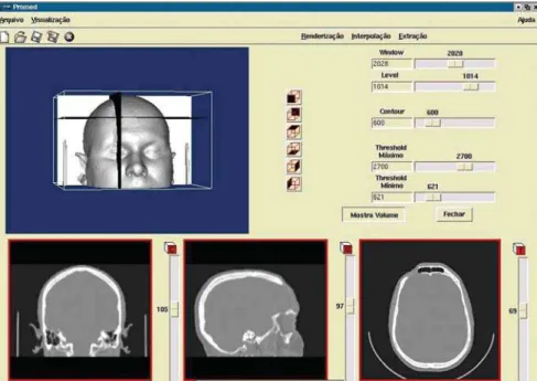
Images manipulation and editing is per-formed with the aid of specific application softwares, and this process requires a close interaction between biomedical specialties and engineering(19). The aim of this pro-cess is the images segmentation for sepa-rating data of interest from the informa-tion set provided by CT. In the case of the prototyping for oralmaxillofacial surgery where the study object is a bone specimen, the images segmentation is aimed at sepa-rating the bony portion from the adjacent tissues (Figures 1 and 2).
Figure 1. Visualization of 3D image, and coronal, sagittal and axial section planes, before segmentation. Note the presence of soft tissues.
Among the tools available for images segmentation, the threshold is fairly uti-lized, and is based on the definition of den-sity intervals expressing, for example, only the voxels corresponding to bone tissues. Should this interval be erroneously deter-mined, the so called dumb-bell effect will occur, with a possible structures suppres-sion or alteration during the process(15). In some cases, the manual images editing is required, with tools such as cut, erase and select. This editing is particularly useful in case of imaging artifacts originating from dental prostheses or restorations.
CenPRA, by means of the project Proto-tipagem Rápida na Medicina (Promed) (Rapid Prototyping in Medicine), has de-veloped the InVesalius free software, a pio-neering application for medical images processing aimed at the production of bio-medical prototypes. The first version of this software is already available to practitio-ners and health institutions in the area of biomedicine (free licenses may be re-quested at: http://www.cenpra.gov.br/ promed). InVesalius, includes some algo-rithms offering3D visualization, segmen-tation, and 2D/3D reformatting resources. Additionally, the software offers a conver-sion process function, allowing images conversion into a format recognized by RP equipment.
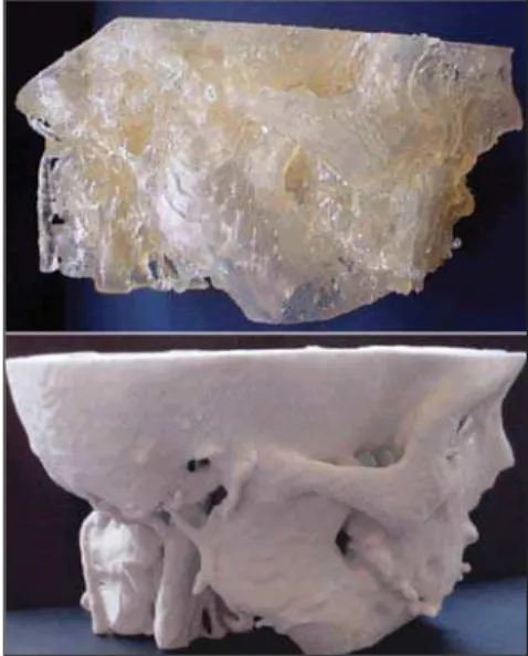
The integration between CAD and medical systems facilitates the manipula-tion and modeling of objects, allowing vir-tual images of segmented structures to be manipulated as if they were pieces of a puzzle or biomechanic prototype. Addi-tionally, CAD systems are appropriate for defining mirroring procedures utilizing the contralateral symmetry of the face, allow-ing the plannallow-ing and development of cus-tomized prosthesis, including by simulat-ing the prosthesis assembly on a 3D model (Figure 3). The construction of the custom-ized prosthesis can be done by modeling the structure which will replace the injured area, or by means of CAD systems opera-tions, generating a 3D model of a mold; then, the mold obtained by RP is utilized to give a shape to the material to be im-planted (typically implantable polymers or ceramic materials). It is important that typi-cal contractions of some of these materi-als are taken into consideration in the molds construction. For implantable me-tallic materials, the mold may be utilized for generating wax models to be utilized, for example, in the microfusion process.
5. Images conversion
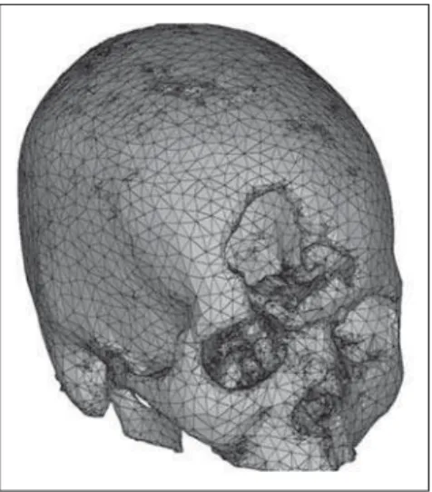
generated by the tomograph is not sup-ported by the prototyping devices; sec-ondly, the tomographic slices thickness usually ranges between 1 mm and 5 mm, and is considerably higher than those uti-lized in RP, about 0.1 mm. Additionally, RP processes utilize data originated from CAD-3D systems modeled by surfaces or solids, while tomographic images are rep-resented by voxels. So, 2D tomographic images must be three-dimensionally refor-matted and converted into a image format utilized in the RP processes; STL is the standard format, the model being
repre-sented by a raw unstructured triangulated surface(20,21) (Figure 4). Then, the STL file is processed by a specific prototyping soft-ware for correction of eventual inconsis-tencies in the surface geometry, triangles vertexes, optimization of the triangles num-ber, as well as in the selection of the con-struction orientation most appropriate to the piece geometry — a phase known as process planning. Following this phase, the virtual STL model is resliced in parallel layers to allow the prototype construction. Hundreds to thousands of triangle fac-ets constitute the model of a complex
struc-ture like the human skull, generating ex-cessively-sized files and difficulting the images processing.
6. Prototype construction
For the prototype construction, the STL files must be sent to the rapid prototyping workstation (usually, the computer where the images are processed is not located at the same place of the prototyping work-station). The files transfer can be accom-plished by a media appropriate for data transmission (CD, e-mail, FTP, local net-work). Once the transfer is completed, the model construction is automatic in the majority of processes. The construction may take several hours to be completed, depending on the number of layers and the prototype height. Table 1 shows an over-view of the advantages and disadvantages of the main rapid prototyping processes utilized in Brazil(20)for biomedical appli-cations. This table is not aimed at under-valuing or otherwise under-valuing equipment of manufacturers, but rather to provide con-cise information to aid health profession-als in their decision making about the pro-cess to be adopted according to their in-vestment requirements and conditions. It is important to take into consideration that the costs for equipment acquisition and operation is high requiring specialized per-sonnel, and not always the return on in-vestment is rewarding. For this reason, the greatest majority of users opt for acquir-ing the services rather than the equipment.
Figure 4. Raw unstructured triangulated surface constructed with the utilization of the marchin cubes algorithm, covering all the surfaces on the image. Figure 3. Development of customized prostheses. Virtual mold at left. At right, prosthesis virtually
posi-tioned in the area where bone tissue is absent.
Figure 5 illustrates biomedical proto-types produced by the processes SLA and SLS and based on a same data set. Some clinical cases may be found at http://www. cenpra.gov.br/promed/casos.htm.
FINAL CONSIDERATIONS
Biomedical prototypes present a high potential in the selection of new, frequently alternative, therapeutic approaches. In Bra-zil, the utilization of such prototypes is still restricted, particularly because of the high production costs involved and poor equip-ment availability in the country. Besides the high cost, the time-consuming nature of the process (from the images acquisi-tion up to the prototype producacquisi-tion) com-plicates the utilization of this method in routine surgical procedures, even if there is an indication. However, it is probable that such limitation will be overcome ei-ther by the technological development, or by the interdisciplinary utilization of this method, increasing the accessibility to bio-medical prototypes.
As far as future prospects are con-cerned, it is important to highlight the rel-evance of this technology in the tissue en-gineering. This is also a multidisciplinary
nique(23,24). The production of a model by rapid prototyping, in a specific shape and with a structure appropriate for adherence and cellular reproduction could be a fea-sible solution for reconstructive treatments in humans(25). An even more spectacular advance is the possibility of directly pro-ducing organs by means of rapid proto-typing-like mechanisms called bioprint-ing(26).
The experience gained by the authors with the obtention of biomodels has al-lowed the researchers to exchange knowl-edge essential for the development of each specific area. CenPRA has experienced the opportunity to have its software utilized by a group of specialists who ultimately con-stitute its target public. Differently from the majority of specialized softwares, the InVesalius was designed for running on personal computers, processing medical images and allowing their dynamic inter-pretation according to the diagnostic re-quirements or even during surgical proce-dures. More than 1500 free licenses have already been granted to specialists of dif-ferent areas of knowledge in Brazil and several other countries. Additionally, CenPRA , in association with partners in the biomedical area, has been developing
Table 1 Comparative chart of RP technologies most frequently utilized for biomedical prototyping.
Process
SLA (3D Systems)
SLS (3D Systems)
FDM (Stratasys)
3DP
(Z Corporation)
Polyjet (Objet)
Advantages
– High dimensional accuracy
– Excellent reproduction of thin structures
– Allows the visualization of inner structures (translucent material) – Excellent screws fixation
– Possibility of constructing color prototypes (two colors)
– Good dimensional accuracy
– Excellent reproduction of thin structures – Excellent screws fixation
– Autoclave sterilizable – Bone verisimilitude
– Good dimensional accuracy – Good reproduction of thin structures – Excellent screws fixation
– Low cost – Rapid construction – Cutting facility
– Possibility of constructing color prototypes (many simultaneous colors)
– Bone verisimilitude
– High dimensional accuracy
– Excellent reproduction of thin structures
– Allows the visualization of inner structures (translucent material) – Excellent screws fixation
Disadvantages
– Difficult surfaces visualization in prototypes constructed of translucent material (Figure 5)
– High cost
– Extremely hard material – High cost
– Extremely hard material – Baixa velocidade de construção – Slow construction
– Reasonable dimensional accuracy – Reasonable reproduction of thin structures – Surface porosity
– Cutting dust release
– Difficult surfaces visualization in prototypes constructed of translucent material
– High cost
domain of the science that combines and apply engineering principles and life sci-ences, in an attempt to develop biological substitutes to restore, maintain and improve functions of a determined tissue or or-gan(22). Studies can already be found in the literature, demonstrating that tissues can really be reproduced using this
methodologies for surgical planning and biomodels construction.
Biomedical prototypes and the InVesalius software have been utilized in the Program of Post-Graduation in Oral-maxillofacial Surgery and Traumatology at PUCRS in the resolution of several surgi-cal ceases. Besides the treatment of facial deformities, these prototypes have been utilized in implantodontics and in the treat-ment of patients with temporomandibular joint ankylosis, giving both the Institution’s patients and graduating professionals the opportunity to access the recent technolo-gies. Additionally PUCRS, in association with CenPRA, has developed researches on the accuracy of rapid prototyping biomodels for surgical planning pur-poses(16) and prototypes construction based on magnetic resonance imaging.
At UFSC, radiologists have been study-ing the relation between CT images acqui-sition parameters and prototypes quality, aiming at a less empirical CT images ac-quisition and processing for biomedical prototypes construction.
The main objective of this undertaking as a whole is to utilize technological re-sources and applied research to offer bet-ter treatments, respecting the dignity of patients who could achieve a better func-tional and aesthetic rehabilitation with these methods.
REFERENCES
1. Peckitt NS. Stereoscopic lithography: customized titanium implants in orofacial reconstruction. Br J Oral Maxillofac Surg. 1999;37:353–69. 2. Sanghera B, Naique S, Papaharilaou Y, et al.
Pre-liminary study of rapid prototype medical mod-els. Rapid Prototyping Journal. 2001;7:275–84. 3. ATLS – Advanced Trauma Life Suport. Student
manual. 6th ed. Chicago: American College of Surgeons; 1997.
4. Ministério da Saúde do Brasil. Morbidade hos-pitalar do SUS por causas externas – por local de internação – Brasil. [Acessado em: 18/5/2007]. Disponível em: http://tabnet.datasus.gov.br/cgi/ deftohtm.exe?sih/cnv/eiuf.def
5. Silva JVL, Yamanaka MC, Saura CE. Rapid pro-totyping: concepts, applications, and potential uti-lization in Brazil. In: 15th International Confer-ence in CAD/CAM Robotics and Factories of the Future; 1999; Águas de Lindóia, SP.
6. Kermer C, Lindner A, Friede I, et al. Preoperative stereolithographic model planning for primary reconstruction in craniomaxillofacial trauma sur-gery. J Craniomaxillofac Surg. 1998;26:136–9. 7. Meurer E. As tecnologias CAD-CAM em
cirur-gia e traumatolocirur-gia bucomaxilofacial. (Tese de Doutorado). Porto Alegre: Pontifícia Universi-dade Católica do Rio Grande do Sul; 2002. Dis-ponível em: http://www.cenpra.gov.br/promed/ PDF/Tese_Meurer.pdf
8. D’Urso PS, Atkinson RL, Lanigan MW, et al. Stereolithographic (SL) biomodelling in cranio-facial surgery. Br J Plast Surg. 1998;51:522–30. 9. James WJ, Slabbekoorn MA, Edgin WA, et al. Correction of congenital malar hypoplasia using stereolithography for presurgical planning. J Oral Maxillofac Surg. 1998;56:512–7.
10. Haex JKT, Poukens JMN. Preoperative planning with the use of stereolithographic model. Phidias Newsletter. 1999(3). [Acessado em: 22/3/2007]. Disponível em: http://193.74.1000.113/Medical/ files/ph3.pdf
11. Petzold R, Zeilhofer HF, Kalender WA. Rapid protyping technology in medicine – basics and applications. Comput Med Imaging Graph. 1999; 23:277–84.
12. Perry M, Banks P, Richards R, et al. The use of computer-generated three-dimensional models in orbital reconstruction. Br J Oral Maxillofac Surg. 1998;36:275–84.
13. Cáceres KPS. Efeitos da variação da espessura do corte tomográfico e da largura do campo de vi-são (FOV) na reprodução de estruturas ósseas finas com a finalidade de prototipagem rápida – estudo in vitro. (Monografia). Florianópolis:
Universidade Federal de Santa Catarina; 2005. 14. Kragskov J, Sindet-Pedersen S, Gyldensted C, et
al. A comparison of three-dimensional computed tomography scans and stereolithographic models
for evaluation of craniofacial anomalies. J Oral Maxillofac Surg. 1996;54:402–11.
15. Choi JY, Choi JH, Kim NK, et al. Analysis of er-rors in medical rapid prototyping models. Int J Oral Maxillofac Surg. 2002;31:23–32. 16. Silva DN. Análise do erro dimensional dos
bio-modelos de sinterização seletiva a laser (SLS) e de impressão tridimensional (3DP), a partir de imagens de tomografia computadorizada, na re-produção da anatomia craniomaxilar: estudo in vitro. (Tese de Doutorado). Porto Alegre: Ponti-fícia Universidade Católica do Rio Grande do Sul; 2004.
17. DICOM Standards Committee. DICOM Home Page. [Acessado em: 22/3/2005]. Disponível em: http://medical.nema.org
18. Kernan BT, Wimsatt JA 3rd. Use of a stereolitho-graphy model for accurate, preoperative adapta-tion of a reconstrucadapta-tion plate. J Oral Maxillofac Surg. 2000;58:349–51.
19. Silva JVL, Meurer E, Zavaglia CAC, et al. Rapid prototyping applications in the treatment of craniomaxillofacial deformities – utilization of bioceramics. Key Engineering Materials. 2003; 254-256:687–90.
20. Volpato N. Prototipagem rápida: tecnologia e aplicações. São Paulo: Edgard Blücher; 2007. 21. Souza MA, Centeno TM, Pedrini H. Integrando
reconstrução 3D de imagens tomográficas e pro-totipagem rápida para a fabricação de modelos médicos. Revista Brasileira de Engenharia Bio-médica. 2003;19:103–15.
22. Langer R, Vacanti JP. Tissue engineering. Science. 1993;260:920–6.
23. Weng Y, Cao Y, Silva CA, et al. Tissue engineered composites of bone and cartilage for mandible condylar reconstruction. J Oral Maxillofac Surg. 2001;59:185–90.
24. Abukawa H, Terai H, Hannouche D, et al. Forma-tion of a mandibular condyle in vitro by tissue en-gineering. J Oral Maxillofac Surg. 2003;61:94– 100.
25. Williams JM, Adewunmib A, Schek RM, et al. Bone tissue engineering using polycaprolactone scaffolds fabricated via selective laser sintering. Biomaterials. 2005;26:4817–27.