REVIEW
Updating technology of shunt valves
Matheus Fernandes de Oliveira,IRenan Muralho Pereira,IIFernando Gomes PintoIII,IV
INeurosurgery Residence Program - Department of Neurosurgery - Hospital do Servidor Pu´blico Estadual de Sa˜o Paulo, Sa˜o Paulo, BrazilIINeurosurgery
League, Faculty of Medicine, University of Sao Paulo and medical student at Universidade Anhembi-Morumbi, Sa˜o Paulo, BrazilIIINeurosurgery Service, Hospital das Clı´nicas, Universidade de Sa˜o Paulo, Sa˜o Paulo, BrazilIVChairman of the Hydrodynamics Group of the Division of Functional Neurosurgery of the Institute of Psychiatry, Hospital das Clı´nicas, Universidade de Sa˜o Paulo, Sa˜o Paulo, Brazil
Cerebrospinal fluid shunts are one of the greatest advances of modern neurosurgery and represent a shift in the treatment of hydrocephalus. The underlying physical principle is quite simple and consists of diverting the flow of cerebrospinal fluid to either intracranial structures, jugular system, right heart atrium, pleura, peritoneum or to other natural cavities, such as the omental bursa and even the bladder. All systems operate by means of a differential pressure between the proximal catheter and distal catheter and are composed of ventricular and distal catheters, and a valve, which is the device that allows unidirectional cerebrospinal fluid flow. Current valve technology allows control of the shunt through regulation of drainage pressure, flow regulation or anti-siphon devices. There are valves with low, medium and high pressure designed to open and allow the flow out of CSF when the intraventricular pressure rises above the opening pressure. In contrast to fixed pressure and programmable pressure, valves with flow regulation attempt to maintain constant flow despite changes in the fluid pressure and patient position. Anti-siphon devices are used to avoid the siphon effect and prevent under- or over-drainage of fluid. We discuss briefly the current aspects of hydrodynamics and update valve technology.
KEYWORDS: hydrocephalus; cerebrospinal fluid shunt; technology.
de Oliveira MF, Pereira RM, Pinto FG. Updating technology of shunt valves. MEDICALEXPRESS. 2014 Aug;1(4):166-169.
Received for publication onApril 07 2014;First review completed onApril 19 2014;Accepted for publication onMay 05 2014
E-mail: [email protected]
B INTRODUCTION
The basic principle in the treatment of hydrocephalus involves performing a bypass from a location upstream to the site of the cerebrospinal fluid (CSF) obstruction to one where it can be better absorbed1,2,3. Such a shunt may be
performed by CSF diversion or by neuroendoscopy. Shunts are the mainstay treatment of hydrocephalus, and even in patients with severe hydrocephalus, shunt insertion can have a dramatic effect on the re-expansion of the cortical mantle, particularly in children1,2,3.
CSF shunts are one of the greatest advances of modern neurosurgery and represent a shift in the treatment of hydrocephalus. It is one of the most widely used neurosurgical procedures and presents high rates of complications.
B HISTORY
The modern era for the treatment of hydrocephalus began with Torkildsen4, who, in 1939, implemented materials and
described the shunt from the lateral ventricles to the cisterna magna, a procedure that still appears in present day textbooks.
Matson5reported the lomboureteral shunt in 1952. Nulsen and Spitz6 introduced the concept of ventricular - jugular bypass as well as described the first valve with a ball and spring, which was later popularized by Hakim7.
Holter developed valve systems made of silicone, which brought a significant improvement to all valve models because silicone is very well tolerated by the human body7,8.
Later, in the 1980s, El - Shafei described the initial experience with a ventriculosinusal shunt9,10. In 1992 a protocol for the ventriculoperitoneal shunt was published by Choux and is widely accepted to this day11.
B HYDRODYNAMICS OF CSF SHUNT
The physical principle underlying the use of CSF shunts is quite simple and consists in diverting the flow of CSF either to intracranial structures, jugular system, right heart atrium, pleura, peritoneum, or to other natural cavities, such as the omental bursa and the bladder10-17.
All systems operate by means of a differential pressure (DP) between the proximal (ventricular) and distal catheter (most commonly peritoneal)10-15. There are several physical factors
involved in cerebrospinal fluid drainage, such as the pressure difference between the catheter tips, the patient’s position, the diameter and length of the tubes, and fluid viscosity10-15. This relationship can be represented by the following equation:
F¼DP=R
DOI:10.5935/MedicalExpress.2014.04.01
166 Copyrightq2014MEDICALEXPRESS.Thisisanopenaccessarticledistributedunderthetermsofthecreativecommonsattribution
Non-CommercialLicense(http://creativecommons.org/licenses/by-nc/4.0/)whichpermitsunrestrictednon-commercialuse,distribution,
where F is the CSF flow, DP is the variation of pressure between the ends of the catheter and R is the resistance of the system. The Hagen Poiseuille equation correlates flow resistance in a tubular system as a function of pressure, radius, length and viscosity of the fluid10-17:
F¼DPpR4=8hL
Where F is the flow,DP is pressure difference, R is radius, his the viscosity of the fluid and L is the length of the tube. Fluid drainage is a function of DP, but all the variable parameters, radius, diameter and viscosity play an import-ant modulating part on shunt operation.
Stevin’s Fundamental Law of Hydrostatic postulates that in connected hydrostatic systems, and respecting the principle of communicating vessels, the pressure at a point varies with the column liquid height10-17. Thus, if two sites are connected by a liquid pipe, the flow will be directed from the site of highest to lowest height.
CSF shunts
As shown in Table 1, there are over 20 choices of CSF shunts3, each with its technical and functional peculiarities. The experience and evolution of surgical technique turned the ventriculo-peritoneal shunt into the preferred technique, due to the low potential for complications and the extensive virtual cavity for CSF reabsorption.
In spite of the market availability of a variety of CSF shunt systems, all have similar features and principles, and are also subject to similar complications. The three main components of a CSF shunt system are: proximal (ventricular) catheter, valve and distal catheter1-3.
Ventricular and distal catheter.Ventricular catheters are made of silastic and inserted through a frontal or parietal-occipital approach, usually on the right (nondominant) hemisphere. A burr is made in the skull and the tip of the catheter is usually placed in the anterior horn of the lateral ventricle. This region is chosen due to its decreased amount of choroid plexus, decreasing the chance of a clogging of the lateral holes in the catheter1-3. A distal catheter is also made of
silastic and its tip is placed in different sites.
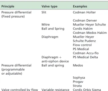
Valves.The second component of the drainage system, the valve, maintains unidirectional flow (craniocaudal) and acts by regulating CSF drainage18-29. This control occurs mainly through pressure regulation (pressure regulators), flow regulation (regulatory flow valves) and anti-siphon mechanisms (anti-siphon devices). Table 2 presents valves currently available.
The pressure at which the valve opens is called the set pressure. There are valves with low, medium and high pressure in each category, referring to opening pressures of about 5, 10, and 15 cm H2O, respectively. Most valves are
designed to open and allow the flow of CSF when the intraventricular pressure rises above the opening pressure18-29. Once the proximal pressure drops below the closing pressure, the valve closes and CSF flow ceases.
In contrast to fixed and programmable pressure valves, flow regulated valves maintain constant flow despite changes in the CSF pressure and patient position18-29.
Anti-siphon devices are used to avoid the siphon effect and its complication, namely over-drainage of CSF. The siphon effect is a phenomenon that occurs due to the increased flow of CSF from the ventricles drained after postural changes such as standing up after sitting. This phenomenon is due to the increased hydrostatic pressure and perfusion pressure of the drainage system. In the vertical position, additional hydrostatic pressure increases the pressure differential and the CSF flow through the valve. One of the critical pressure regulators is that they are subject to this phenomenon in the vertical position18-29.
A recent advance in bypass valves technology has been the introduction of programmable valves. Programmable valves can be adjusted externally using a special magnetic device that alters the position of an internal rotor and thereby modifies the opening pressure of the valve. This removes the need for a surgical procedure when the patient requires a valve with a different pressure. This type of valve tends to be well suited for handling difficult cases of over-drainage or under-drainage of CSF. It is unclear whether the benefits outweigh the increased costs of such devices in all patients18-29.
Since the programmable valve contains a magnet, most valves need to be reprogrammed immediately after all magnetic resonance imagings (MRI). However, a program-mable valve that is not altered by a magnetic field is also available. It “locks” the configuration and can be changed only with a specific magnetic programmer. Routine house-hold equipment such as mobile phones and computers are not strong enough to affect the valve, but special care should be taken when patients are around strong magnetic sources18-29. Such devices include a programmable valve, Medos (Medos Codman, Le Cocle, Switzerland), the adjustable Table 1 -Possible distal sites for ventricular shunts
1. EXTERNAL
2. INTERNAL INTRACRIANIAL Subarachnoid space/ Subdural space/ Superior sagittal sinus
EXTRACRANIAL Subgaleal space/ Mastoid antrum/ Duct of salivary gland CERVICAL Duct of salivary gland/
Common facial vein THORACIC Right atrium/ Superior vena
cava/ Pleural cavity/ Thoracic duct / Spinal epidural space/ Bone marrow
ABDOMINAL Peritoneal cavity/ Omental bursa/ Stomach/ Gallbladder/ Urinary bladder/ Ureter/ Ileum/ Uterine tube
Table 2 -Available valve types
Principle Valve type Examples
Pressure differential (Fixed pressure)
Slit Codman Holter
Codman Denver Mitre Mueller Heyer Schulte Ball and Spring Cordis Hakim
Codman Medos Hakim Diaphragm Mueller Heyer
Schulte Pudenz Flow control PS Medical Codman Accu-flo Diaphragmþ
anti-siphon device
PS Medical Delta
Pressure differential (programmable or adjustable)
Ball and spring Medos
Sophysa Progav Strata
Valve controlled by flow Variable resistance Cordis Orbis Sigma
MEDICALEXPRESS 2014 August;1(4):166-169 Updating technology of shunt valves
de Oliveira MF et al.
valve, Sophysa (Sophysa, Orsay, France), the Strata valve (Medtronic, USA), and the Progav valve (Aesculap, Berlin, Germany). The adjustment after implantation is accomplished through the aid of radiography (Medos) or a compass held over the device (Sophysa, Strata and Progav)18-29.
In order to avoid the occurrence of the siphoning effect, a variety of valve models have been developed. However, all of them generally act through additions of resistances to the drainage system, reducing in this way the flow of CSF during postural changes. The device, which is subcutaneously placed in series with the valve, holds a movable membrane that moves in response to changes in pressure across it. The outer surface is theoretically atmospheric pressure. When the pressure in the bypass drops, the diaphragm moves to occlude the lumen of the shunt. Such devices are available as separate components to insert below the valve in bypass, or can be incorporated in the valve itself, as in the Delta valve (PS Medical Corporation, California, USA) and the Sphera valve (HpBio, Sa˜o Paulo, Brazil) that combine a diaphragm valve or ball-spring, respectively, and a control device siphon membrane in the body of the valve18-29.
A different approach to the problem of the siphoning effect is seen in the Orbis Sigma valve (Cordis Corporation). In contrast to pressure regulating valves, this valve is designed to be a flow regulating device allowing a fairly constant flow rate over a wide range of differential pressures30-34.
Several level I studies have demonstrated a significant improvement in over-drainage complications with anti-siphon devices or the application of programmable valves or flow-regulated valves30-34.
B CONCLUSIONS
Shunt technology is advancing rapidly. New materials allowing better biocompatibility and even impregnated antibiotics in catheters are increasing the options for CSF shunts. Other concepts of valve systems, designed in accordance to hydrodynamic principles, are also being developed.
Neurosurgeons must implement the latest technology and highest quality care in order to insure better control of hydrocephalus and decrease complications. However one must also be aware of the scientific background and evidence for each valve.
A growing issue that also needs attention is the bias present in scientific publications. This bias can be selection, analysis and management. Many studies are sponsored by large corporations and the authors may have conflicts of interest. In a scenario where large investments are imperative to apply appropriate treatment, an independent and lucid evaluation, though challenging, is necessary.
B CONFLICTS OF INTEREST
Authors declare no conflicts of interest.
B RESUMO
Derivaco˜es liquo´ricas sa˜o um dos maiores avancos da neurocirurgia moderna e representam uma mudanca no tratamento da hidrocefalia. O princı´pio fı´sico ba´sico e´ muito simples e consiste em desviar o fluxo do lı´quido cefalorraquidiano, para estruturas intracranianas, sistema
jugular, a´trio direito do coraca˜o, pleura, peritoˆnio ou para outras cavidades naturais, tais como a bolsa omental e ate´ a bexiga. Todos os sistemas funcionam por meio da pressa˜o diferencial entre o cateter proximal e o distal e sa˜o compostas de cate´teres ventricular e distal, ale´m de uma va´lvula, que e´ o dispositivo que garante fluxo unidirecional de lı´quido cerebrospinal. A tecnologia atual compreende va´lvulas de controle do shunt atrave´s de regulaca˜o da pressa˜o, do fluxo, ale´m de dispositivos anti-sifa˜o. Existem va´lvulas de baixa, me´dia e alta pressa˜o concebidas para abrir e permitir o fluxo de FCS quando a pressa˜o intraventricular sobe acima da pressa˜o de abertura. Em contraste dispositivos de pressa˜o fixa ou de pressa˜o programa´vel, as va´lvulas de regulaca˜o de fluxo funcionam para manter constante o fluxo apesar de variaco˜es na pressa˜o de fluido e posica˜o do paciente. Dispositivos anti-sifa˜o sa˜o utilizados para evitar o efeito de sifa˜o e evitar sub- ou sobre-drenagem de fluido. Discutimos brevemente os aspectos atuais da hidrodinaˆmica e tecnologia de va´lvulas de atualizaca˜o.
B REFERENCES
1. Rekate HL. The definition and classification of hydrocephalus: a personal recommendation to stimulate debate. Cerebrospinal Fluid Res. 2008;5:2. 2. Thomson D. Hydrocephalus and Shunts. Neurosurgery Springer
Specialist Surgery Series, 2005; p. 425-42.
3. Drake JM. The surgical management of pediatric hydrocephalus. Neurosurgery. 2008;62(Suppl 2):633-640, discussion 640-2.
4. Torkildsen A. A new palliative operation in cases of inoperable occlusion of the Sylvian aqueduct. Acta Chir Scand. 1939;82:117-24.
5. Matson DD. A new operation for the treatment of communicating hydrocephalus. J Neurosurg. 1949;6(3):238-47.
6. Nulsen FE, Spitz EB. Treatment of hydrocephalus by direct shunt from ventricle to jugular vein. Surg Forum. 1951;30:399-403.
7. Wallman LJ. Shunting for hydrocephalus. An oral history. Neurosurgery. 1982;11(2):308-13.
8. Missori P, Paolini S, Curra` A. From congenital to idiopathic adult hydrocephalus: a historical research. Brain. 2010;133(Pt 6):1836-49. 9. Maset AL, Castro SC, Camilo JR. Consideraco˜es hidrodinaˆmicas sobre a
derivaca˜o liquo´rica. Parte I: Efeitos do cateter peritoneal. Arq Bras Neurocir. 2005;24(1):9-16.
10. Maset AL, Camilo JR, Duarte KP, Vieira ER. Consideraco˜es hidrodinaˆ-micas sobre a derivaca˜o liquo´rica. Parte III: Distu´rbios hidrodinaˆmicos em sistemas de drenagem externa. Proposta de soluca˜o. Arq Bras Neurocir. 2006;25(2):100-11.
11. Maset AL, Camilo JR, Vieira DDR. Consideraco˜es hidrodinaˆmicas sobre a derivaca˜o liquo´rica. Parte II: O efeito sifa˜o em sistemas de drenagem externa. Arq Bras Neurocir. 2005;24(1):45-51.
12. El-Shafei IL, El-Shafei HI. The Retrograde Ventriculovenous Shunts: The El-Shafei Retrograde Ventriculojugular and Ventriculosinus Shunts. Pediatr Neurosurg. 2010;46(3):160-71.
13. El-Shafei IL, El-Shafei HI. The retrograde ventriculo-sinus shunt (El Shafei RVS shunt). Rationale, evolution, surgical technique and long-term results. Pediatr Neurosurg. 2005;41(6):305-17.
14. Choux M, Genitori L, Lang D, Lena G. Shunt implantation: reducing the incidence of shunt infection. J Neurosurg. 1992;77(6):875-80.
15. Bouzerar R, Tekaya I, Bouzerar R, Bale´dent O. Dynamics of hydrocepha-lus: a physical approach. J Biol Phys. 2012;38(2):251-66.
16. Penn RD, Linninger A. The physics of hydrocephalus. Pediatr Neurosurg. 2009;45(3):161-74.
17. Symss NP, Oi S. Theories of cerebrospinal fluid dynamics and hydrocephalus: historical trend. J Neurosurg Pediatr. 2013;11(2):170-7. 18. Cinalli G, Maixner WJ, Sainte-Rose C. Pediatric hydrocephalus. 1st Ed:
Springer-Verlag; 2005.
19. Bergsneider M, Miller C, Vespa PM, Hu X. Surgical management of adult hydrocephalus. Neurosurgery. 2008;62(Suppl 2):643-659, discussion 659-60. 20. Kurtom KH, Magram G. Siphon regulatory devices: their role in the
treatment of hydrocephalus. Neurosurg Focus. 2007;22(4):E5.
21. Drake JM, Sainte-Rose C. The Shunt book. Cambridge, USA: Blackwell Science; 1995.
22. de Oliveira MF, Saad F, Reis RC, Rotta JM, Pinto FC. Programmable valve represents an efficient and safe tool in the treatment of idiopathic normal-pressure hydrocephalus patients. Arq Neuropsiquiatr. 2013;71(4):229-36. 23. Zemack G, Romner B. Adjustable valves in normal-pressure
hydrocepha-lus: a retrospective study of 218 patients. Neurosurgery. 2002;51 (6):1392-1400, discussion 1400-2.
Updating technology of shunt valves
de Oliveira MF et al. MEDICALEXPRESS 2014 August;1(4):166-169
24. Vanneste J, Augustijn P, Dirven C, Tan WF, Goedhart ZD. Shunting normal-pressure hydrocephalus: do the benefits outweigh the risks? A multicenter study and literature review. Neurology. 1992;42(1):54-9. 25. Hanlo PW, Cinalli G, Vandertop WP, Faber JA, Bøgeskov L, Børgesen SE,
et al., Treatment of hydrocephalus determined by the European Orbis Sigma Valve II survey: a multicenter prospective 5-year shunt survival study in children and adults in whom a flow-regulating shunt was used. J Neurosurg. 2003;99(1):52-7.
26. Pinto FC, Saad F, Oliveira MF, Pereira RM, Miranda FL, Tornai JB, et al., Role of endoscopic third ventriculostomy and ventriculoperitoneal shunt in idiopathic normal pressure hydrocephalus: preliminary results of a randomized clinical trial. Neurosurgery. 2013;72(5):845-53, discussion 853-4.
27. Pinto FC, Pereira RM, Saad F, Teixeira MJ. Performance of fixed-pressure valve with antisiphon device SPHERA(w) in hydrocephalus treatment
and overdrainage prevention. Arq Neuropsiquiatr. 2012;70(9):704-9. 28. Gomes Pinto FC, de Oliveira MF. Response to journal club: role of
endoscopic third ventriculostomy and ventriculoperitoneal shunt in idiopathicnormal pressure hydrocephalus: preliminary results of a randomized clinical trial. Neurosurgery. 2013;73(5):911-2.
29. Lemcke J, Meier U, Mu¨ller C, Fritsch MJ, Kehler U, Langer N, et al., Safety and efficacy of gravitational shunt valves in patients with idiopathic
normal pressure hydrocephalus: a pragmatic, randomised, open label, multicentre trial (SVASONA). J Neurol Neurosurg Psychiatry. 2013;84(8):850-7.
30. Boon AJ, Tans JT, Delwel EJ, Egeler-Peerdeman SM, Hanlo PW, Wurzer HA, et al., Dutch Normal-Pressure Hydrocephalus Study: randomized comparison of low- and medium-pressure shunts. J Neurosurg. 1998;88(3):490-5.
31. Eide PK, Brean A. Cerebrospinal fluid pulse pressure amplitude during lumbar infusion in idiopathic normal pressure hydrocephalus can predict response to shunting. Cerebrospinal Fluid Res. 2010;7:5.
32. Oi S, Di Rocco C. Proposal of “evolution theory in cerebrospinal fluid dynamics” and minor pathway hydrocephalus in developing immature brain. Childs Nerv Syst. 2006;22(7):662-9.
33. Mattei TA, Morris M, Nowak K, Smith D, Yee J, Goulart CR, et al., Addressing the siphoning effect in new shunt designs by decoupling the activation pressure and the pressure gradient across the valve. J Neurosurg Pediatr. 2013;11(2):181-7.
34. RMattei TA, Nair K, Morris M, Cole D, Flatt M, Goulart CR, et al., Design and benchmark testing of a bicorporal pump for the treatment of normal-pressure hydrocephalus and idiopathic intracranial hypertension. J Neurosurg Pediatr. 2013;11(2):188-97.
MEDICALEXPRESS 2014 August;1(4):166-169 Updating technology of shunt valves
de Oliveira MF et al.