Sao Paulo Med J. 2011; 129(1):51-3
51
Case report
Ileal perineurioma as a cause of intussusception
Perineurioma ileal como uma causa de intussuscepção
Sheila Cristina Lordelo Wludarski
I, Isabel Irene Rama Leal
II, Herbeth Franco Queiroz
III,
Tulio Marcos Rodrigues da Cunha
IV, Carlos Eduardo Bacchi
VPathology Reference Laboratory, Botucatu, São Paulo, Brazil
IMD. Associate pathologist, Pathology Reference Laboratory, Botucatu, São Paulo, Brazil. IIMD. Pathologist, Hospital Regional da Asa Norte (HRAN), Brasília, Federal District, Brazil. IIIMD. Urologist, Hospital Regional da Asa Norte (HRAN), Brasília, Federal District, Brazil. IVMD. General surgeon, Hospital Regional da Asa Norte (HRAN), Brasília, Federal District, Brazil. VMD, PhD. Chief pathologist, Pathology Reference Laboratory, Botucatu, São Paulo, Brazil.
ABSTRACT
CONTEXT: Perineuriomas are rare tumors composed of cells resembling those of the normal perineurium. It usually occurs in subcutaneous, soft-tissue or intraneural locations. Very few reports in the literature have described perineuriomas in the gastrointestinal tract, including the stomach, colon and jejunum.
CASE REPORT: We report the clinicopathological and immunohistochemical features of a case of ileal perineurioma that was manifested clinically as intestinal obstruction due to intussusception. Ileal perineurioma has not previously been reported at this anatomical location.
RESUMO
CONTEXTO: Perineurioma é uma rara neoplasia composta de células que lembram aquelas do perineuro normal e geralmente ocorre no subcutâneo, tecidos moles ou em localização intraneural. Poucos relatos na literatura descrevem perineuriomas no trato gastrointestinal incluindo estômago, cólon e jejuno.
RELATO DE CASO: Os autores apresentam as características clinicopatológicas e imunoistoquímicas de um caso de perineurioma ileal apresentando-se clinicamente por obstrução intestinal decorrente de intussuscepção. Perineurioma ileal não havia sido descrito até o momento nessa localização anatômica.
KEY WORDS: Ileum.
Immunohistochemistry. Nerve sheath neoplasms. Gastrointestinal tract. Intussusception.
PALAVRAS-CHAVE: Íleo.
Imunoistoquímica. Neoplasias da bainha neural. Trato gastrointestinal. Intussuscepção.
INTRODUCTION
Perineuriomas are uncommon benign neoplasms that occur main-ly in the subcutis, but also arise in soft tissues or intraneural locations. he neoplastic cells comprising perineuriomas resemble those of the normal perineurium. hese neoplasms were irst described by Lazarus and Trombetta in 1978.1 An unusual variant of soft-tissue perineu-rioma has been characterized by Fetsch and Miettinen as a scleros-ing variant.2 Perineuriomas have also been reported in unusual lo-cations such as the kidneys3 and the paratesticular region.4 Very few reports5-10 have described perineuriomas in the gastrointestinal tract, and these have mainly been in the stomach, colon and jejunum. We report the clinicopathological and immunohistochemical features of a case of ileal perineurioma, of soft-tissue type, manifested clinical-ly through intestinal obstruction due to intussusception. To the best of our knowledge, perineurioma has not been previously reported in this anatomical location.
CASE REPORT
A previously healthy 25-year-old white male presented with com-plaints of abdominal pain for two weeks. he pain was mainly located
in the periumbilical area and was associated with nausea. here was no fever. he pain progressively increased and the patient started pre-senting episodes of vomiting. Abdominal radiographs and ultrasound scans revealed indings of intussusception. Laparotomy was performed and ileal intussusception was found 60 cm from the ileal-cecal valve, caused by a 5-cm tumor involving the intestinal wall of the ileum. he tumor was surgically removed and the patient’s postoperative evolu-tion was uneventful.
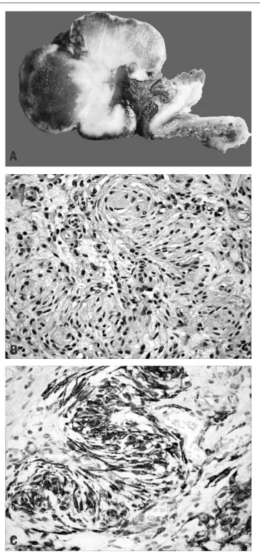
Clinical information was obtained from the patient’s records. he gross pathological examination revealed an ulcerated polypoid tumor of the ileum measuring 5 cm across the greatest diameter. he cut sur-face showed a bright whitish tumor mass with areas of hemorrhage.
Sao Paulo Med J. 2011; 129(1):51-3 Wludarski SCL, Leal IIR, Queiroz HF, Cunha TMR, Bacchi CE
52
he expression of epithelial membrane antigen (EMA), claudin-1, S-100 protein, CD117, CD34, cytokeratin and smooth-muscle actin and desmin was investigated by means of immunohistochemistry us-ing a standard avidin-biotin method. Immunohistochemical analysis revealed difuse expression of EMA (Figure 1B) and claudin-1 by the neoplastic cells, but the cells were negative for S-100 protein, CD117, CD34, cytokeratin, smooth muscle actin and desmin.
DISCUSSION
Perineuriomas are rare benign peripheral nerve sheath tumors arising mainly in the subcutis, but they have also been described in soft tissue and intraneural locations.11 hey occur in adults and are not associated with neuroibromatosis types 1 and 2. he most common morphological ind-ing from soft-tissue perineuriomas is the presence of neoplastic spindle-cell populations composed of slender ibroblastic-like spindle-cells arranged in a vague fascicular, storiform or whorl-forming pattern. Perineuriomas are very rare outside of the subcutis, soft tissue and intraneural locations.
hey have been reported in the kidneys3 and the paratesticular re-gion.4 Very few reportsof perineuriomas arising in the gastrointestinal tract have been published.5-10 Hornick et al.5 irst described riomas of the intestine. hey presented 10 cases of intestinal perineu-riomas, of which nine were located in the colon and one in the jeju-num, with no cases located in the ileum. Additional reports of gastroin-testinal perineuriomas have included rare cases of perineurioma of the stomach,6-8 a case of perineurioma of the esophagus9 and a case of be-nign hybrid perineurioma-schwannoma of the colon.10 In the gastroin-testinal tract, perineuriomas usually present as asymptomatic intramu-cosal lesions, which are incidentally detected by screening tests. Here, as shown in Table 1, we described the irst case of ileal perineurioma of a patient who presented with signs and symptoms relating to intussuscep-tion. Most of the morphological features seen in this case were also ob-served by Hornick et al.,5 including the presence of uniform bland spin-dle cells with ovoid to elongated nuclei and pale indistinct eosinophilic cytoplasm, myxoid or collagenous stroma, and rare mitotic igures. he result from immunophenotyping our case consisted of indings that typ-ically occur in perineuriomas, i.e. strong expression of EMA and clau-din-1 with no expression of S-100 protein. It has been stated that virtu-ally all perineuriomas express EMA,12,13 while they are also positive for claudin-1 in about 90% of the cases. Both of these markers reveal mem-branous patterns through immunostaining. Moreover, perineuriomas have been found to be negative for S-100 protein.
he most important diferential diagnoses in this clinical pathologi-cal context (tumors involving the intestinal wall) are with gastrointestinal stromal tumor (GIST) and schwannoma.6,7 GISTs are usually composed of bland spindle and/or epithelioid cells without the uniquely perivascu-lar location of the tumor cells that was seen in the present case, and over 95% of GISTs are positive for CD117 (KIT), while often negative for EMA. Schwannomas are neoplasms that rarely occur in the gastrointes-tinal tract, with the exception of the stomach. In these rare cases located in the gastrointestinal tract, they show distinct morphological indings in-cluding highly cellular areas, presence of thick wall vessels, inlammatory
Table 1. Literature review* on ileal perineurioma
Electronic
databases Search strategies
Results
Found Related
PubMed Perineurioma AND Ileum
Limits: Case Reports
63 0
SciELO Perineurioma 4 0
Lilacs Perineurioma 23 0
Scirus Perineurioma AND Ileum 15 0
*Performed on August 20, 2010.
Figure 1. A: Macroscopic indings from ileal perineurioma. B: Hematoxylin
and eosin. Proliferation of bland spindle cells with ovoid to elongated nuclei and indistinct cytoplasm. Note the tendency for the tumor cells to be located around vessels in whorls of striking appearance. C: Immunohistochemical expression of epithelial membrane antigen (EMA) by the neoplastic cells.
A
B
Ileal perineurioma as a cause of intussusception
Sao Paulo Med J. 2011; 129(1):51-3
53
iniltrate and sometimes germinal center formation. Immunohistochemi-cal analysis can easily separate schwannomas from perineuriomas, since the former are positive for S-100 protein and glial ibrillary acidic protein and the latter are positive for EMA. Other diferential diagnoses in such cases that are worth mentioning are with leiomyomas and leiomyosarco-mas. hese tumors have a more fascicular pattern of growth with no ten-dency for the tumor cells to be located around small vessels. Leiomyomas and leiomyosarcomas characteristically demonstrate expression of muscle markers such as desmin and smooth muscle actin, and our case was nega-tive for both of these markers.
According to Hornick et al.,5 it seems that perineuriomas of the gastrointestinal tract probably have a benign clinical course. his case was very challenging to diagnose, because it was a rare tumor in an un-usual location.
REFERENCES
1. Lazarus SS, Trombetta LD. Ultrastructural identiication of a benign perineurial cell tumor. Cancer. 1978;41(5):1823-9.
2. Fetsch JF, Miettinen M. Sclerosing perineurioma: a clinicopathologic study of 19 cases of a distinctive soft tissue lesion with a predilection for the ingers and palms of young adults. Am J Surg Pathol. 1997;21(12):1433-42.
3. Kahn DG, Duckett T, Bhuta SM. Perineurioma of the kidney. Report of a case with his-tologic, immunohistochemical, and ultrastructural studies. Arch Pathol Lab Med. 1993;117(6):654-7.
4. Fagerli JC, Hasegawa SL, Schneck FX. Paratesticular perineurioma: initial description. J Urol. 1999;162(3 Pt 1):881-2.
5. Hornick JL, Fletcher CD. Intestinal perineuriomas: clinicopathologic deinition of a new ana-tomic subset in a series of 10 cases. Am J Surg Pathol. 2005;29(7):859-65.
6. Agaimy A, Wuensch PH. Perineurioma of the stomach. A rare spindle cell neoplasm that should be distinguished from gastrointestinal stromal tumor. Pathol Res Pract. 2005;201(6):463-7. 7. Chetty R. Myxoid perineurioma presenting as a gastric polyp. Ann Diagn Pathol.
2010;14(2):125-8.
8. Agaimy A, Märkl B, Kitz J, et al. Peripheral nerve sheath tumors of the gastrointestinal tract: a multicenter study of 58 patients including NF1-associated gastric schwannoma and unu-sual morphologic variants. Virchows Arch. 2010;456(4):411-22.
9. Kelesidis T, Tarbox A, Lopez M, Aish L. Perineurioma of esophagus: a irst case report. Am J Med Sci. 2009;338(3):230-2.
10. Emanuel P, Pertsemlidis DS, Gordon R, Xu R. Benign hybrid perineurioma-schwannoma in the colon. A case report. Ann Diagn Pathol. 2006;10(6):367-70.
11. Hornick JL, Fletcher CD. Soft tissue perineurioma: clinicopathologic analysis of 81 cases including those with atypical histologic features. Am J Surg Pathol. 2005;29(7):845-58. 12. Ariza A, Bilbao JM, Rosai J. Immunohistochemical detection of epithelial membrane antigen
in normal perineurial cells and perineurioma. Am J Surg Pathol. 1988;12(9):678-83. 13. Theaker JM, Gatter KC, Puddle J. Epithelial membrane antigen expression by the perineurium
of peripheral nerve and in peripheral nerve tumours. Histopathology. 1988;13(2):171-9.
Conlict of interest: Not declared
Sources of funding: Not declared
Date of irst submission: August 11, 2009
Last received: August 20, 2010
Accepted: October 26, 2010
Address for correspondence:
