http://dx.doi.org/10.1590/0037-8682-0164-2014
INTRODUCTION
Address to: Dr. Parviz Parvizi. Molecular Systematic Laboratory/Parasitology Department /Pasteur Institute of Iran. 69 Pasteur Ave, Tehran, Iran.
Phone/Fax: 98 21 6649-6414 e-mail: [email protected] Received 9 September 2014 Accepted 4 October 2014
The existence of only one haplotype of
Leishmania major
in the main and potential reservoir hosts of
zoonotic cutaneous leishmaniasis using different
molecular markers in a focal area in Iran
Narmin Najafzadeh
[1], Mohammad Mehdi Sedaghat
[2], Syed Shuja Sultan
[3], Adel Spotin
[1],[4],
Alireza Zamani
[1], Roozbeh Taslimian
[1], Amir Yaghoubinezhad
[1],[5]and Parviz Parvizi
[1][1]. Molecular Systematics Laboratory, Parasitology Department, Pasteur Institute of Iran, Tehran, Iran. [2]. Department of Medical Entomology and Vector Control, School of Public Health, Tehran University of Medical Sciences, Tehran, Iran. [3]. South Tehran Health Center, Tehran University of Medical Sciences, Tehran, Iran. [4]. Department of Medical Parasitology & Mycology, School of Medicine, Tabriz University, Medical Sciences, Tabriz, Iran. [5]. Department of Cellular and Molecular Biology, Higher Education Institute of Rab-Rashid, Tabriz, Iran.
ABSTRACT
Introduction: Leishmania major is the causative agent of zoonotic cutaneous leishmaniasis (ZCL), and great gerbils are the main reservoir hosts in Iran. Abarkouh in central Iran is an emerging focal point for which the reservoir hosts of ZCL are unclear. This research project was designed to detect any Leishmania parasites in different wild rodent species. Methods: All rodents
captured in 2011 and 2012 from Abarkouh district were identifi ed based on morphological characteristics and by amplifi cation of
the rodent cytochrome b (Cyt b) gene. To detect Leishmania infection in rodents, deoxyribonucleic acid (DNA) of each ear was extracted. Internal transcribed spacer-ribosomal deoxyribonucleic acid (ITS-rDNA), microsatellites, kinetoplast deoxyribonucleic acid (kDNA) and cytochrome b genes of Leishmania parasites were amplifi ed by polymerase chain reaction (PCR). Restriction
fragment length polymorphism (RFLP) and sequencing were employed to confi rm the Leishmania identifi cation. Results: Of 68 captured rodents in the region, 55 Rhombomys opimus were identifi ed and nine Leishmania infections (9/55) were found. In addition, eight Meriones libycus and two Tatera indica were sampled, and one of each was confi rmed to be infected. Two Meriones
persicus and one Mus musculus were sampled with no infection. Conclusions: The results showed that all 11 unambiguously
positive Leishmania infections were Leishmaniamajor. Only one haplotype of L. major (GenBank access No. EF413075) was found and at least three rodents R. opimus, M. libycus and T. indica—appear to be the main and potential reservoir hosts in this ZCL focus. The reservoir hosts are variable and versatile in small ZCL focal locations.
Keywords: Leishmania parasite. Zoonotic cutaneous leishmaniasis. Rodents. Haplotype. Iran.
Leishmaniasis is one of the nine emerging individual infectious diseases that have been largely neglected around the world and in the Middle East1.
In Iran, leishmaniasis is observed in three clinical forms: zoonotic cutaneous leishmaniasis (ZCL), anthroponotic cutaneous leishmaniasis (ACL) and zoonotic visceral leishmaniasis (ZVL). ZCL caused by Leishmania major has a health as well as socioeconomic impacts in Iran. ZCL has been reported in rural regions of Iran in 15 of 31 Provinces,including: Bushehr, Hormozgan and Fars in the south2,3; Ilam and
Khuzestan in the southwest and west4,5; Golestan, Khorasan
and Semnan in the northeast and north4-6; and Isfahan in the
central region7.
During the life cycle of ZCL, which depends on the
geographical location of the disease, sandfl ies act as vectors of
Leishmania, Bartonella bacilliformis and some arboviruses,
and wild rodents are considered to be the reservoir hosts8. Many
investigations have been conducted on different aspects of ZCL in naturally important foci in Iran9-11, although some areas
of neighboring Provinces have been neglected for unknown reasons . Predisposing factors, such as increasing migration of patients from endemic foci to potential areas, irregular
construction and urbanization and changing sandfl y fauna in the
region affect the distribution and survival of ZCL9,12.
Yazd Province in central Iran is one of these regions, and the number of cases of ZCL has been increasing since 198110.
The offi cial reports from the health center in Yazd Province has
demonstrated that the number of cases of CL in the Ardakan area (which is an important potential focus in southwestern Yazd) increased from 1996 to 1997 (total: 372 cases), which
Rhombomys opimus
Meriones libycus
T atera indica
Abarkub District
Abarkub
District Yazd Province
L. Major
L. Tropica
L. infantum
C F B A D E
A
Khoram abad
B
Abarghasar
C
Chahgir
D
Abarkouh
E
Gonbad
F
Harooni
Village name
N
E W
S NW NE
SW SE
may lead to monitoring and surveillance activities in this district13. A few studies have examined some cities in Yazd
Province (Ardakan, Taft, Bafgh, and Khatam cities); however, no molecular-epidemiologic investigations have been performed in Abarkouh district yet. Abarkouh has many historical places (such as Abarkouh's Cedar) and tourism values therefore, tourists play an important role in spreading the infection to other
Provinces. Thus, the isolation, detection and identifi cation of
Leishmania spp. in rodents are essential for disease prognosis,
diagnosis methods, the monitoring of clinical outcomes, epidemiological perspectives and treatment program planning. Some reports have demonstrated that Rhombomys opimus and
Meriones libycus are the most important reservoir hosts of ZCL
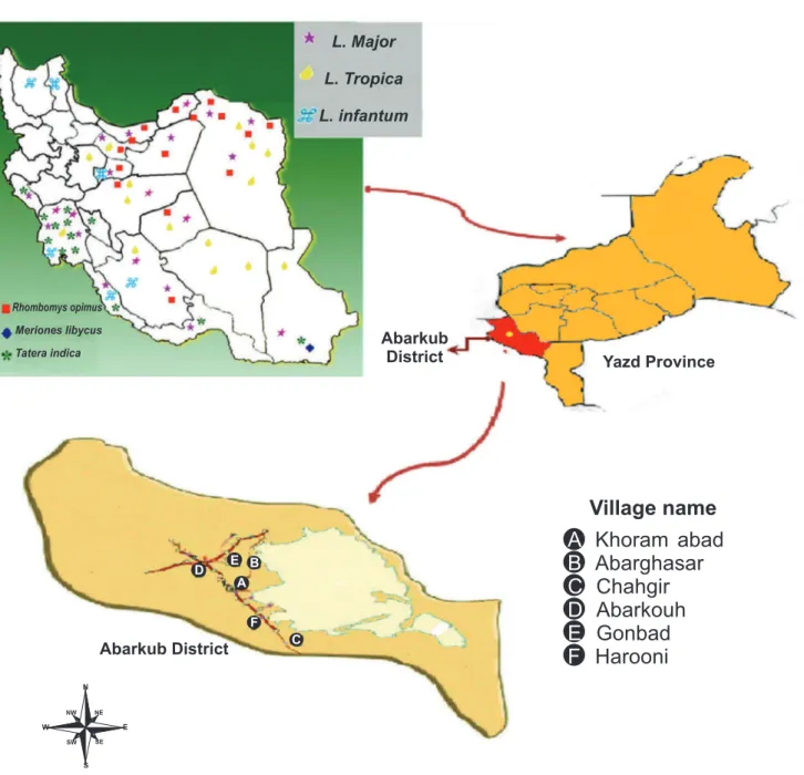
FIGURE 1 -Map ofAbarkouh showing sampling regions and the geographical distribution of reservoir hosts of ZCL in different regions of Iran. L: Leishmania; ZCL: zoonotic cutaneous leishmaniasis.
in the neighboring Provinces of Isfahan and Sh iraz14,15. The
geographical distribution of reservoir hosts of ZCL in different regions of Iran is shown in Figure 111.
For population genetic studies and species identifi cation, we
METHODS
Study area, sampling and laboratory methods
This cross-sectional/descriptive study was performed in 2011 and 2012, and rodent samples were obtained from 5 villages across Abarkouh district, Yazd Province, central Iran, including: Abarghasr, Haroni, Khorram abad, Gonabad and Chahgir.
Abarkouh district is situated between Fars (southern Iran) and Isfahan (central Iran) Provinces (Figure 1). These Provinces are considered hyper-endemic regions that are important sites for ZCL7. Abarkouh district, with an altitude of 1,510 meters
above sea level (a.s.l.) (4,954 feet), geographic coordinates of "31°07′44″N 53°16′57″E31.13°N 53.28°E" (Figure 1) and a population of approximately 21,818 people, is located in Yazd Province in central Iran. Due to its hot and dry climate and its proximity to Isfahan and Fars Provinces, Abarkouh is considered a new and emerging focus of ZCL as well.
The rodents were sampled in Abarkouh area 120km southwest of Yazd Province, using wooden and wire traps. To identify active colonies of rodents within a diameter of 1-1.5km around villages in Abarkouh, approximately 30-40 live traps were used, and the rodents were captured monthly by baiting with dates and cucumbers. The genus and species of each rodent were determined based on external features, including: ears, color, tail, body measurements, teeth, feet and cranium15,24,25.
Each protocol and method applied in this survey was conducted according to the principles expressed in the Declaration of Helsinki and was approved by the Human and Animal Research Ethics Committee of the Pasteur Institute of Iran.
contains nearly 10,000 small circular DNAs (kDNA minicircle). This minicircle comprises a variable region (600bp) and a conserved region (120bp). Microsatellite markers in Leishmania parasites are co-dominant and allelic and combine 1-7 nucleotide units into short, tandemly repeated DNA sequences19,20.
Currently, multilocus microsatellite typing (MLMT) is being used widely in population genetic studies in different species of
Leishmania parasites21,22. Minicircle kDNA and microsatellite
ITS-rDNA are also well known as molecular markers for the detection of Leishmania infections7,21.
The Cyt b gene encodes the central catalytic subunit of an enzyme present in the respiratory chain of mitochondria and exists in almost all organisms. This gene has been broadly
used for phylogenetic studies and identifi cation of animals and
plants23. The Cyt b gene of the genus Leishmania consists of two
regions: the edited region (the most 5′ region of 23bp), which
undergoes ribonucleic acid (RNA) editing, and the non-edited
region (the 3′ region of 1,056bp) 23.
Since the rodent fauna, the Leishmania species and their infection rate in Abarkouh district of Yazd Province, Iran, haves not been elucidated completely; we have designed this study in order to investigate these aspects of ZCL.
Two impression touch slides were obtained from both ears of each rodent by scratching. For brief microscopic observation, rodent samples were collected from the ears after removing the hair and making small scratches from which to extract serous
fl uid, which was then fi xed on a microscopic slide with methanol
and stained for 30min with Giemsa diluted 1:10. The slides were then observed under a light microscope to detect the presence of Leishman bodies.
Furthermore, serous fl uid from the rodents' ears was injected
into Balb/C mice to monitor for the appearance of Leishmania
infection lesions. Prepared serous fl uid from infected Balb/C mice accompanied by serous fl uid from scratches from each
ear of rodents was cultured in Novy-MacNeal-Nicolle (NNN) medium. Subsequently, the cultures were incubated at 22°C for 6 weeks. The cultures were checked at two-day intervals until they reached the growth phase (log phase) based on observation
using an inverted microscope. Positive cultures were confi rmed
by the presence of promastigotes, which were sub-cultured into restriction fragment length polymorphism (RFLP) medium weekly.
The harvested promastigotes from the early stationary phase (approximately 2×10⁶ promastigotes/ml) and serous fl uid from
each ear of the rodent were injected subcutaneously into the base tail of a Balb/C mouse. Inoculated Balb/C mice were examined weekly for the appearance of lesions at the injection site for 6 months. Samples from infected Balb/C mice with cutaneous lesions were used for DNA extraction.
Molecular methods
Smears prepared from infected Balb/C mice, with serous
fl uid and/or cuts from each rodent ear, were kept in separate 1.5ml microtubes containing 100μl phosphate-buffered saline (PBS) and then centrifuged briefl y three times at 13,000rpm.
The PBS was discarded. Each rodent ear was placed in a 1.5ml microtube and placed in liquid nitrogen 3 times for 3min each. The genomic DNA of each rodent and any parasite within was extracted using the ISH-Horovize method and a GeNet BIO kit (Global Gene Network South Korea); these procedures were carried out in the systematic molecular laboratory of the
Pasteur Institute in a room where amplifi ed and cloned DNAs
were never processed11,25.
The DNA samples extracted from rodent tissues were used in polymerase chain reaction (PCR) to amplify a 624bp fragment of the cyto chrome b gene (Cyt b) from the mitochondrial DNA to accurately identify the rodent species. We followed the protocol of Kent and Norris (Table 1)26.
The internal transcribed spacer-ribosomal deoxyribonucleic acid gene was amplified for the detection of Leishmania infection using ITS1-5.8SrRNA-ITS2 fragments, with ITS1F as the forward primer and ITS2R4 as the reverse primer (Table 1)7.
To perform RFLP analysis, the PCR products were blunt digested using endonuclease BsuR1 (HaeIII) (Fermentas, Life
Sciences, Germany) in the recognition site pattern GG↓CC,
Genes Primer name Primer sequence Fragment size (bp)
Annealing temperature &
cycle number
ITS-rDNA
ITS1F (F) 5' GCAGCTGGATCATTTTCC 3'
462 58°C, 37 ITS2R4 (R) 5' ATATGCA GAA GAGAGG AGG C 3'
Microsatellite DNA
ITSmF1 (F) 5´ GTGTGGAAGCCAAGAGGAGG 3´
160 58°C, 37 ITSmR2 (R) 5´ GCAAGCACCAGAGAGGAGT 3´
kDNA
LINR4 (F) 5′ GGGGTTGGTGTAAAATAGGG 3′
650
S
em
i-ne
st
ed
P
CR
1st step:
52°C, 17 LIN 17 (fi rst-step R) 5′ TTTGAACGGGATTTCTG 3′
2nd step:
58°C, 33 LIN19 (second-step R) 5′ CAGAACGCCCCTACCCG 3′
Leishmania Cytochrome b
LCBF1 (F) 5′ GGTGTAGGTTTTAGTTTAGG 3′
880 50°C, 39 LCBR2 (R) 5′ CTACAATAAACAAATCATAATATACAATT 3′
Rodent Cytochrome b*
UNFOR403 (F) 5′ TGAGGACAAATATCATTCTGAGG 3′
624 58°C, 35 UNREV1025 (R) 5′ GGTTGTCCTCCTCCAATTCATGTTA 3′
Bp: Base pairs; ITS-rDNA: internal transcribed spacer-ribosomal deoxyribonucleic acid; DNA: deoxyribonucleic acid; kDNA: kinetoplast deoxyribonucleic acid; (F): Forward primer; (R): Reverse primer; *Rodent cytochrome b genes were not used for the detection of Leishmania infection but only for rodent species identifi cation.
TABLE 1 - Primer sequences and conditions used for all employed genes for the identifi cation of Leishmania parasites within rodents.
The primer sets LINR4 (forward), LIN17 (fi rst-step reverse)
and LIN19 (second-step reverse) were used in the semi-nested PCR for the minicircle kDNA gene16. The primers anneal
within the conserved area of the minicircle and are based on the conserved sequence blocks recognized by Brewster and Baker (Table 1)28.
The third method used for Leishmania infection identifi cation was microsatellite ITS-rDNA analysis; the protocol used in this assay was designed by Parvizi et al.25. The primers were ITSMF1
(forward) and ITSMR2 (reverse) (Table 1).
To detect Leishmania infection, we also used a fragment of the cytochrome b gene from mitochondrial DNA, and the
primers used in this amplifi cation were LCBF1 (forward) and
LCBR2 (reverse) (Table 1)17.
After amplification, the DNA samples were excised,
purifi ed and sequenced using an ABI PRISM TM310 automated
sequencer (Applied Biosystems, USA). The sequences obtained were edited and aligned with database sequences using SequencherTM v. 4.4 software to identify unique sequences
(= haplotypes), which were analyzed phylogenetically using MEGA5.05 software29,30.
Ethical considerations
This study was approved by the D epartment of Parasitology, Pasteur Institute of Iran, Tehran, Iran.
RESULTS
In total, 68 rodents were captured in five villages in Abarkouh district (Figure 1; Table 2). Thirty-three of 68 rodents were alive and transported to the Pasteur Institute of Iran, Tehran, for further studies using conventional and molecular
methods. Thirty-fi ve rodents were dead after being caught by
wire and wooden live traps at sampling sites. The ears of these dead rodents were used only for molecular methods. Based on morphological characteristics and rodent molecular markers (Cyt b sequences), fi ve species were identifi ed. The most abundant rodent was R. opimus (55/68: 81%). The frequencies and abundances of the other rodent species were as follows:
M. libycus (8/68: 12%), Meriones persicus (2/68: 3%), Tatera
indica (2/68: 3%) and Mus musculus (1/68: 1%), respectively
(Table 2).
Eleven of 68 (16%) rodents were found to be infected with
Leishmania parasites using molecular methods (3/68 (4.4%)
using ITS-rDNA, 10/68 (14%) using microsatellites, 3/68 (4.4%) using minicircle kDNA and 4/68 (5.8%) by amplifying Cyt b from Leishmania parasites). At least 3 of 5 rodent species
had Leishmania infections (Figure 2). Only two of 33 (6.06%)
Rhombomys opimus 81% 0/10 0/10 1/4 8/26 0/6 1/55 1/55 2*/55 8(3*)/55 2(1*)/55 3*/55 9* / 55
(13.04%)
Meriones libycus 12% 0 0 0/1 0/3 1/4 1/8 1/8 1/8 1**/8 1**/8 1**/8 1/8
(12.5%)
Mus musculus 1% 0 0 0 0 0/1 0/1 0/1 0/1 0/1 0/1 0/1 0/1
(0%)
Meriones persicus 3% 0 0 0 0/1 0/1 0/2 0/2 0/2 0/2 0/2 0/2 0/2
(0%)
Tatera indica 3% 0 0 0 0 1/2 0/2 0/2 0/2 1/2 1*/2 0/2 1/2
(50%)
Total 100% 0/10 0/10 1/5 8/30 2/14 2/68 2/68 3/68 10 /68 3/68 4/68 11 /68
(0%) (0%) (20%) (27%) (14%) (2.94%) (2.94%) (4.4%) (14%) (4.4%) (5.8%) (16%) Villages Species F re que nc
y of c
apt ure d rode nt s H arooni A ba rgha sr K hora m a ba d G onba d Cha hgi r M ic ros copi c O bs erva ti on of L ei shm ani a pa ra si te s BA L B/ C Inj ec ti on L ei shm ani a m aj or (+ve ) by IT S -rD N A P CR L ei shm ani a m aj or (+ve ) by m ic ros at el li te P CR L ei shm ani a m aj or (+ve ) by kD N A P CR L ei shm ani a m aj or (+ve ) by L ei shm ani a Cyt b P CR T ot al
ITS-rDNA: internal transcribed spacer-ribosomal deoxyribonucleic acid; kDNA: kinetoplast deoxyribonucleic acid. +ve: Positive sample;
PCR: polymerase chain reaction; Cyt b: Cytochorome b. *3 of 13 positive samples were also tested via one additional gene and were confi rmed
to have Leishmania major infection; **The positive microscopic and BALB/C injected samples also tested positive in molecular tests (ITS-rDNA, microsatellite and kDNA gene amplifi cation).
Conventional Molecular
methods methods
TABLE 2 -Leishmania infections among different rodents captured in Abarkouh district in Yazd Province, Iran, using conventional and molecular methods.
the ear, light microscope observation, culturing in NNN and inoculating in Balb/C mice (Table 2).
The most interesting result was that despite the low number
of captured rodents, fi ve different rodent species were collected and identifi ed. Leishmania infection was detected from three of
these species, and for the fi rst time, T. indica was captured in
Abarkouh district and identifi ed both morphologically through
diagnostic keys and molecularly by sequencing of the Cyt b gene. In addition, one of the two T. indica specimens was infected with L. major.
To fi nd identify additional Leishmania parasite infections and molecular variation rates among collected samples, different genes were employed. Standard and semi-nested PCR were used to amplify ITS-rDNA, microsatellites, kDNA and Cyt b genes from Leishmania parasites (Figure 2).
All 11 Leishmania-positive samples were analyzed using
RFLP and sequencing to defi nitively identify Leishmania species (Figure 2). With RFLP, which allows for the differentiation of
each species unambiguously, two fragments of 120 and 310bp belonging to L. major were obtained (Figure 2).
All sequences from positive samples by ITS-rDNA gene
were blasted and confi rmed to be most similar to L. major, and only L. major with one common haplotype (GenBank accession No. EF413075) was found after direct sequencing, editing, aligning and comparing with the sequences submitted to GenBank using Sequencher TM v. 4.4 and phylogenetic analysis by MEGA5.05 software.
DISCUSSION
In our study, only the L. major parasite with one common
haplotype (GenBank access No. EF413075) was fi rmly identifi ed
in three rodent species. Leishmania parasites have been isolated from all three species in other ZCL foci in Iran11,15,25,31. This is
M +Ve AB01 AB02 AB03 AB04 AB05 AB06 -Ve AB07
880bp
M -Ve +Ve +Ve (enz) AB12 Ab32
460
310
120
A
B
FIGURE 2 - A:Electrophoresis image of Cyt b gene amplifi cation in Leishmania infection among different rodent species of Abarkouh district, Yazd Province, Iran. B: RFLP of ITS-rDNA gene electrophoresis image after digestion with BsuR1 (HaeIII) enzyme of PCR products using In-Silico software (CLC bio A/s, Aarhus, Denmark) of Leishmania infection among different rodent species of Abarkouh district, Yazd Province, Iran (Bordbar and Parvizi 2013) (+Ve contains Leishmania major parasite PCR product without the enzyme effect, and +Ve (enz) is a Leishmania major
parasite PCR product with the enzyme effect). Cyt b: cytochrome b; RFLP: Restriction fragment length polymorphism; ITS-rDNA:
internal transcribed spacer-ribosomal deoxyribonucleic acid; PCR:
polymerase chain reaction. +ve: positive sample, enz: with enzyme.
in P. papatasi, a proven vector of ZCL in Iran, in the same area
in Abarkouh where L. major was isolated in rodents7,10,32.
Finding additional Leishmania infections in different
rodent species compared with only one sandfl y species can be explained by the fact that among sandfl ies, we mainly examined
P. papatasi, and only a small number of other sandfl y species
were tested and found to be Leishmania negative, which did
not provide suffi cient for a precise result10. In addition, only
P. papatasi is able to develop L. major in its midgut and transfer
the parasite to salivary glands to cause ZCL33. However, we
analyzed all the captured rodent samples from different species, and therefore, we were able to identify Leishmania infections in at least three rodent species. The Leishmania infection rate in rodents as the reservoir host of ZCL is much higher
than in sandfl ies as vectors, and in some cases, more than 50%
of the samples were found to be infected with Leishmania parasites14.
Based on our experience in different ZCL foci in Iran, we expected to identify more Leishmania infections in reservoir hosts in Abarkouh district and to observe at least a small amount of variation in the ITS-rDNA gene of L. major in rodents. However, only one haplotype was found, and approximately 16% (11/68) of the tested rodents were infected with Leishmania parasites11,15,25. After sequencing, only one haplotype of L. major,
which is also the common haplotype present in Iran, including Fars and Isfahan Provinces, was detected from Abarkouh rodent samples (GenBank accession No. EF413075).
The low density of sampled rodents as well as Leishmania parasites may be due to a control program of the health care authorities of Abarkouh district that uses zinc phosphate poison and the destruction of rodents' barrowers to control ZCL.
According to reports of different ZCL foci in Iran, many haplotypes of L. major have been identifi ed in sandfl ies, rodents and humans7,11,15,27. The objective of the present study was to
use molecular methods and different genes to identify additional
Leishmania infections and various haplotypes; to this end, four
different genes were employed to detect Leishmania infections in rodents and/or the numbers of haplotypes circulating in the area, but this method relies on a few sequences from all of the
genes from our samples, and no variations were identifi ed. We
also used routine laboratory (conventional) methods, such as NNN cultures, microscopic observation and Balb/C mouse injection. Because most captured rodents died before being transferred to our lab, only a few live rodents were used for the conventional methods, and the infection rate was low. Only 2 infected samples were found by microscopic observation of the presence of amastigotes on slides and the appearance of a lesion after Balb/C mouse injection. Because the NNN cultures
were prepared in the fi eld and due to fungal infection in some
cultures, no growth was shown in any of the cultures.
We employed fi ve different genes during this investigation;
rodents (8/68) was detected using the microsatellite ITS-rDNA gene because of its short tandemly repeated DNA sequence
fragments and because it is highly specifi c. A comparison of
the rest of the genes demonstrated that Cyt b as a mitochondrial gene is more sensitive for Leishmania detection (4/68) because of its high copy numbers per cell; however, nuclear genes are
more specifi c, and of those, the ITS-rDNA gene (3/68), because
it is homogenous and highly conserved with few intracellular polymorphisms, a linear genome and has readable sequences, is considered a suitable gene for sequencing, genus, species, strain and/or even haplotype detection.
Our Leishmania infection data in rodents are similar to
the results of a parallel study among sandfl ies and suspected
patients that was carried out near the time of our investigation10
(Parvizi P et al: unpublished data)..
In previous investigations, reservoir hosts of ZCL have been distributed in different regions. R. opimus and M. libycus are dominant in the northeastern and central regions; M. libycus
and T. indica in the central and southwestern regions and
T. indica in southwestern and southern Iran11,34. R. opimus and
M. libycus have previously been found to be infected with
L. major parasites from Golestan and Isfahan Provinces11,14,15,25.
In addition, T. indica was found to be infected with L. major in Fars Province, Iran31. Interestingly, we were able to identify
L. major infections in all three of these rodents within Abarkouh
district of Yazd Province in central Iran.
In this survey, T. indica was captured for the fi rst time in Abarkouh district; the existence of this rodent in the area may be explained by the fact that Abarkouh neighbors Fars Province, which is a known habitat for T. indica31, and the rodents can
be transported and/or migrate to Abarkouh from Fars and vice versa. The simultaneous existence of T. indica along with
R. opimus and M. libycus as main and potential reservoir hosts
of ZCL in Abarkouh district and the fact that Abarkouh has been largely neglected as an important ZCL focus gives this district an important role in the ZCL life cycle, epidemiology, prognosis and disease-control programs.
Leishmania major was firmly identified in R. opimus,
M. libycus and T. indica, which indicates that at least these three
rodent species can be incriminated as reservoir hosts of ZCL in this location. R. opimus was abundant andhad a greater rate
of L. major infection and should be incriminated as the main
reservoir host of ZCL35.
ACKNOWLEDGMENTS
The authors declare that there is no confl ict of interest.
CONFLICT OF INTEREST
FINANCIAL SUPPORT
REFERENCES
The authors would like to thank the authorities Abarkouh Health Care for their help and support during this investigati on.
We thank Mehdi Baghban for helping with the fi eld work and
Elnaz AlaeeNovin for assistance in the Molecular Systematic Laboratory. A part of this research was funded through MSc studentships to Alireza Zamani based at the Pasteur Institute of Iran, Tehran, and registered for Islamic Azad University, Science and Research Branch of Fars, Iran.
This study was supported by the Pasteur Institute of Iran grant 501, awarded to Dr. Parviz Parvizi.
1. World Health Organization Control of Leishmaniasis. Technical report series 949 of WHO Expert Committee. Geneva: WHO; 2010.
2. Zare S, Baghestani S. Cutaneous Leishmaniasis in Hormozgan, Iran. Int J Dermatol 2001; 40:29-631.
3. Maraghi S, Samarbaf Zadeh A, Sarlak AA, Ghasemian M, Vazirianzadeh B. Identifi cation of Cutaneous Leishmaniasis Agents by Nested Polymerase Chain Reaction (Nested-PCR) in Shush City, Khuzestan Province, Iran. Iran J Parasitol 2007; 2:13-15.
4. Jahani MR, Gharavi MJ, Hadi Shirzad H. Passive Detection of Cutaneous Leishmaniasis in Police Personnel Deployed in the Provinces of Isfahan, Ilam, Bushehr, Khorasan and Khuzestan, Iran. Iran J Public Health 2003; 32:23-27.
5. Sharbatkhori M, Spotin A, Taherkhani H, Roshanghalb M, Parvizi P.
Molecular variation in Leishmania parasites from sandfl ies species of
a zoonotic cutaneous leishmaniasis in northeast of Iran. J Vector Borne Dis 2013; 51:16-21.
6. Alavinia SM, Arzamani K, Reihani MH, Jafari J. Some epidemiological aspects of cutaneous leishmaniasis in Northern Khorasan Province, Iran. Iran J Arthropod Borne Dis 2009; 3:50-54.
7. Parvizi P, Ready PD. Nested PCRs and sequencing of nuclear ITS
rDNA fragments detect three Leishmania species of gerbils in sandfl ies
from Iranian foci of zoonotic cutaneous leishmaniasis. TM & IH 2008; 13:1159-1171.
8. Ready PD. Biology of Phlebotomine sand fl ies as vectors of disease
agents. Ann Rev Entomol 2013; 58:227-250.
9. Parvizi P, Bordbar A, Najafzadeh N. Detection of Wolbachia pipientis,
including a new strain containing the wsp gene, in two sister species
of Paraphlebotomus sandfl ies, potential vectors of zoonotic cutaneous
leishmaniasis. Mem Inst Oswaldo Cruz 2013; 108:414-420.
10. Jafari R, Najafzadeh N, Sedaghat MM, Parvizi P. Molecular characterization of sandfl ies and Leishmania detection in main vector of zoonotic cutaneous leishmaniasis in Abarkouh district of Yazd province, Iran. Asian Pac J Trop Med 2013; 6:792-797.
11. Mirzaei A, Rouhani S, Kazerooni PA, Farahmand M, Parvizi P. Molecular detection and conventional identifi cation of Leishmania
species in reservoir hosts of zoonotic cutaneous leishmaniasis in Fars province, South of Iran. Iran J Parasitol 2013; 8:280-288.
12. Ready PD. Leishmaniasis emergence and climate change. Rev Sci Tech 2008; 27:399-412.
13. Safari Z, Saadati M, Doroudian M, Hossaini SM, Hashemi M, Rezai zarchi S. Cutaneous leishmaniasis characterization in the Center Part of Iran; is that an emerging disease? Adv Environ Biol 2011; 5:3464-3470. 14. Akhavan AA, Yaghoobi-Ershadi MR, Mirhendi H, Alimohammadian MH, Rassi Y, Shareghi N, Jafari R, Arandian MH, Abdoli H, Ghanei M, Jalali-zand N, Khamesipour A. Molecular epizootiology of Rodent leishmaniasis
in hyperendemic area of Iran.Iran J Public Health 2010; 39:1-7.
15. Mirzaei A, Rouhani S, Taherkhani H, Farahmand M, Kazemi B,
Hedayati M, et al. Isolation and detection of Leishmania species among
naturally infected Rhombomys opimus, a reservoir host of zoonotic
cutaneous leishmaniasis in Turkemen Sahara, North East of Iran.
16. Parvizi P, Mauricio I, Aransay AM, Miles MA, Ready PD. First detection
of Leishmania major in peridomestic Iranian sandfl ies: comparison of
nested PCR of nuclear ITS ribosomal DNA and semi-nested PCR of minicircle kinetoplast DNA. Acta Trop 2005; 93:75-83.
17. Luyo-Acero GE, Uezato H, Oshiro M, Takei K, Kariya K, Katakura K, et al. Sequence variation of the Cytochrome b gene of various human infecting members of the genus Leishmania and their phylogeny. Parasitol 2004; 128:483-491.
18. Maleki-Ravasan N, Oshaghi MA, Javadian E, Rassi Y, Sadraei J, Mohtarami F. Blood meal identifi cation in fi eld-captured sandfl ies: comparison of PCR RFLP and ELISA assays. Iran J Arthropod Borne Dis 2009; 3:8-18.
19. Oryan A, Shirian S, Tabandeh MR, Hatam GR, Randau G, Daneshbod Y. Genetic diversity of Leishmania major strains isolated from different clinical forms of cutaneous leishmaniasis in southern Iran based on minicircle kDNA. Infect Genet Evol 2013;19:226-31.
20. Jamjoom MB, Ashford RW, Bates PA, Kemp SJ, Noyes HA. Polymorphic microsatellite repeats are not conserved between Leishmania donovani
and Leishmania major. Mol Ecol Notes 2002; 2:104-106.
21. Kuhls K, Chicharro C, Canavate C, Cortes S, Campino L, Haralambous C, et al. Differentiation, Gene Flow, among European populations of
Leishmania infantum MON-1.PLoS Negl Trop Dis 2008; 2:261.
22. Russell R, Iribar MP, Lambson B, Brewster S, Blackwell JM, Dye C,
et al. Intra and inter-specifi c microsatellite variation in the Leishmania
subgenus Viannia. Mol Biochem Parasit 1999; 103:71-77.
23. Degli esposti M, De Vries S, Crimi M, Ghelly A, Patarnello T, Meyer A. Mitochondrial cytochrome b: evolution and structure of the protein. Biochim Biophys Acta 1993; 1143:243-271.
24. Ziaei H. A fi eld guide for identifying of Iranian desert mammalians. 1st ed. Tehran: Iranian Environment Organization; 1996.
25. Parvizi P, Moradi G, Akbari G, Ready PD, Farahmand M, Piazak N, et al. PCR Detection and sequencing of parasite ITS rDNA gene from
reservoirs host of zoonotic cutaneous leishmaniasis in central of Iran. Parasitol Res 2008; 103:1273-1278.
26. Kent R, Norris DE. Identifi cation of mammalian blood meals in mosquitoes by a multiplex polymerase chain reaction targeting
cytochrome b. Am J Trop Med Hyg 2005; 73:336-342.
27. Bordbar A, Parvizi P. High infection frequency, low diversity of
Leishmania major and fi rst detection of Leishmania turanica in human
in northern Iran. Acta Trop 2014; 133:69-72.
28. Brewster S, Barker DC. Analysis of minicircle classes in Leishmania
(Viannia) species. T Roy Soc Trop Med H 2002; 96 (suppl I):55-63.
29. Parvizi P, Alaeenovin E, Kazerooni PA, Ready PD. Low diversity of
Leishmania parasites in sandfl ies and the absence of the great gerbil in
foci of zoonotic cutaneous leishmaniasisin Fars province, southern Iran. T Roy Soc Trop Med H 2013; 107:356-362.
30. Tamura K, Peterson D, Peterson N, Stecher G, Nei M, Kumar S, et al. MEGA5: Molecular evolutionary genetics analysis using maximum likelihood, evolutionary distance, and maximum parsimony methods. Mol Biol Evol 2011; 10:2731-2739.
31. Mehrabani D, Motazedian MH, Hatam GR, Asgari Q, Owji SM,
Oryan A. Leishmania Major in Tatera indica in Fasa, Southern Iran,
microscopy, culture, isoenzyme, PCR and morphologic study. Asian J Anim Vet Adv 2011; 6:255-264.
32. Killick-Kendrick R. Phlebotomine vectors of the leishmaniasis: A review. Med Vet Entomol 1990; 4:1-24.
33. Volf P, Benková I, Myšková J, Sádlová J, Campino L, Ravel C. Increased transmission potential of Leishmania major/Leishmania infantum
hybrids. Int J Parasitol 2007; 37:589-593.
34. Rouhani S, Mirzaei A, Spotin A, Parviz P. Novel identifi cation of
Leishmania major in Hemiechinus auritus and molecular detection of
this parasite in Meriones libycus from an important foci of zoonotic
cutaneous leishmaniasis in Iran. J Infect Public Health 2014; 7:210-217.
35. Killick-Kendrick R, Ward RD. Ecology of the Leishmania. Parasitol