http://dx.doi.org/10.1590/0037-8682-0135-2014
Major Article
INTRODUCTION
Address to: Dra Maria do Rosário Rodrigues Silva. IPTSP/UFG. Rua 235 s/n, Setor Universitário, 74605-050 Goiânia, GO, Brasil.
Phone: 55 62 3209-6127; Fax: 55 62 3209-6363 e-mail: [email protected]
Received 12 August 2014 Accepted 10 October 2014
Pimenta pseudocaryophyllus
inhibits virulence factors and
promotes metabolic changes in
Candida
yeast
Flávio Ezeddinne El Assal
[1], Joelma Abadia Marciano Paula
[2], Leonardo Silva Capeletti
[2],
Fernando Yano Abrão
[1], Fábio Silvestre Ataídes
[1], Fabyola Amaral da Silva Sá
[1],
Carolina Rodrigues Costa
[1], Orionalda de Fátima Lisboa Fernandes
[1],
Lúcia Kioko Hasimoto Souza
[1]and Maria do Rosario Rodrigues Silva
[1][1]. Instituto de Patologia Tropical e Saúde Pública, Universidade Federal de Goiás, Goiânia, GO. [2]. Unidade de Ciências Exatas e Tecnológicas, Universidade Estadual de Goiás, Anápolis, GO.
ABSTRACT
Introduction: This is the fi rst study to examine the in vitro susceptibility and the expression of virulence factors in Candida
species in the presence of Pimenta pseudocaryophyllus (Gomes) L.R. Landrum (Myrtaceae), a Brazilian plant known as pau-cravo. Additionally, the mechanisms of action of the crude ethanol extract and the ethyl acetate and aqueous fractions of this
plant were investigated. Methods: The in vitro susceptibility of Candida was tested using the broth microdilution method,
whereas an XTT reduction assay was used for biofi lms. Adherence was determined by counting the number of yeast cells that adhered to 100 oral epithelial cells, and hyphal formation was verifi ed in the hyphal induction medium M199. Flow cytometry
with propidium iodide and FUN-1 was performed to assess the mechanism of action. Results: The results revealed that the crude
ethanol extract and the ethyl acetate and aqueous fractions of P. pseudocaryophyllus inhibited the growth of Candida isolates at a
minimal inhibitory concentration (MIC) ranging from 64 to 256µg/mL, whereas the 50% sessile minimal inhibitory concentration (SMIC50) ranged from 512 to >1,024µg/mL. Adherence and hyphal formation were signifi cantly reduced in the presence of the crude ethanol extract and both fractions. Although cell membrane injury was detected, the predominant mechanism of action
appeared to be the alteration of yeast metabolism, as demonstrated by fl ow cytometry. Conclusions: Our results indicated that
antifungal activity reduced the expression of virulence factors in yeast via the alteration of yeast metabolism, suggesting that the crude extract of P. pseudocaryophyllus and its fractions may contain novel antifungal agents.
Keywords: Pimenta pseudocaryophyllus. Candida spp.Antifungal activity.Virulence factors. Metabolic changes.
Fungal infections have increased signifi cantly in recent years due to an increase in the number of immunocompromised individuals. Candida species are responsible for the majority of
yeast infections in humans1. Resistance in yeast and many side
effects associated with antifungal agents have been reported2,3,
and thus, alternative therapeutics lacking these limitations are under investigation. In particular, medicinal plants are being
explored for novel treatments. Pimenta pseudocaryophyllus
(Gomes) L.R. Landrum (Myrtaceae), popularly known as
pau-cravo, craveiro-do-mato, louro-cravo or chá-de-bugre, is found in the Brazilian Atlantic Forest and Cerrado regions, especially in cerradão; it is a tree with a height of approximately four
meters or a small bush in some regions, and it displays antifungal
activity in vitro. According to Paula et al.4,5, the crude extract of
this plant exerts antiedematogenic and antinociceptive effects on mice.
Biofilm formation, adherence to epithelial cells and
fi lamentation arevirulence mechanisms associated with yeast
species of the Candida genus6. The ability to inhibit or reduce
the expression of virulence factors is a desirable characteristic of an antifungal agent7.
In the present study, we examined the in vitro susceptibility,
biofi lms, adherence to epithelial cells and hyphal formation of
Candida species in the presence of the crude extract and the
ethyl acetate and aqueous fractions of P. pseudocaryophyllus.
Furthermore, we investigated the basic mechanisms of action of these extracts against these yeast species.
METHODS
Plant material and preparation of the Pimenta pseudocaryophyllusextract and fractions
The leaves of P. pseudocaryophyllus were collected in
and the voucher specimen was deposited in the herbarium of the
Universidade Federal de Goiás (UFG) under number 27159. The
crude ethanol extract and fractions obtained from the leaves of
P. pseudocaryophyllus (citral chemotype) were prepared
according to Paula et al5. Four fractions were obtained: hexane,
dichloromethane, ethyl acetate, and aqueous fractions. Previous experiments had demonstrated that the crude ethanol extract and the ethyl acetate and aqueous fractions provide the best results based on in vitro susceptibility tests against Candida yeast species.
Therefore, these extracts were selected for the present study. Fluconazole (Pfi zer Pharmaceutical Group, New York, NY) was used in all assays.
Chemical components of the extract
High-performance liquid chromatography (HPLC) was performed to characterize and quantify the primary constituents of the crude extract. Quercitrin (quercetin 3-O-α-L-rhamnopyranoside; Sigma), catechin (Sigma), and gallic acid (Sigma) were used as reference standards.
Organisms
Bioassays were performed on 12 Candida isolates obtained
from blood and nail samples and stored at -70°C in yeast
extract potato dextrose (YEPD) agar (Difco®) containing 10% glycerol in the Laboratory of Mycology at the Institute of Tropical Diseases and Public Health of the Federal University of Goiás. These isolates were obtained from a previous study that was approved by the Bioethics Committee of the Hospital das Clínicas in Goiânia, Goiás. Candida parapsilosis ATCC 22019 was used as the standard strain.
Antimicrobial activity
Antifungal susceptibility tests using the crude extract and
the ethyl acetate and aqueous fractions of P. pseudocaryophyllus
leaves were performed using the broth microdilution method according to the Clinical and Laboratory Standards Institute
(CLSI) document M27-A38. The crude extract and the
fractions were dissolved in 0.1% dimethyl sulfoxide (DMSO) (0.1mL DMSO in of 9.9mL Roswell Park Memorial Institute [RPMI] medium), which exerted a negligible effect on the
growth kinetics of the yeast species9. Then, the extract and
the fractions were diluted in RPMI medium buffered to pH 7.0 using morpholinepropanesulfonic acid (MOPS). The fi nal concentrations of these samples ranged from 2.0 to 1,024µg/mL.
The inoculum of Candida isolates was suspended in
sterile saline (0.85%) that was adjusted to a cell density of 0.5 McFarland standards using a spectrophotometer at a wavelength of 530nm. This suspension was diluted 1:50, followed by a 1:20 dilution in RPMI-1640 medium to obtain a fi nal concentration
from 1 × 10³ to 5 × 10³ cells/mL. Aliquots of 100μL of the fungal
suspension and 100μL of plant extract were inoculated in the wells of a microtiter plate. To determine the minimal inhibitory concentration (MIC), the plates were incubated at 35ºC and examined after 72h. Each assay was performed in duplicate. The MICs for the P.pseudocaryophyllus crude extractand fractions were defi ned as the lowest concentrations at which there was an absence of fungal growth. The MIC of fl uconazole was defi ned
as the lowest concentration that achieved 80% growth inhibition compared to the growth of the drug control.
The minimal fungicidal concentrations (MFCs)of the
P. pseudocaryophyllus crude extract and its fractions were
determined as described by Torres-Rodriguez et al10. Briefl y,
10µL of the samples were collected from optically clear wells and seeded on Sabouraud dextrose agar. The plates were incubated at 35oC for 72h, and the MFC was defi ned as the
lowest concentration at which no fungal colony growth was detected.
In vitro susceptibility of Candidabiofi lms
The biofi lm was formed in pre-sterilized polystyrene 96-well
microtiter plates as described by Pierce et al.11. Briefl y, 100µL
of the standardized suspension (RPMI-1640 broth) containing
106 cells/mL (counted using a hemocytometer) of C. albicans
(n=6) or C. parapsilosis (n=6) isolates were placed in the selected
wells of the microtiter plates and incubated for 48h at 37°C. The crude extract and the ethyl acetate and aqueous fractions of P. pseudocaryophyllus, which varied from 1,024 to 1µg/mL
(according to two-fold serial dilutions), were added to the formed biofi lms and incubated for 48h at 35°C. The 50% and 80% sessile minimal inhibitory concentrations (SMIC50 and SMIC80, respectively) relative to the control were determined using a 2,3-bis(2-methoxy-4-nitro-5-sulfo-phenyl)-2H-tetrazolium-5-carboxanilide (XTT) reduction assay and were measured spectrophotometrically using a microtiter plate reader at 550nm.
Yeast adherence
The adherence of Candida isolates was conducted according
to Kimura and Pearsall12, with some modifi cations: 0.5mL of
the yeast suspension (10 isolates) containing 2.5 x 10⁷ yeast/
mL with or without the crude extract or the ethyl acetate or aqueous plant fraction at a concentration of 1/4, 1/2 or 1 x MIC (as previously determined) was mixed with 0.5mL of a pooled oral epithelial cell suspension collected from healthy adult volunteers. Adherence was determined by counting the number
of yeast cells that adhered to 100 epithelial cells via microscopy13.
Hyphal induction
The hyphal induction assay was performed according to
a previously published method by Ha and White14. Candida
albicans isolates (n=5) grown in YEPD medium for 48h were
RESULTS
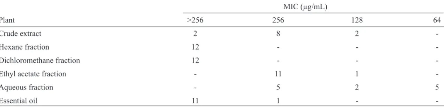
TABLE 1 - Inhibitory activity of Pimenta pseudocaryophyllus against 12 yeast isolates of the genus Candida.
MIC (µg/mL)
Plant >256 256 128 64
Crude extract 2 8 2
-Hexane fraction 12 - -
-Dichloromethane fraction 12 - -
-Ethyl acetate fraction - 11 1
-Aqueous fraction - 5 2 5
Essential oil 11 1 -
-MIC: minimal inhibito ry concentration.
Mechanism of action
The mechanisms of action of the crude ethanol extract and
the ethyl acetate and aqueous fractions against C. parapsilosis
(ATCC 22019) and two clinical isolates of C. albicans were
analyzed via flow cytometry (Accuri C6 cytometer, Becton Dickinson) according to the approach developed by Vale-Silva et al.15 and Pina-Vaz and Rodrigues16. Two markers were used:
propidium iodide (PI), which is a marker of membrane integrity due to its ability to permeate only damaged membranes, and 2-chloro- 4-(2,3-dihydro-3-methyl-[benzo-1,3-thiazol-2-yl]methylidene)-1-phenylquinolinium iodide (FUN-1), which is an indicator of yeast
metabolic activity. The inoculum of 400µL containing 2x10⁶ cells/
mL was added to 100µL of the crude ethanol extract or either fraction at a concentration of 1/4, 1/2, 1, 2 or 4 x MIC. Amphotericin B (2mg/ mL) was used as a control. Controls included the following: an autofl uorescence control, consisting of untreated yeast, and a labeled cell control, consisting of yeast that were treated with markers but not exposed to the crude ethanol extract or either fraction.
Data analysis
All the assays were performed in triplicate. Microsoft® Excel 2007 was used to analyze the data, and statistical analyses were performed using the Statistical Package for the Social Sciences (SPSS®) for Windows® version 16.0. The infl uence of the crude ethanol extract and the ethyl acetate and aqueous fractions of P. pseudocaryophyllus on Candida adhesion and hyphal formation were evaluated using the Mann-Whitney U-test. P values <0.05 were considered to be signifi cant.
Comparison of the retention times (RT), which was based on the HPLC profi les, between the reference standards and the samples revealed the presence of catechin and quercetin in the crude ethanol extract of P.pseudocaryophyllus. These substances were identifi ed in the extracts and fractions obtained from the
leaves of the citral chemotype of P.pseudocaryophyllus.
The in vitro antifungal susceptibility of the crude extract
of P. pseudocaryophyllus leaves, as well as the ethyl acetate
and aqueous fractions, inhibited the growth of Candida at a
MIC range of 64 to 256µg/mL (Table 1) and at a MFC range
of 256 to >1,024µg/mL. The aqueous fraction inhibited the
growth of 41% of the Candida isolates at a MIC of 64µg/mL
and a MFC of 256µg/mL.
Of the 12 Candida isolates, biofi lm formation was only
detected in 10 isolates (6 C. albicans and 4 C. parapsilosis). In vitro susceptibility assays demonstrated high SMIC values
for the crude ethanol extract and the ethyl acetate and aqueous fractions of P. pseudocaryophyllus, as well as fl uconazole,
against the yeast in the biofi lms. The SMIC50 values ranged from
512 to >1,024µg/mL and the SMIC80 values were greater than
1.02µg/mL for all of the Candida isolates evaluated.
The crude extract and the ethyl acetate and aqueous fractions of P. pseudocaryophyllus, at their respective MIC, signifi cantly
reduced the number of Candida yeast that adhered to oral
epithelial cells compared to the control. The aqueous fraction was the most active and displayed a level of performance similar to that of fl uconazole in terms of the MIC. The number of yeast cells that adhered at 1/2 x MIC of the aqueous fraction was similar to that in the presence of 1 MIC of the crude extract. The means and
standard deviations (SDs) of adherence are shown in Table 2.
The crude extract and fractions of P. pseudocaryophyllus, at their respective MIC, signifi cantly reduced hyphal formation compared to the control. At a concentration of 1/2 x MIC, the crude extract and the aqueous fraction also caused a signifi cant
reduction in hyphal formation (Table 2).
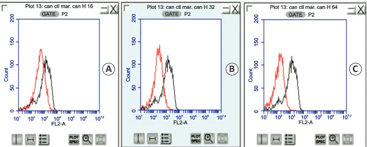
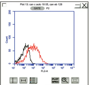
Flow cytometric analysis demonstrated that the crude extract and the ethyl acetate and aqueous fractions affected the
metabolism of C. albicans and C. parapsilosis, while the crude
extract exhibited an additional capacity to injure the membranes of these cells. The metabolism of the yeast treated with these components was impaired in a dose-dependent manner compared to the control yeast, and this effect was intensifi ed at higher concentrations. Figure 1 shows the alterations in Candida
metabolism in the presence of various concentrations of the
aqueous fraction of P. pseudocaryophyllus. The crude extract and
TABLE 2 - The number of Candida isolates (n=10) that adhered to 100 epithelial cells and the number of hyphae in isolates of Candida albicans (n=5) in the presence of the crude extract or the ethyl acetate or aqueous fraction of Pimenta pseudocaryophyllus
compared to the control.
Adherence Hyphae
mean ± SD P mean ± SD P
Control 503.05 ± 75.80 200.00 ± 6.12
Crude extract
1 MIC 118.00 ± 16.53 <0.001* 134.60 ± 33.76 0.008*
1/2 x MIC 387.75 ± 58.47 0.002* 174.60 ± 13.18 0.016*
1/4 x MIC 445.05 ± 69.49 0.165 186.40 ± 9.74 0.056
Ethyl acetate fraction
1 MIC 204.60 ± 29.96 <0.001* 185.40 ± 9.66 0.032*
1/2 x MIC 478.10 ± 72.52 0.436 192.80 ± 5.89 0.095
1/4 x MIC 498.10 ± 76.68 0.739 196.00 ± 4.18 0.222
Aqueous fraction
1 MIC 34.00 ± 4.68 <0.001* 124.80 ± 19.89 0.008*
1/2 x MIC 122.15 ± 15.56 <0.001* 171.80 ± 16.18 0.016*
1/4 x MIC 498.10 ± 76.68 0.315 195.00 ± 9.35 0.421
*Statistically signifi cant as evaluated by the Mann-Whitney U Test. MIC: minimal inhibito ry concentration.
Plot 13: can ctl mar. can H 16 Plot 13: can ctl mar. can H 16 Plot 13: can ctl mar. can H 16
Plot 13: can ctl mar. can H 16 Plot 13: can ctl mar. can H 32Plot 13: can ctl mar. can H 32Plot 13: can ctl mar. can H 32Plot 13: can ctl mar. can H 32 Plot 13: can ctl mar. can H 64Plot 13: can ctl mar. can H 64Plot 13: can ctl mar. can H 64Plot 13: can ctl mar. can H 64 GATE P2
GATE P2 GATE P2
GATE P2 GATE P2GATE P2GATE P2GATE P2 GATE P2GATE P2GATE P2GATE P2
FL2-A FL2-A FL2-A
FL2-A FL2-AFL2-AFL2-AFL2-A FL2-AFL2-AFL2-AFL2-A
101 2 3 4 5 6 7.2
10 10 10 10 10 10
101 2 3 4 5 6 7.2
10 10 10 10 10 10
101 2 3 4 5 6 7.2
10 10 10 10 10 10
101 2 3 4 5 6 7.2
10 10 10 10 10 10 101 2 3 4 5 6 7.2
10 10 10 10 10 10
101 2 3 4 5 6 7.2
10 10 10 10 10 10
101 2 3 4 5 6 7.2
10 10 10 10 10 10
101 2 3 4 5 6 7.2
10 10 10 10 10 10 101 2 3 4 5 6 7.2
10 10 10 10 10 10
101 2 3 4 5 6 7.2
10 10 10 10 10 10
101 2 3 4 5 6 7.2
10 10 10 10 10 10
101 2 3 4 5 6 7.2
10 10 10 10 10 10
200200200 200200200200 200200200200
150150150 150150150150 150150150150
100100100 100100100100 100100100100
505050 50505050 50505050
CountCountCount CountCountCountCount CountCountCountCount
000 0000 0000
FIGURE 1 - Histograms showing the fl uorescence in relative units (orange fl uorescence: FL2 channel, FL2-A log). Metabolism in yeast (Candidaparapsilosis ATTC 22019) treated with the aqueous fraction (AF) of Pimentapseudocaryophyllus (in red) at 1/4 x MIC (A),
1/2 x MIC (B) and 1 x MIC (C) compared to the control (in black).MIC: minimal inhibitory concentration.
DISCUSSION
Plot 13: can c auto 18 05, can eb 128 Plot 13: can c auto 18 05, can eb 128 Plot 13: can c auto 18 05, can eb 128 Plot 13: can c auto 18 05, can eb 128
200200200200
150150150150
100100100100
50505050
CountCountCountCount
0000
FL2-A FL2-A FL2-A FL2-A
101 102 103 104 105 106 107.2
101 102 103 104 105 106 107.2
101 102 103 104 105 106 107.2
101 102 103 104 105 106 107.2
GATE P2 GATE P2 GATE P2 GATE P2
FIGURE 2 -Histogram showing the red fl uorescence in relative units (red fl uorescence: FL3 channel, FL3-A log) of Candida parapsilosis
(ATCC 22019) stained with propidium iodide. Control consisting of stained untreated cells(in black). Stained cells treated with the crude extract of the leaves of Pimentapseudocaryophyllus(in red).
Medicinal plants are emerging as sources of new antifungal drugs that display greater efficiency, wider availability
and lower toxicity17,18. The growth inhibition of yeast
caused by P. pseudocaryophyllus and the antiedematogenic
and antinociceptive effects observed in mice in previous
investigations by our group4,5 have increased interest in this
plant. According to Scorzoni et al19, plant compounds displaying
antifungal activity at an MIC of 250µg/mL are considered to be useful for therapeutic purposes. Paula et al.5 determined
that the MIC of the crude extract and the ethyl acetate and
aqueous fractions of P. pseudocaryophyllus was <256µg/mL
for isolates of Candida spp. and was ≤128µg/mL for isolates
of Cryptococcus neoformans complex species. In the current
study, the MIC of the aqueous fraction was 64µg/mL for 42% of the examined isolates.
The antifungal activity of P. pseudocaryophyllus may be
attributed to fl avonoids, which are constituents of the crude ethanol extract of this plant and some of its fractions. Narayana et al.20 have reported the biological activity of fl avonoids,
specifi cally quercetin, against yeast. Furthermore, the antifungal
activity of catechin has been demonstrated against C. albicans
and T. mentagrophytes. Masatomo and Kazuko21 demonstrated
the inhibitory activity of catechins against C. albicans, and
Toyoshima et al.22 examined the mechanism underlying the
effects of the green tea of catechin on T. mentagrophytes and
suggested that catechin attacked the cell membrane and caused the lysis of conidia and hyphae.
Antifungal agents that act by reducing the expression of yeast virulence factors are expected to be more effi cient.
According to Zuzarte et al.23, inhibition of fi lamentation appears
be suffi cient to treat candidiasis. In the present study, the
P. pseudocaryophyllus crude extract and aqueous fraction (which
displayed performance similar to that of the fl uconazole control) reduced hyphal formation and decreased adherence to buccal
epithelial cells, signifi cantly affecting these Candida virulence
mechanisms. Similar results obtained by Zuzarte et al.23 and
Vale-Silva et al.24 verifi ed that hyphal formation in Candida was
reduced in the presence of essential oils from Thymus x viciosoi
and Lavandula multifi da L. Our results indicate that further studies of P. pseudocaryophyllus should be conducted to assess
its therapeutic potential for the treatment of Candida infections.
Elucidating the mechanism of action of each antifungal agent is an important step toward their appropriate use and may help to reduce toxicity. In the present study, the examined fractions of P. pseudocaryophyllus altered the metabolism of the target
yeasts, as shown in Figure 1. The results obtained by Evensen
and Braun25 indicated that polyphenols from Camellia sinensis
inhibited biofi lm formation by C. albicans via the alteration of
yeast protein metabolism. In addition to changes in metabolism, we observed injury to the fungal cell membrane in response to the P. pseudocaryophyllus crude extract and ethyl acetate
fraction. It is diffi cult to explain why the aqueous fraction was not able to injure the yeast membranes. However, we suggest that this fi nding may be due to the absence of polar compounds from the aqueous fraction, causing it to act preferentially on fungal metabolism but preventing it from damaging the cell membrane. High concentrations (2 x MIC) of the ethyl acetate fraction were necessary to damage the cell membrane; however, at a concentration equivalent to 1 x MIC, the crude extract caused similar damage. These results indicate that the crude extract likely contains constituents that act synergistically. Although both mechanisms of action have been observed in the crude ethanol extract as well as the ethyl acetate and aqueous fractions of P. pseudocaryophyllus, the metabolic effects on yeast appear
to predominate. The altered metabolism and plasma membrane injury of yeast observed in this study indicated that the constituents of P. pseudocaryophyllus might inhibit yeast development
and most likely induce the destruction of the microorganism. These results demonstrated that the antifungal activity of the crude extract and the ethyl acetate and aqueous fractions of P. pseudocaryophyllus of the citral chemotype inhibited Candida virulence mechanisms, including adherence and
hyphal induction (Table 2). Flavonoids, such as quercetin
and catechin, found in P. pseudocaryophyllus are considered to be antimicrobials and explain this antifungal activity. In conclusion, our results indicate that the crude extract and the
ethyl acetate and aqueous fractions of P. pseudocaryophyllus
The authors declare that there is no confl ict of interest. CONFLICT OF INTEREST
FINANCIAL SUPPORT
The authors acknowledge the financial support of the National Council of Technological and Scientifi c Development (CNPq).
REFERENCES
1. Silva S, Negri M, Henriques M, Oliveira R, Williams DW, Azeredo J.
Candida glabrata, Candida parapsilosis and Candida tropicalis:
biology, epidemiology, pathogenicity and antifungal resistance. FEMS
Microbiol Rev 2012; 36:288-305.
2. Pappas PG, Kauffmans CA, Andes D, Benjamin Jr DK, Calandra TF, Edwards Jr JE, et al. Clinical Practice Guidelines for the management of Candidiasis (2009) update by the Infectious Diseases Society of America. Clin Infect Dis 2009; 48:503-535.
3. Arnold TM, Dotson E, Sarosi GA, Hage CA. Traditional and emerging antifungal therapies. Proc Am Thorac Soc 2010; 7:222-228.
4. Paula JAM, Paula JR, Bara MTF, Rezende MH, Ferreira HD. Pharmacognostic study about Pimenta pseudocaryophyllus (Gomes)
L.R. Landrum leaves - Myrtaceae. Rev Bras Farmacogn2008; 18: 265-278.
5. Paula JAM, Silva MRR, Costa MP, Diniz DGA, Sá FAS, Alves SF, et al. Phytochemical analysis and antimicrobial, antinociceptive, and anti-infl ammatory of two chemotypes of Pimenta pseudocaryophyllus
(Myrtaceae). Evid Based Complement Alternat Med 2012; 2012:1-15. 6. Calderone RA, Fronzi WA. Virulence factors of Candida albicans.
Trends Microbiol 2001;9:327-335.
7. Espinel-Ingrof A. Novel antifungal agents, targets or therapeutic strategies for the treatment of invasive fungal diseases: a review of the literature (2005-2009). Rev Iberoam Micol 2009; 26:15-22.
8. Clinical and Laboratory Standards Institute (CLSI). Reference method for broth dilution antifungal susceptibility testing of yeasts, 3rd ed.
Approved standard. CLSI M27-A3 (28). Wayne, PA: CLSI; 2008.
9. Rodriguez-Tudela JL, Cuenca-Estrella M, Diaz-Guerra TM, Mellado E.
Stan-dardization of Antifungal Susceptibility Variables for a Semiautomated Methodology. J Clin Microbiol 2001; 39:2513-2517.
10. Torres-Rodriguez JM, Alvarado-Ramirez E, Murciano F, Sellart M.
MICs and minimum fungicidal concentrations of posaconazole, voriconazole and fluconazole for Cryptococcus neoformans and
Cryptococcus gattii. J Antimicrob Chemother 2008; 62:205-210.
11. Pierce CG, Uppuluri P, Tristan AR, Wormley Jr FL, Mowat E, Ramag G, et al.
A simple and reproducible 96-well plate-based method for the formation of fungal biofilms and its application to antifungal susceptibility testing. Nature Protocols 2008; 3:1494-1500.
12. Kimura LH, Pearsall NH. Adherence of Candida albicans to human
buccal epithelial cells. Infect Immun1978; 21:64-68.
13. Biasoli SM, Tosello ME, Magaró HM. Adherence of Candida strains
isolated from the human gastrointestinal tract. Mycoses 2002; 45:465-469.
14. Ha KC, White TC.Effects of azole antifungal drugs on the transition from yeast cells to hyphae in susceptible and resistant isolates of the pathogenic yeast Candida albicans. Antimicrob Agents Chemother
1999; 43:763-768.
15. Vale-Silva LA, Buchta V, Vokurková D, Pour M. Investigation of the mechanism of action of 3-(4-bromophenyl)-5-acyloxymethyl-2,5-dihydrofuran-2-one against Candida albicans by fl ow cytometry.
Bioorg Med Chem Lett 2006; 16:2492-2495.
16. Pina-Vaz C, Rodrigues AG. Evaluation of Antifungal Susceptibility using fl ow citometry. Cap. 21. In: Sharon A, editor. Molecular and
Cell Biology Methods for Fungi, methods in molecular biology. 2010;
638:281-289.
17. Singh G, Kumar P, Joshi SC. Treatment of dermatophytosis by a new antifungal agent ‘apigenin’. Mycoses 2014; 57:497-506.
18. Danielli LJ, Reis M, Bianchini M, Camargo GS, Bordignon SAL, Guerreiro IK, et al. Antidermatophytic activity of volatile oil and nanoemulsion of Stenachaenium megapotamicum (Spreng.) Baker.
Ind Crops and Prods 2013; 50:23-28.
19. Scorzoni L, Benaduci T, Almeida AMF, Silva DHS, Bolzani VS, Gianinni MJSM. The use of standard methodology for determination of antifungal activity of natural products against medical yeasts Candida
sp and Cryptococcus sp. Braz J Microbiol 2007; 38:391-397.
20. Narayana KR, Reddy MS, Chaluvadi MR, Krish DR. Biofl avonoids
classifi cation, pharmacological, biochemical effects and therapeutic potential. Indian J Pharmacol 2001; 33:2-16.
21. Masatomo H, Kazuko T. Multiple effects of green tea catechin on the antifungal activity of antimycotics against Candida albicans.
JAC2004; 53:225-229.
22. Toyoshima Y, Okubo S, Hara Y, Shimamura T. Effect of catechin on
the ultrastructure of Trichophyton mentagrophytes. Kansenshogaku
Zasshi 1993; 68:295-303.
23. Zuzarte M, Vale-Silva LA, Gonçalves MJ, Cavaleiro C, Vaz S, Canhoto J,
et al. Antifungal activity of phenolic-rich Lavandula multifi da L. essential oil. Eur J Clin Microbiol Infect Dis 2012; 31:1359-1366. 24. Vale-Silva LA, Gonçalves MJ, Cavaleiro C, Salgueiro L, Pinto E.
Antifungal Activity of the Essential Oil of Thymus x viciosoi against
Candida, Cryptococcus, Aspergillus and Dermatophyte species.
Planta Med 2010; 76:882-888.
25. Evensen NA, Braun PC. The effects of tea polyphenols on Candida
albicans: inhibition of biofi lm formation and proteasome inactivation.