Address to: Prof. José Antonio Marin-Neto. Divisão de Cardiologia/HC/
FM-RP/USP. Av. dos Bandeirantes 3900, 14048-900 Ribeirão Preto, SP, Brasil.
Phone/Fax: 55 16 3633-4010/3633-0869 e-mail: [email protected]
Received 7 February 2013
Accepted 26 June 2013
Pathogenesis of chronic Chagas cardiomyopathy:
the role of coronary microvascular derangements
José Antonio Marin-Neto
[1],
Marcus Vinicius Simões
[1]and Anis Rassi Junior
[2][1].Divisão de Cardiologia, Hospital das Clínicas, Faculdade de Medicina de Ribeirão Preto, Universidade de São Paulo, Ribeirão Preto, SP. [2]. Hospital Anis Rassi, Goiânia, GO.
ABSTRACT
There is ample experimental and clinical evidence of functional and structural microvascular abnormalities occurring in patients
with Chagas cardiomyopathy, possibly due to the infl ammatory process and/or autonomic disturbances caused by Trypanosoma cruzi infection. Those microvascular derangements are likely to constitute at least an ancillary factor that potentiates and amplifi es
the chronic infl ammation in myocardial tissue. It is possible to devise appropriate therapeutic interventions aimed at reverting
or slowing the progression of the microvascular abnormalities to positively affect the natural history of Chagas cardiomyopathy. Keywords: Chagas disease. Microvascular coronary disease. Pathogenesis.
INTRODUCTION
One hundred years after the discovery of Chagas disease, the pathogenesis of chronic Chagas cardiomyopathy (CCC) is still open to debate1. Nevertheless, there is both experimental
and clinical evidence of coronary microvascular disturbances leading to ischemic myocardial damage in animals infected with Trypanosoma cruzi and in patients with chronic Chagas heart disease2.
EVIDENCE FROM HISTOPATHOLOGY STUDIES IN HUMANS
Necroscopic studies of patients who died in the acute phase
of Chagas disease showed perivascular infl ammatory changes
that were considered to be involved in causing myocardial damage3. Further necropsy studies of chronic Chagas patients
also described extensive vascular lesions, with obstruction of the small intramural coronary artery branches due to intimal
proliferation and arteriole wall thickening. In addition, the myocardial fi bers topographically related to these vascular
lesions presented myocytolytic necrosis, a type of cell death related to ischemic injury4. The authors concluded that
microvascular ischemia is directly involved in the genesis of the fundamental histopathologic pattern of CCC, consisting of diffuse focal myocarditis associated with myocytolysis
and subsequent reparative fi brosis5. Another type of structural
derangement is related to the rarefaction of the microvessels into the myocardium. This process of decapillarization was thought
to be related to extraluminal compression and was shown to be topographically related to foci of myocytolysis, as demonstrated by a necroscopic radiological study of Chagas disease patients using a coronary injection of an opaque medium6. Vascular
changes characterized by a decreased density of coronary vessels
were confi rmed in another postmortem radiological study of
patients with proven Chagas disease7.
Microvascular abnormalities due to the extensive basement membrane thickening of capillaries were also thought to be associated with the focal diffuse myocytolysis observed in both necroscopic studies and biopsy specimens8. Other independent investigations confi rmed the occurrence of structural changes
in the microvascular coronary tree, with both abnormal dilation and constriction leading to myocardial damage in patients with Chagas disease9-12.In addition, considering that the focal
distribution of cell necrosis and subsequent reparative interstitial
fi brosis observed in Chagas disease follows a pattern similar
to that described in experimental models of ischemia and reperfusion, transient microvascular ischemic disturbances of low intensity and short duration have been postulated to cause ischemic myocardial damage and to ultimately lead to the development of cardiomyopathy13,14.Coalescent
microinfarctions have also been implicated in the genesis of the typical Chagas disease-related ventricular aneurysms that are postulated to occur in watershed coronary areas15. Further
necropsy studies in humans correlated the presence of an apical aneurysm with scores proportional to the myocardial mass and coronary perfusion and concluded that ischemic factors could be involved in the genesis of the peculiar lesion of Chagas cardiomyopathy16. Following the demonstration
lead to overactivity of the sympathetic system, causing chronic myocardial ischemia17. These investigators also performed a
macroscopic study in necropsied patients with Chagas disease and described an increased capacity of the coronary artery bed. The
authors concluded that these fi ndings would lend further support
to the neurogenic pathogenesis concept18. However, this issue is
controversial because an in vivo study using quantitative coronary angiography in Chagas heart disease patients with chest pain and no obstructive coronary artery disease (CAD) in the subepicardial vessels showed that the baseline coronary artery diameter and tonus were not related to the degree of autonomic dysfunction19.
EVIDENCE FROM ANIMAL MODELS OF T. CRUZI INFECTION AND IN VITRO STUDIES
Early studies in the 1970s used histopathologic and histoenzymologic methods to compare the myocardial lesions found in Wistar rats chronically infected with T. cruzi with those lesions detected in other rats submitted to repeated injections of isoproterenol. The researchers found that there was marked histoenzymatic similarity between the lesions found in both experimental models and concluded that the myocardial damage described in the rats with T. cruzi infection could be caused by ischemia triggered by excessive stimulation with catecholamines20. Using histochemical methods in
mice chronically infected with T. cruzi, several independent investigators have described microcirculatory derangements that cause ischemia associated with occlusive platelet thrombi in small epicardial and intramural coronary arteries21,22. In the
same experimental model, other microvascular derangements were detected, such as focal areas of vascular constriction and endothelial microvascular proliferation23. Similar alterations
have been found in other models of experimental T. cruzi infection, including the development of typical ventricular aneurysms24. The mechanisms responsible for occlusive platelet
thrombosis and microcirculatory spasm were directly related to the T. cruzi infection causing endothelial cell damage25 and
indirectly related to the interaction of the endothelium with immune effector cells26,27. In vitro studies of T. cruzi infection
of human umbilical vein endothelium cells showed increased production of endothelin. This potent vasoconstrictor mediated arteriolar spasm and impaired cyclic adenosine monophosphate (AMP) metabolism, thus impairing AMP’s protective role against platelet adhesion to and aggregation along the vascular wall28,29. Trypomastigote forms of T. cruzi also produce a
neuraminidase that may remove sialic acid from the surface of mammalian myocardial and endothelial cells30. The loss
of this protective component of the endothelial surface may be associated with platelet aggregation and microvascular thrombosis. These abnormal findings were corroborated by increased production of thromboxane-A2 and enhanced platelet adherence and aggregation in an experimental murine model of T. cruzi infection31. The increased production of cytokines by infl ammatory cells is also likely to contribute to
microvascular abnormal reactivity in experimental models of Chagas heart disease32. These abnormalities have been shown
to be reverted by verapamil, a vasodilatory calcium channel blocker with antiplatelet properties, thus attenuating myocardial aggression and, more importantly, leading to increased survival
of mice acutely infected with T. cruzi . These fi ndings were also corroborated by direct in vivo visualization of the murine cremaster vascular bed. This model showed that animals infected with T.cruzi had arterioles with segmental areas of spasm and reduced flow velocity; these abnormalities, which were not observed in control mice, were also antagonized by treatment with verapamil34. Finally, disturbed molecular mechanisms are
thought to modify the function of the microcirculation in Chagas cardiomyopathy. For example, calreticulin, a calcium-binding protein that is also present in the parasite, was shown to interfere with modulation of the complement system and inhibit angiogenesis35.
EVIDENCE FROM CLINICAL STUDIES
Myocardial ischemic manifestations are commonly detected in patients with Chagas heart disease and may contribute to the pathogenesis of the disease’s complex pathophysiology. Atypical angina pectoris is a common complaint in T. cruzi -infected patients36. The characteristics of the chest pain
are slightly consistent with ischemic cardiac pain (location and character) but typically have no relation to effort and no relief by the use of nitrates. Most of these patients have
concomitant transient or defi nite ST-T changes on a 12-lead
electrocardiogram (ECG) and abnormal Q waves compatible with electrically inactive left ventricular (LV) areas37 ,38. More
rarely, patients may present with a clinical picture that mimics an acute coronary syndrome37. Similar to coronary heart disease,
segmental LV wall motion abnormalities are common in patients with chronic Chagas disease and are even occasionally detected when the LV chamber is already globally dilated 39,40. Despite
the occurrence of all of these manifestations, suggesting the presence of myocardial ischemia, catheter-based coronary angiography in Chagas’ cardiomyopathy nearly invariably
demonstrates the absence of signifi cant obstructive coronary
disease at the epicardial level36-41.
CORONARY MACROVASCULAR DYSFUNCTION
Although the angiographic appearance of the coronary arteries is normal in most patients with chest pain and documented T. cruzi infection, impairment of the regulation of the coronary circulation may occur at the macrovascular level. The possibility of coronary spasm was postulated as causing acute myocardial infarction in Chagas disease patients with normal coronary arteries42. However, other investigators showed
no evidence of coronary artery spasm in any of 31 consecutive patients with Chagas' disease who exhibited precordial pain severe enough to warrant cardiac catheterization, despite the
specifi c stimulus of the forced hyperventilation test
Consistent with the experimental studies discussed above, impairment of endothelium-dependent coronary vasodilatation in response to acetylcholine has been reported in patients with Chagas’ heart disease, but these patients had a preserved response to the non-endothelium-dependent effect of adenosine43. In another study, Chagas patients with angina that
and non-endothelial dysfunction in Chagas patients with chest pain and angiographically normal epicardial coronary arteries. However, there are no conclusive data showing that these functional macrovascular derangements have any relevant impact on the pathogenesis of Chagas cardiomyopathy.
CORONARY MICROVASCULAR DYSFUNCTION
Since the 1970s, the pioneering work of Emilio Kuschnir and his collaborators in Córdoba, Argentina, employing primitive nuclear medicine methods (the measurement of global myocardial
fl ow with 86-rubidium), showed that chronic Chagas patients,
similar to patients with CAD, exhibited a reduction in myocardial perfusion at rest and during physical exercise in comparison with normal individuals45-47. Several other investigations have
subsequently described striking myocardial perfusion abnormalities in Chagas disease patients with angiographically normal
coronary arteries, thus supporting the concept of abnormal fl ow
regulation that essentially occurs at the microvascular level41,48,4 9.
Using 201-thallium stress-redistribution myocardial perfusion scintigraphy (MPS), one of these investigations demonstrated at least one perfusion defect in all 23 chagasic patients studied48. Fixed defects, connoting myocardial fi brosis, were found
in myocardial segments exhibiting akinesis or dyskinesis, and stress-induced, reversible myocardial ischemia was detected in LV segments with less severe wall motion impairment in 8 (35%) patients48. These fi ndings were confi rmed and extended
by a further study of 37 patients with various stages of Chagas heart disease49. Regional MPS was also assessed by the use of
201-thallium with single-photon emission computed tomography (SPECT), this time focusing on the topographic correlation between segmental myocardial perfusion abnormalities and
regional LV dysfunction. Perfusion defects (fi xed, paradoxical
and reversible) were observed in most (78%) of the patients, and a
signifi cant topographic correlation was observed between perfusion
disturbances and wall motion abnormalities predominating in the apical and inferior-posterior LV segments. Reversible, stress-induced myocardial perfusion defects were detected in segments exhibiting normal wall motion in 5 (42%) of 12 Chagas disease patients who otherwise had no evidence of myocardial disease (possibly with the indeterminate form of the disease)49. Of note,
these reversible ischemic defects were mostly observed in the apical and inferior-posterior LV segments, i.e., the same regions in which regional contractile dysfunction is more frequently found in later stages of Chagas cardiomyopathy49. An additional intriguing fi nding was that marked sympathetic denervation, as
evaluated by myocardial scintigraphy with
iodine-123-meta-iodobenzylguanidine (MIBG), was demonstrated in topographic
association with the presence of perfusion abnormalities in patients with less advanced deterioration of LV function49. This fi nding suggests that the sympathetic denervation and
perfusion derangements may be pathophysiologically linked and that these events occur at early stages in the natural course of Chagas cardiomyopathy, before the installation of typical segmental contractile dysfunction. This suggestion may indeed be the case based on a more recent study of 36 patients with serologically proven Chagas disease who were initially evaluated
with Tc-99m-Sestamibi MPS at rest and during physical exercise and who had the nuclear examination repeated after a mean period of 5.6 years50. Of the 36 patients, 20 (56%) exhibited reversible
ischemic myocardial perfusion defects involving an average of 10.2% ± 9.7% of the LV area, as determined by the SPECT technique. A topographic association between the presence of ischemia in the initial evaluation and the development of
myocardial fi brosis was suggested by the results, as among the
47 segments presenting reversible perfusion defects in the initial study, 32 (68%) progressed to perfusion defects at rest, and of the 469 segments not showing reversibility in the initial study, only 41 (8.7%) had the same progression. The individual increase in the perfusion defect area at rest (possibly indicating the presence
of fi brosis) was signifi cantly correlated with the corresponding
reduction in LV ejection fraction (with a mean decrease over time in the entire group of patients from 55% ± 11% to 50% ± 13%). Hence, in this longitudinal study, the deterioration of LV systolic function over time was associated with both the presence of reversible ischemic defects at the initial assessment and an increase in the extent of irreversible perfusion defects during follow-up50. In patients with Chagas cardiomyopathy selected for malignant
ventricular arrhythmia, perfusion defects were also predominant in areas from which the reentrant circuits frequently arise51.
Although the mentioned studies have shown that the detection of microvascular myocardial ischemia is associated with a worse outcome regarding ventricular function, the role of MPS in the detection of early myocardial involvement in patients with the indeterminate form of chronic Chagas disease is more unclear. A recent study found normal myocardial perfusion in all 18 patients with the indeterminate form of Chagas disease who were submitted to stress-rest MPS with Tc-99m- Sestamibi52.
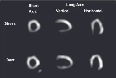
Because the tracer used in most previously mentioned studies41,48,49 was 201-thallium (Figure 1 depicts an illustrative example), its accumulation and retention could be infl uenced by
FIGURE 1 - Representative tomographic slices of stress-rest 201-thallium myocardial perfusion scintigraphy of a Chagas disease patient (male, 48 years old) presenting chest pain and normal epicardial coronary arteries. A reversible perfusion defect is observed in the inferior wall and in part of the apical, septal and posterior-lateral walls. The left ventricular ejection fraction was 36%, and hypokinesia of the inferior and posterior-lateral walls was detected by echocardiogram.
Stress
Rest
Short Axis
Long Axis
cardiomyocyte metabolic derangements related to the underlying
infl ammatory process. Hence, the scintigraphic defects described
in the previous studies might not represent true perfusion abnormalities. This alternative possibility is supported by the frequent occurrence of reverse redistribution defects predominating in LV regions with normal or mildly impaired wall motion in those studies. However, myocardial perfusion was also evaluated in 18 chronic Chagas’ heart disease patients with recurrent chest pain that was severe enough to warrant coronary angiography using an exclusive perfusion tracer, 99m-technetium-labeled microspheres, injected into the LV cavity44. All patients presented normal epicardial
coronary arteries. Ten of the 18 patients exhibited extensive
perfusion defects in 49 (39%) of 126 LV segments. In total,
19 (40%) of these segments with perfusion defects had severely impaired wall motion, most likely corresponding to extensive
fi brosis. The remaining 60% of the segments with perfusion
defects had normal or mildly reduced wall motion. Thus, this investigation clearly showed marked resting myocardial perfusion abnormalities preceding the appearance of any wall motion impairment44
More recently, the use of magnetic resonance imaging
(MRI) to examine patients with CCC has been reported53,54. In one study, 51 patients with CCC were investigated by MRI, including techniques of late contrast enhancement, for the detection of myocardial fi brosis. Myocardial fi brosis was
found in 20% of 15 patients with the indeterminate form and in 84.6% of 26 patients with overt Chagas heart disease. The
distribution of fi brosis was atypical for CAD and predominated
in the midwall and subepicardial portions of the LV walls, encompassing multiple coronary territories. This pattern was
compatible with myocardial fi brosis associated with myocardial
ischemia secondary to microvascular derangement and chronic myocarditis, as also documented in hypertrophic and dilated cardiomyopathy55,56. Similar fi ndings and interpretation were
reported by another study, which investigated 67 Chagas disease patients. Late enhancement was detected in 23.9% of the patients, with a highly heterogeneous distribution: subendocardial in 26.8%, midwall in 14%, subepicardial in 22.6% and transmural in 36%54.
PATHOPHYSIOLOGICAL CONSEQUENCES OF CORONARY MICROVASCULAR PERFUSION
DISTURBANCES AND THERAPEUTIC PERSPECTIVES ON CHAGAS DISEASE
On the basis of these clinical investigations and the background information from the pathological and experimental studies discussed, it is reasonable to hypothesize that myocardial perfusion derangements could contribute to the development of the characteristic segmental LV dysfunction observed in the chronic phase of Chagas heart disease. This pathophysiological mechanism may be similar to the chronic hyperperfusion that causes hibernating myocardium in patients with longstanding coronary artery disease. A corollary is that at least in the early stages of the disease, wall motion abnormalities due to ischemia may be reversible in Chagas patients. This concept is compatible with evidence from studies showing improvement
in LV function in Chagas heart disease patients who received acute or chronic administration of dipyridamole57 and isosorbide
dinitrate58. However, these studies obtained no direct evidence
indicating that the improvement in LV function was due to the
relief of myocardial ischemia. In fact, the associated changes
in ventricular loading conditions caused by the administration of dipyridamole and nitrate might have contributed to the improvement in ventricular performance. Only one study directly addressed the question of hibernating myocardium in CCC. The study was performed using post-extrasystolic potentiation, which is commonly observed during radiological-contrast left ventriculography, and showed that most myocardial areas with impaired wall motion indeed had a contractile reserve; that is, these areas were viable59. However, the hypothesis that
hypokinetic areas of viable myocardium, unmasked through post-extrasystolic potentiation, would be predominant in regions exhibiting scintigraphic, reversible (ischemic) perfusion defects,
was not confi rmed by the results of the study60.
Thus, although scientifi cally attractive, the hypothesis of
chronic myocardial ischemia in Chagas’ cardiomyopathy awaits
a direct demonstration of benefi t to be derived from the long-term
administration of drugs acting upon the microcirculation, with a
signifi cant impact on the outcome of Chagas disease patients with
angina-like symptoms. One such study is currently ongoing, with the primary objective of testing the hypothesis that the prolonged administration of verapamil and acetylsalicylic acid to Chagas patients with normal epicardial coronary arteries, chest pain and reversible ischemic perfusion defects at baseline examination will lead to a reduction in the extent and intensity of the perfusion abnormalities and a mitigation of the angina-like symptoms61.
Finally, as the pathological changes found in the microvasculature of patients with chronic Chagas disease are linked either directly or indirectly to the persistence of T. cruzi, it is reasonable to speculate that therapy aimed at eliminating the parasite could prevent or ameliorate the microcirculatory disturbances. This hypothesis is reasonable, despite recent guidelines being evasive in recommending trypanocidal treatment for patients in the chronic phase of Chagas disease62.
1. Marin-Neto JA, Cunha-Neto E, Maciel BC, Simões MV. Pathogenesis of chronic Chagas heart disease. Circulation 2007; 115: 1109-1123.
2. Rossi MA, Tanowitz HB, Malvestio LM, Celes MR, Campos EC, Blefari V, et al. Coronary microvascular disease in chronic Chagas cardiomyopathy Including an overview on history, pathology, and other proposed pathogenic mechanisms. PLoS Negl Trop Dis 2010; 4:e674.
3. Vianna G. Contribuição para o estudo da anatomia patológica da moléstia de Chagas. Mem Inst Oswaldo Cruz 1911; 3:276-294.
4. Torres CM. Arteriosclerosis of the fi ne arterial branches of the myocardium (Chagas' coronaritis) & focal myocytolysis in chronic Chagas' heart disease. Hospital (Rio de Janeiro) 1958; 54:597-610.
REFERENCES
5. Torres CM. Miocitólise e fi brose do miocárdio na doença de Chagas. Mem Inst Oswaldo Cruz 1960; 58:161-182.
6. Jörg ME. Tripanosomiasis cruzi; anarquia angiotopográfi ca por descapilarización mesenguimorreactiva: cofactor patogénico de la miocardiopatia crónica. Pren Med Argent 1974; 61:94-106.
7. Ferreira CS, Lopes ER, Chapadeiro E, Silva Neto IJ, Oliveira Almeida H, Souza WF. Coronariografi a post mortem na cardite chagásica crônica: correlação anátomo-radiológica. Arq Bras Cardiol 1980; 34:81-86. 8. Ferrans VJ, Milei J, Tomiata Y, Storino RA. Basement membrane
thickening in cardiac myocytes and capillaries in chronic Chagas’ disease. Am J Cardiol 1988; 61:1137-1140.
9. Laranja FS, Dias E, Nóbrega C, Miranda A. Chagas’ disease: a clinical, epidemiological and pathological study. Circulation 1956; 1035-1060.
10. Köberle F. Cardiopatia Chagásica. Hospital 1958; 53:311-346.
11. Ferreira CS, Lopes ER, Chapadeiro E, Almeida HO, Souza WF, Silva-Neto JJ. Coronariografi a post-mortem na cardite chagásica crônica. Correlação anátomo-radiológica. Arq Bras Cardiol 1980; 34:81-86. 12. Higuchi ML, Brito T, Parzianello LC, Fukasawa S, Ramires JA. Severe
arteriolar dilatation and ischemic lesions in chronic Chagas’ cardiopathy: a 3D confocal laser microscopic study. J Am Coll Cardiol 1998; 31:382.
13. Morris AS, Tanowitz HB, Wittner M, Bilezikian JP. Pathophysiological insights into the cardiomyopathy of Chagas’ disease. Circulation 1990; 82:1900-1909.
14. Rossi MA. Microvascular changes as a cause of chronic cardiomyopathy in Chagas’disease. Am Heart J 1990; 120:233-236.
15. Higuchi ML, Fukasawa S, De Brito T, Parzianello LC, Bellotti G, Ramires JA. Different microcirculatory and interstitial matrix patterns in idiopathic dilated cardiomyopathy and Chagas’disease: a three dimensional confocal microscopy study. Heart 1999; 82:279-285.
16. Oliveira JAM. Heart aneurysm in Chagas’ disease. Rev Inst Med Trop S Paulo 1998; 40:301-307.
17. Oliveira JSM, Mello de Oliveira JA, Frederigue Jr U, Lima Filho EC. Apical aneurysm of Chagas’s heart disease. Br Heart J 1981; 46:432-437.
18. Oliveira JSM, Santos JCM, Muccillo G, Ferreira AL. Increased capacity of the coronary arteries in chronic Chagas’heart disease: further support for the neurogenic pathogenesis concept. Am Heart J 1985; 109: 304-308.
19. Simões MV, Bromberg-Marin G, Antloga CM, Maciel BC, Marin-Neto JA. Tonus e diâmetro arterial coronário não se correlacionam com o grau de denervação autonômica em pacientes com cardiopatia chagásica crônica. Rev Bras Cardiol Invas 2008; 16:70-76.
20. Mello de Oliveira JA. Observações sobre a histoquímica de algumas enzimas do miocárdio na fase crônica da infecção experimental pelo
Trypanosoma cruzi, Chagas, 1909, em ratos Wistar. Rev Goiana Med
1976; 22:1-65.
21. Rossi MA, Gonçalves S, Ribeiro-dos-Santos R. Experimental Trypanosoma
cruzi cardiomyopathy in BALB/c mice: the potential role of intravascular
platelet aggregation in its genesis. Am J Pathol 1984; 114:209-216.
22. Rossi MA, Carobrez SG. Experimental Trypanosoma cruzi cardiomyopathy in BALB/c mice: histochemical evidence of hypoxic changes in the myocardium. Br J Exp Pathol 1985; 66:155-160.
23. Factor SM, Cho S, Wittner M, Tanowitz HB. Abnormalities of the coronary microcirculation in acute murine Chagas’ disease. Am J Trop Med Hyg 1985; 34:246-253.
24. Figueiredo F, Marin-Neto JA, Rossi MA. The evolution of experimental
Trypanosoma cruzi cardiomyopathy in rabbits: further parasitological,
morphological and functional studies. Int J Cardiol 1986; 10:277-290. 25. Rossi MA. Aortic endothelial cell changes in the acute septicemic
phase of experimental Trypanosoma cruzi infection in rats: scanning and transmission electron microscopy study. Am J Trop Med Hyg 1997; 57:321-327.
26. Andrade ZA, Andrade SG, Correa R, Sadigursky M, Ferrans VJ. Myocardial changes in acute Trypanosoma cruzi infection. Ultrastructural
evidence of immune damage and the role of microangiopathy. Am J Path 1994; 144:1403-1411.
27. Scharfstein J, Andrade D. Infection-associated vasculopathy in experimental chagas disease pathogenic roles of endothelin and kinin pathways. Adv Parasiol 2011; 76:101-27.
28. Morris SA, Tanowitz HB, Makman M. Trypanosoma cruzi: alteration of cAMP metabolism following infection of human endothelial cells. Exp Parasit 1992; 74:69-76.
29. Tanowitz HB, Huang H, Jelicks LA, Chandra M, Loredo ML, Weiss LM, et al. Role of endothelin 1 in the pathogenesis of chronic chgasic heart disease. Infec Immun 2005; 73:2496-2503.
30. Libby P, Alroy J, Pereira MEA. A neuraminidase from Trypanosoma
cruzi removes sialic acid from the surface of mammalian myocardial and
endothelial cells. J Clin Invest 1986; 77:127-135.
31. Tanowitz HB, Burns ER, Sinha KA, Kahn NN, Morris SA, Factor SM, et al. Enhanced platelet adherence and aggregation in Chagas’ heart disease: a potential pthogenic mechanism for cardiomyopathy. Am J Trop Med Hyg 1990; 43:274-281.
32. Huang H, Calderon TM, Berman JW, Braunstein VL, Weiss LM, Wittner M, et al. Infection of endothelial cells with Trypanosoma cruzi activates NF-kappaB and induces vascular adhesion molecule expression. Infec Immun 1999; 67:5434-5440.
33. Morris SA, Weiss LM, Factor SM, Bilezikian JP, Tanowitz H, Wittner M, et al. Verapamil ameliorates clinical, pathological and biochemical manifestations of the experimental chagasic cardiomyopathy in mice. J Am Coll Cardiol 1989; 14:782-789.
34. Tanowitz HB, Kaul DK, Chen B, Morris SA, Factor SM, Weiss LM, et al. Compromised microcirculation in acute murine Trypanosoma cruzi
infection. J Parasit 1996; 82:124-130.
35. Ferreira V, Molina MC, Schwaeble W, Lemus D, Ferreira A. Does
Trypanosoma cruzi calreticulin modulate the complement system and
angiogenesis? Trends Parasitol 2005; 21:169-174.
36. Marin-Neto JA, Rassi Jr A, Simões MV, Maciel BC, Schmidt A. Chagas heart disease. In: Yusuf S, Cairns JA, Camm AJ, Fallen EL, Gersh BJ, editors. Evidence-Based Cardiology. 3rd edition. Chapter 51; 2010.
p. 823-841.
37. Feit A, El-Sherif N, Korostoff S. Chagas’ disease masquerading as coronary artery disease. Arch Intern Med 1983; 143:144-145.
38. Simões MV, Mello-Ayres E, Attab-Santos JL, Schmidt A, Pintya AO, Maciel BC, et al. Detection of myocardial ischemia in Chagas’ heart disease patients with angina-like symptoms by the effort test and holter monitoring. Arq Bras Cardiol 1993; 60:315-319.
39. Hammermeister KE, Caeiro T, Crespo E, Palmero H, Gibson DG. Left ventricular wall motion in patients with Chagas’ disease. Br Heart J 1984; 51:70-76.
40. Arreaza N, Puigbó JJ, Acquatella H, Casal H, Giordano H, Valecillos R, et al. Radionuclide evaluation of left ventricular function in chronic Chagas’ cardiomyopathy. J Nucl Med 1983; 24:563-567.
41. Hagar JM, Rahintoola SH. Chagas’ heart disease in the United States. N Engl J Med 1991; 325:763-768.
42. Vianna LG, Campos GP, Magalhães AV. Myocardial infarct without coronary obstruction associated with chronic Chagas cardiopathy. Arq Bras Cardiol 1979; 33:41-47.
43. Torres FW, Acquatella H, Condado JA, Dinsmore R, Palácios IF. Coronary vascular reactivity is abnormal in patients with Chagas’ disease. Am Heart J 1995; 129:995-1001.
44. Marin-Neto JA, Simões MV, Ayres-Neto EM, Attab-Santos JL, Gallo Jr L, Amorim DS, et al. Studies of the coronary circulation in Chagas’ heart disease. São Paulo Medical Journal 1995; 113:826-834.
46. Kuschnir E, Kustich F, Epelman M, Santamarina N, Podio RB. Valoration de fl uxo miocardico con Rb 86, en pacientes con cardiopatia chagasica, con insufi ciencia coronaria y en controles normales. Parte 2 Respuesta al ejercicio y a la cardiotonifi cación aguda. Arq Bras Cardiol 1974; 27:721-732. 47. Castro R, Kuschnir E, Sgammini H. Evaluacion de la performance
cardíaca y perfusion miocárdica com radiotrazadores en la cardiopatia chagásica crônica. Rev Fed Arg Cardiol 1988; 17:226-231.
48. Marin-Neto JA, Marzullo P, Marcassa C, Gallo-Junior L, Maciel BC, Bellina CR, et al. Myocardial perfusion abnormalities in chronic Chagas’disease as detected by thallium-201 scintigraphy. Am J Cardiol 1992; 69:780-784.
49. Simões MV, Pintya AO, Bromberg-Marin G, Sarabanda AV, Antloga CM, Pazin-Filho A, et al. Relation of regional sympathetic denervation and myocardial perfusion disturbance to wall motion impairment in Chagas’ cardiomyopathy. Am J Cardiol 2000; 86:975-981.
50. Hiss FC, Lascalla TF, Maciel BC, Marin-Neto JA, Simões MV. Changes in myocardial perfusion correlate with deterioration of left ventricular systolic function in chronic Chagas’cardiomyoparthy. J Am Coll Cardiol Img 2009; 2:164-172.
51. Sarabanda AVL, Sosa E, Simões MV, Figueiredo GL, Pintya AO, Marin-Neto JA. Ventricular tachycardia in Chagas’ disease: a comparison of clinical, angiographic, electrophysiologic and myocardial perfusion disturbances between patients presenting with either sustained or non-sustained forms. Int J Cardiol 2005; 102:9-19.
52. Abuhid IM, Pedroso ERP, Rezende NA. Scintigraphy of the detection of myocardial damage in the indeterminate form of Chagas disease. Arq Bras Cardiol 2010; 95:30-34.
53. Rochitte CE, Oliveira PF, Andrade JM, Ianni BM, Parga JR, Ávila LF, et al. Myocardial delayed enhancement by magnetic resonance Imaging in patients with Chagas’ disease - A marker of disease severity. J Am Coll Cardiol 2005; 46:1553-1558.
54. Regueiro A, García-Álvarez A, Sitges M, Ortiz-Pérez JT, De Caralt MT, Pinazo MJ, et al. Myocardial involvement in Chagas disease: insights from cardiac magnetic resonance. Int J Cardiol 2011; 165:107-112.
55. Sotgia B, Sciagrà R, Olivotto I, Casolo G, Rega L, Betti I, et al. Spatial relationship between coronary microvascular dysfunction and delayed contrast enhancement in patients with hypertrophic cardiomyopathy. J Nucl Med 2008; 49:1090-1096.
56. Wang L, Yan C, Zhao S, Fang W. Comparison of (99m)Tc-MIBI SPECT/18F-FDG PET imaging and cardiac magnetic resonance imaging in patients with idiopathic dilated cardiomyopathy: assessment of cardiac function and myocardial injury. Clin Nucl Med 2012; 37:1163-1169.
57. Kuschnir E, Sgammini H, Castro R, Evequoz C, Ledesma R. Miocardiopatia chagásica crônica: effectos del dipiridamol sobre la dinâmica ventricular. Arq Bras Cardiol 1983; 41:373-378.
58. Marin-Neto JA, Souza ACS, Maciel BC, Gallo-Junior L, Iazigi N. Avaliação angiocardiográfi ca nuclear do efeito do dinitrato de isosorbitol em pacientes chagásicos. Arq Bras Cardiol 1988; 51:367-371.
59. Brandão JMM, Miziara A, Figueiredo GL, Lima Filho MO, Ayres-Neto EM, Marin-Neto JA. Potenciação pós-extrassistólica na cardiopatia chagásica crônica. Estudo com ventriculografi a de contraste radiológico. Arq Bras Cardiol 2005; 84: 376-380.
60. Miziara A, Marin-Neto JA, Marchini JFM, Figueiredo GL, Pintya AO, Simões MV, et al. Discinesia ventricular esquerda reversível identifi cada por potenciação pós-extrassistólica em miocardiopatia chagásica crônica não é causada por hibernação miocárdica. Rev Bras Cardiol Invasiva 2009; 17:358-368.
61. Macedo LGR, Lemos DC, Lago IM, Figueiredo GL, Lima Filho MO, Schmidt A, et al. Base racional e plano de estudo prospectivo para avaliar o efeito de terapêutica antiplaquetária e vasodilatadora microcirculatória em pacientes com cardiopatia chagásica crônica e distúrbios microvasculares coronários. Rev Bras Cardiol Invasiva 2012; 20:82-88.