Case Report
Major Article
http://dx.doi.org/10.1590/0037-8682-0154-2013
INTRODUCTION
Address to: Dr. Wei Zhao. Center of Scientifi c Technology/Ningxia Medical University. 750004 Yinchuan, Ningxia Hui Autonomous Region, China. Phone: 86 27 6889-4893
e-mail: [email protected] Received 6 August 2013 Accepted 3 October 2013
Analysis of the chemical components of hydatid fl uid
from
Echinococcus granulosus
Juyi Li
[1],[2],
Yan Ju
[1],[3],
Xiufang Wang
[4],
Zhaoqing Zhang
[4],
Junliang Li
[1],[3],
Mingxing Zhu
[1],[3]and Wei Zhao
[3],[5][1]. School of Basic Medical Sciences, Ningxia Medical University, Yinchuan, Ningxia Hui Autonomous Region, China. [2]. Department of Pharmacy, The Third Hospital of Wuhan, Wuhan, Hubei Province, China. [3]. Cystic Hydatid Disease Laboratory, Ningxia Medical University, Yinchuan, Ningxia Hui Autonomous Region, China. [4]. Department of Rehabilitation, The Third Hospital of Wuhan, Wuhan, Hubei Province, China. [5]. Center of Scientifi c Technology, Ningxia Medical University, Yinchuan, Ningxia Hui Autonomous Region, China.
ABSTRACT
Introduction: The aim of this study was to explore the environment of Echinococcus granulosus (E. granulosus) protoscolices and their relationship with their host. Methods: Proteins from the hydatid-cyst fl uid (HCF) from E. granulosus were identifi ed by proteomics. An inductively coupled plasma atomic emission spectrometer (ICP-AES) was used to determine the elements, an automatic biochemical analyzer was used to detect the types and levels of biochemical indices, and an automatic amino acid analyzer was used to detect the types and levels of amino acids in the E. granulosus HCF. Results: I) Approximately 30 protein
spots and 21 peptide mass fi ngerprints (PMF) were acquired in the two-dimensional gel electrophoresis (2-DE) pattern of hydatid fl uid; II) We detected 10 chemical elements in the cyst fl uid, including sodium, potassium, calcium, magnesium, copper, and zinc; III) We measured 19 biochemical metabolites in the cyst fl uid, and the amount of most of these metabolites was lower than that in normal human serum; IV) We detected 17 free amino acids and measured some of these, including alanine, glycine, and valine. Conclusions: We identifi ed and measured many chemical components of the cyst fl uid, providing a theoretical basis for
developing new drugs to prevent and treat hydatid disease by inhibiting or blocking nutrition, metabolism, and other functions of the pathogen.
Keywords: Echinococcus granulosus. Hydatid fl uid. Two-dimensional gel electrophoresis. Peptide mass fi ngerprints. MASCOT software.
Echinococcosis, also called hydatid disease, is a zoonos is caused by the larval stage of Echinococcus. Echinococcosis affects humans and other mammals, such as sheep, dogs, rodents, and horses1. Once Echinococcus infects a host, the oncosphere of Echinococcus will develop into a cyst. The cyst forms a relatively stable internal environment to avoiddamage to the larvae from the host immune system. Hydatid cyst fl uid (HCF) is an important component of the internal environment and fi lls the entire cyst. HCF is a clear or clear yellow liquid with antigenic properties. HCF provides needed nutrition forlarval growth, playing an important role in their lifecycle of Echinococcus.
Only a few comprehensive studies of the chemical composition of HCF in human liver have been reported. Previous studies focused on livestock such as sheep and cattle. Capron and Yarzabal discovered antigen 5 and antigen B in HCF through sodium dodecyl sulfate-polyacrylamide gel
electrophoresis (SDS-PAGE) and western blot analysis2,3. Zhu used improved two-dimensional polyacrylamide electrophoresis to identify 111 proteins in the liver HCF of infected sheep 4. Chemale attempted to analyze proteins in liver HCF of cattle but failed to establish a 2-DE database because of the effect of highly abundant albumin and immunoglobulin5. Forty-eight E. granulosus proteins were identifi ed by Aziz6; however, many previously identifi ed components of HCF were not included. HCF proteins are composed of 44% albumin, 39% α-globulin and β-globulin, and 17% γ-globulin7. Li determined that liver HCF and lung HCF from sheep and yak contained 17 amino acids, but the total protein level was very low, equivalent to a level of approximately 1-2% in serum8. Similarly, the cholesterol level was also found to be low (approximately 12% in serum). Polysaccharides, together with proteins and lipids, were present in sheep liver HCF9. Other researchers also detected urea, uric acid, proteins and amino acids, lipids, electrolytes, glucose, glycogen, many trace elements, and proteases, for example, in the HCF10-13. In summary, these studies established a baseline assessment of the chemical constituents of HCF.
RESULTS METHODS
the essential components of parasite growth and, potentially, in developing novel methods for preventing E. granulosus infection.
Purifi cation of hydatid-cyst fl uid
Human HCF was collected after the surgical removal of fertile cysts from patients with cystic hydatid disease in The General Hospital of Ningxia Medical University. In total, 21 cysts of different sizes were isolated in a germ-free environment, and 55ml of cyst fl uid was aspirated from these cysts using sterile needles under aseptic conditions and centrifuged at 10,000g for 15min at 4oC to remove particles. The supernatant fl uid was stored at -80oC until use.
Electrophoresis and in-gel digestion
First, 20ml of cyst liquid was pre-frozen at -85oC for 5h and then placed in the freeze-dryer (-60oC, vacuum) overnight. After 24h, the cyst liquid was freeze-dried into a powder. The powder was lysed using lysis buffer [9mol/l urea, 4% 3-(3-cholamidopropyl) dimethyl-ammonio-1-propane sulfonate (CHAPS), 1% dithiothreitol (DTT), and 0.5% protease-inhibitor cocktail], full y oscillated and blended at 4oC for 1h, and centrifuged at 12,000g at 4oC for 30min. The fi nal supernatant fl uid was stored at -85oC until use. To remove salt, lipids, and undesired detergents, cleanup was performed with the ReadyPrepTM 2-D Cleanup Kit (BIO-RAD) California (USA). Next, the AurumTM Serum Protein Mini Kit (BIO-RAD) was used to remove the most abundant proteins. The protein pellets were re-solubilized in immobilized pH gradient (IPG) rehydration buffer (7M urea, 2M thiourea, 2% CHAPS, 50mM DTT, 0.2% Bio-Lyte ampholyte, 0.001% bromophenol blue) and then centrifuged. The supernatant was held at 4oC. The total protein concentration for each sample was determined using the Bradford assay14. Areas of the 17cm IPG strips (BIO-RAD) were wetted with the above sample in a rehydration tray, and mineral oil was added to prevent evaporation. The isoelectric focusing (IEF) program was set as follows: step 1: 250V (linear) for 30min; step 2: 500V (rapid) for 1h; step 3: 4,000V (linear) for 4h; step 4: 4,000V (rapid) for 30,000Vh; and step 5: 500V (rapid) for 20h (holding step). A total of 2ml of equilibration buffer I [6mol/l urea, 0.375mol/l Tris-HCL (pH 8.8), 20% glycerol, 2% SDS, 2% DTT] was added to the top of the strip in an 11-cm equilibration tray, followed by gentle rocking for 15min. Equilibration buffer I was discarded and replaced with equilibration buffer II [6mol/l urea, 0.375mol/l Tris-HCL (pH 8.8), 20% glycerol, 2% SDS, 2.5% iodoacetamide], followed by gentle rocking for 15min. IPG was loaded onto an SDS-PAGE gel composed of a 12% separation gel, and the proteins were stained using Coomassie Brilliant Blue. Each protein spot was sliced and destained 3 to 4 times by incubation in 50% acetonitrile and 25mM NH4HCO3 for 10min. After destaining, the gel pieces were placed in 10mM DTT/100 mM ammonium bicarbonate solution and deoxidated for 1h at 65oC. The DTT solution was removed after cooling to room temperature. Then,
the samples were alkylated with 55mM iodoacetamide/100mM ammonium bicarbonate solution for 45 min at room temperature in the dark and then dried in a vacuum centrifuge. The dried gel pieces were subsequently rehydrated with 20μl of 100mM NH4HCO3 containing 12.5ng/µl trypsin (Sigma) for 16h at 37oC and then ultrasonically treated in 15µl of 0.1% trifl uoroacetic acid (TFA). The extracts were analyzed by matrix-assisted laser desorption/ionizationtime-of-fl ight mass spectrometry (MALDI-TOF-MS).
Analysis of inorganic elements and biochemical parameters in hydatid-cyst fl uid
Cyst fl uid was added to an equal volume of acid digestion mixture (nitric acid and perchloric acid at a ratio of 4:1). Samples were heated in an automatic electric digestive device. After cooling, a clear solution formed and was diluted to 10ml with 1% nitric acid. An equal volume of acid digestion mixture treated the same way served as the control. The blank control, the standard for each element, the elements in the controls, and the elements in the samples were sequentially determined.
The automatic biochemical analyzer (AU-400, Olympus) was calibrated and controlled with quality-control liquid and standard liquid (Olympus) and then used to analyze cyst fl uid. The results are shown as the mean ± standard deviation.
Free amino acid analysis of hydatid-cyst fl uid Cyst fluid was mixed with an equal volume of 10% sulfosalicylic acid and centrifuged at 10,000g for 15min. The supernatant was passed through a 0.45-µm fi lter, and amino acids were detected by the auto amino-acid analyzer (HITACHI L-8900). The fl uid was separated using 4.6mm×60mm sulfonic acid cation resin in the lithium citrate buffer (PH 2.8-4.1) at 0.3ml/min for 30min. The reaction temperature was 135oC. The fl ow rate of the color development reagent ninhydrin was 0.25ml/min. The results are shown as the average amino acid concentrations ± standard deviation.
Composition of parasite proteins in hydatid-cyst fl uid We identifi ed 30 protein spots using PD Quest 8.0 2D analysis software (Figure 1). The molecular weight of most of these
proteins ranged from 43 to 97kDa. The isoelectric points of the 21 proteins identifi ed in the cyst fl uid ranged from 5 to 9. The data were uploaded to the swiss pro program and searched by Mascot
(Table 1). Protein scores greater than 75 were considered signifi cant
(p<0.05). We found three proteins, namely β-hemoglobin (Nº 3), albumin (Nº 12), and serum transferrin (Nº 18), in the cyst fl uid.
Inorganic element content and biochemical properties of hydatid-cyst fl uid
TABLE 1 - Proteins identifi ed by peptide mass fi ngerprints. Peptide mass fi ngerprints from the protein spots cut from the gel (Figure 1) used in the MASCOT search.
Spot number Mass Isoelectric point Score Expected Matches Protein name and species 1 15,763 7.52 27 1.1e+03 5 GRPE_BACFN 2 15,680 7.02 36 1.4e+02 3 RS5_CALS8 3 16,102 6.75 80 0.0047 9 HBB_ HUMAN 4 25,238 5.57 28 7.9e+02 6 AHPD_ACIBL 5 42,350 6.12 34 2e+02 9 MINN_BACCN 6 58,634 4.78 30 5.4e+02 9 ACPS_DESDG 7 58,837 4.66 49 7.1 10 PYREL_METBU 8 57,985 6.87 63 0.25 21 SYP_HELPH 9 58,953 9.41 53 2.4 20 PURA_METMA 10 73,357 5.72 59 0.68 36 ALBU_HUMAN 11 73,286 5.80 30 5.8e+02 10 HBBY_MESAU 12 71,317 5.92 121 4.2e-07 33 ALBU_HUMAN 13 75,309 5.93 42 30 10 CMOA_AERS4 14 75,388 6.25 41 45 9 LDHD_STAAC 15 75,462 6.26 32 3.2e+02 3 Y195_BPT7 16 75,394 6.41 51 3.8 14 Y262_STAES 17 75,410 6.41 51 3.8 14 Y262_STAES 18 71,317 5.92 106 1.3e-05 36 ALBU_HUMAN 19 98,230 6.05 25 1.5e+03 4 MT2_YARLI 20 98,782 5.95 35 1.5e+02 9 CB061_MOUSE 21 16,570 9.47 46 13 14 DOT1_EMENI
FIGURE 1 - 2-DE gel of HCF protein extracts that were fi rst run on a 17cm pH 3-10 IPG strip in the fi rst dimension and then run on a 20×20-cm 12% SDS-PAGE gel in the second dimension; 400µg of total proteins was loaded. 2-DE: two-dimensional gel electrophoresis; HCF: hydatid-cyst fl uid; IPG: immobilized pH gradient; SDS-PAGE: sodium dodecyl sulfate-polyacrylamide gel electrophoresis; IP: isoelectric point; MW: molecular weight.
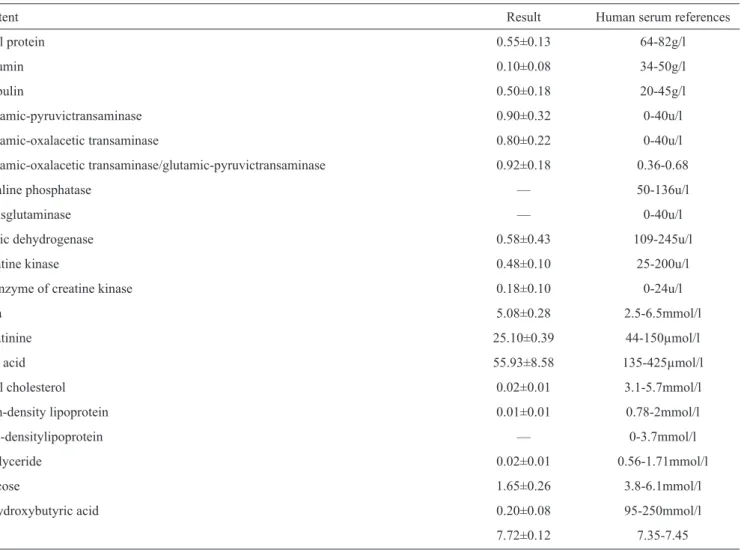
acid, triglycerides, uric acid, and creatine kinase, are lower than those in normal human serum, with each component typically constituting a serum equivalent of approximately 1% of the HCF. The cholesterol level is also lower in cyst fl uid; however,
TABLE 2 - The mineral element composition of hydatid fl uid. The elements were determined using the inductively coupled plasma atomic emission spectrometer.
Element χ±s (mg/l) Analytical line (nm) Calcium 113.93±34.05 315.88 Potassium 216.76±2.85 766.49 Magnesium 44.21±4.05 285.21 Ferrum — 238.20 Copper 0.01±0.01 324.75 Chromium — 267.71 Cadmium — 214.44 Zinc 0.12±0.05 213.86 Selenium — 196.03 Sodium 1554.46±61.09 589.59
MW
4 5
8 6
7
9 10
18
20 11
19
12 13
17 14 15 16 IP 3-10
3
2 1
the ratio of oxalacetic transaminase to glutamic-pyruvictransaminase is higher than that in normal human serum, and the levels of both enzymes are relatively high. The levels of urea, low-density lipoprotein, glutamic-pyruvic transaminase, the isoenzyme of creatine kinase, transglutaminase, and glutamic-oxalacetic transaminase are similar in cyst fl uid and in normal human serum.
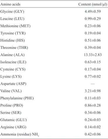
Analysis of amino acids in hydatid-cyst fl uid
The results of the amino acid analysis in the HCF of E. granulosus are shown in Table 4. We measured the levels of 17 amino acids in the cyst fluid. The level of alanine was highest, followed by glycine. We failed to detect aspartic acid, most likely due to its low concentration.
TABLE 3 - Biochemical indices of the hydatid-cyst fl uid of Echinococcus granulosus. Biochemical indexes of hydatid-cyst fl uid detected by
the automatic biochemical analyzer (AU-400, Olympus).
Content Result Human serum references
Total protein 0.55±0.13 64-82g/l
Albumin 0.10±0.08 34-50g/l
Globulin 0.50±0.18 20-45g/l
Glutamic-pyruvictransaminase 0.90±0.32 0-40u/l Glutamic-oxalacetic transaminase 0.80±0.22 0-40u/l Glutamic-oxalacetic transaminase/glutamic-pyruvictransaminase 0.92±0.18 0.36-0.68
Alkaline phosphatase — 50-136u/l
Transglutaminase — 0-40u/l
Lactic dehydrogenase 0.58±0.43 109-245u/l
Creatine kinase 0.48±0.10 25-200u/l
Isoenzyme of creatine kinase 0.18±0.10 0-24u/l
Urea 5.08±0.28 2.5-6.5mmol/l
Creatinine 25.10±0.39 44-150µmol/l
Uric acid 55.93±8.58 135-425µmol/l
Total cholesterol 0.02±0.01 3.1-5.7mmol/l High-density lipoprotein 0.01±0.01 0.78-2mmol/l Low-densitylipoprotein — 0-3.7mmol/l Triglyceride 0.02±0.01 0.56-1.71mmol/l
Glucose 1.65±0.26 3.8-6.1mmol/l
Α-h ydroxybutyric acid 0.20±0.08 95-250mmol/l
Ph 7.72±0.12 7.35-7.45
DISCUSSION
Three proteins, β-hemoglobin, albumin, and serum transferrin, were found in the cyst fl uid. Transferrins are iron-binding blood-plasma glycoproteins that control the level
of free iron in biological fl uids and participate in the body's resistance to infection. Thus, the transferrin in cyst fl uid is likely able to transport the iron required for the growth of E. granulosus. Albumin is the most abundant protein in plasma. Its main function in mammals is to maintain oncotic pressure. It is also a transport protein for, e.g., fatty acids, unconjugated bilirubin, and thyroid hormones. Albumin is also a nutrient for cells. Human albumin in the cyst fl uid provides energy to the larvae. Hemoglobin is a composite protein containing iron, which is composed of ferroheme and globin and plays an important role in transporting oxygen and carbon dioxide. β-hemoglobin is a subtype of hemoglobin that is detected in cyst fl uid. β-hemoglobin provides necessary energy to larvae by transporting oxygen and carbon dioxide.
TABLE 4 - Analysis of amino acids in the hydatid-cyst fl uid of
Echinococcus granulosus. Analysis of amino acids in hydatid-cyst
fl uid detected by the auto-amino-acid analyzer (Hitachi L-8900). Amino acids Content (nmol/µl) Glycine (GLY) 4.49±0.59 Leucine (LEU) 0.99±0.29 Methionine (MET) 0.23±0.06 Tyrosine (TYR) 0.19±0.04 Histidine (HIS) 0.51±0.06 Threonine (THR) 0.39±0.04 Alanine (ALA) 13.33±2.83 Isoleucine (ILE) 0.63±0.15 Cysteine (CYS) 0.17±0.04 Lysine (LYS) 0.77±0.02 Aspartate (ASP) — Valine (VAL) 3.21±0.98 Phenylalanine (PHE) 0.11±0.03 Proline (PRO) 0.86±0.28 Serine (SER) 0.34±0.06 Glutamic (GLU) 0.24±0.03 Arginine (ARG) 0.14±0.02 Ammonia (residue) NH3 0.42±0.06
of these elements into the cyst is strictly controlled to meet the requirements of parasite growth.
We found that the levels of both glutamic-oxalacetic transaminase and glutamic-pyruvictransaminase are high in cyst fl uid (Table 4), suggesting a high level of transamination in larvae. High activity of these enzymes may also be related to a liver disorder caused by liver echinococcosis. The level of uric acid in cyst fl uid is also very high. Uric acid and ascorbic acid are strong reducing agents (electron donors) and potent antioxidants. In humans, over half the antioxidant capacity of blood plasma is derived from uric acid16, and plasma uric acid levels correlate with longevity in primates and other mammals17. Thus, uric acid may play a protective role during the growth of protoscolexes.
Alanine is enriched in cyst fl uid compared with other amino acids. High levels of alanine were also reported in cyst fl uid in other studies18. In our assays, aspartic acid was shown to be absent from cyst fl uid. The intake of amino acids in hydatid disease could occur in two steps: amino acids fi rst cross the cyst wall by free diffusion and then enter the cyst fl uid through free diffusion and active transport mediated by specifi c receptors in the germinal layer19. The absence of aspartic acid suggests that the cyst membrane does not contain aspartic acid receptors and that the larvae cannot synthesize this amino acid. Therefore, the
larvae must obtain aspartic acid through transamination, as is consistent with the high levels of transaminases found in the HCF. The levels of amino acids vary remarkably in cyst fl uid from different hosts (pigs, sheep, cattle, and humans)20,21.
These results indicate that the entrance of all organic and inorganic chemicals depends on parasite requirements. The chemical composition of cyst fl uid plays an important role in protecting against hydatid disease and providing nutritional material. Knowledge of parasite nutrition can aid in identifying new ways to prevent hydatid disease by changing the nutrient composition of cyst fl uid or blocking nutrition and metabolic pathways.
The authors declare that there is no confl ict of interest. CONFLICT OF INTEREST
FINANCIAL SUPPORT
REFERENCES
This work was supported by grants from the National Natural Science Foundation of China (Nº 30960360).
1. Alvaro D, Cecilia C, Florencia I, Gerardo L, Jose OP, Fernando F. Understanding the laminated layer of larval Echinococcus. I: structure. Cell
2011; 27:204-213.
2. Capron A, Biguet J, Vernes A, Afchain D. Antigenic structure of helminthes.
Immunological aspects of the host-parasite relationship. Pathol Biol 1968; 16:121-138.
3. Yahzabal LA, Dupas H, Bout D, Naquira F, Capron A. Echinococcus granulosus: the distribution of hydatid fl uid antigens in the tissues of the
larval stage. II. Localization of the thermostable lipoprotein of parasitic origin (antigen B). Exp Parasitol 1977; 42:115-120.
4. Zhu CL, Ye BH, Zhu XL, Zhao XZ, Huang J, Zhang JY. 2-D electrophoresis preliminary research of hydatid cyst fl uid, scolex and cyst wall. Chinese J Zoonoses 1989; 5:27-29.
5. Chemale G, van Rossum AJ, Jefferies JR, Barrett J, Brophy PM, Ferreira HB, et al. Proteomic analysis of the larval stage of the parasite
Echinococcus granulosus: Causative agent of cystic hydatid disease.
Proteomics 2003; 3:1633-1636.
6. Aziz A, Zhang W, Li J, Loukas A, McManus DP, Mulvenna J. Proteomic characterisation of Echinococcus granulosus hydatid cyst fl uid from
sheep, cattle and humans. Proteomics 2011; 21:1-13.
7. Zhao WX. Human Parasitology. People’s Medical Publishing House; 1987; p. 514.
8. Li ZH, Zhu XQ, Sun J. Basic composition analysis of cyst fl uid in
livestocks. Chinese J Vet Sci Technol 1985; 11:36-37.
9. Kang JF, Fu YC, Feng XH. Preliminary analysis of hydatid cyst fl uid and antigen. J Xing Jiang Med College 1986; 9:290-292.
10. Frayha GJ, Haddad R. Comparative chemical composition of protoscolices and hydatid cyst fl uid of Echinococcus granulosus (Cestoda). Int J
Parasitol 1980; 10:359-364.
11. Goodchild CG, Kagan IG. Comparison of proteins in hydatid fl uid and serum by means of electrophoresis. J Parasitol 1961; 47:175-180. 12. Kagan IG, Norman L. Antigenic analysis of Echinococcus antigens by
13. Kagan IG, Goodchild CG. Paper electrophoresis of sera from man and experimental animals infected with various helminths. J Parasitol 1961b;
47:373-377.
14. Assady M, Farahnak A, Golestani A, Esharghian M. Superoxide dismutase (SOD) enzyme activity assay in Fasciola spp. parasites and liver tissue extract. Iran J Parasitol 2011; 6:17-22.
15. Rayha GJ, Haddad R. Comparative chemical composition of protoscolices and hydatid cyst fl uiColtortid of Echinococcus granulosus (Cestoda). Int J Parasitol 1980; 10:359-364.
16. Maxwell SRJ, Thomason H, Sandler D, Leguen C, Baxter MA, Thorpe GHG, et al. Antioxidant status in patients with uncomplicated insulin-dependent and non-insulin-insulin-dependent diabetes mellitus. European J Clin Investigation 1997; 27:484-490.
17. Cutler RG. Urate and ascorbate: their possible roles as antioxidants in determining longevity of mammalian species. Arch Gerontol Geriatrics 1984; 3:321-348.
18. Chen PH, Yang XL. Research of free amino acid in pig hydatid cyst fl uid. Chinese J Parasitol Parasitic Dis 1990; 8:181-184.
19. Chen PH, Wang FY, Zhang ZM. Albendazole effect on amino acid from pig hydatid cyst fl uid in vitro. J Parasites Med Insect 1994; 1:27-31. 20. Jeffs SA, Arme C. Echinococcus granulosus (Cestoda): uptake of L-amino
acids by secondary hydatid cysts. Parasitol 1988; 96:145-156.