INTRODUCTION
Address to: Drª Eliana Maria Zanotti-Magalhães. Deptº Biologia Animal/IB/ UNICAMP. Rua Monteiro Lobato 255, 13083-862 Campinas, SP, Brasil.
Phone: 55 19 3521- 6303
e-mail: [email protected]
Received 26 June 2013
Accepted 27 September 2013
Susceptibility of Argentinean Biomphalaria tenagophila
and Biomphalaria straminea to infection by
Schistosoma mansoni and the possibility of geographic
expansion of mansoni schistosomiasis
Luciana Franceschi Simões
[1],
Eliana Anunciato Franco Camargo
[1],
Leticia Duart Bastos
[1],
Maria Francisca Neves
[1],
José Ferreira de Carvalho
[2],
Luiz Augusto Magalhães
[1]and Eliana Maria Zanotti-Magalhães
[1][1]. Departamento de Biologia Animal, Instituto de Biologia, Universidade Estadual de Campinas, Campinas, SP. [2]. Statistika Consultoria, Campinas, SP.
ABSTRACT
Introduction:Human migration and the presence of natural vectors (mollusks) of Schistosoma mansoni are the primary causes of the expansion of mansoni schistosomiasis into southern areas of South America. Water conditions are favorable for the expansion of this disease because of the extensive hydrographic network, which includes the basins of the Paraná and Uruguay rivers and favors mollusk reproduction. These rivers also aid agriculture and tourism in the area. Despite these favorable conditions, natural
infection by S. mansoni has not yet been reported in Argentina, Uruguay, or Paraguay. Methods: Two species of planorbid from
Argentina, Biomphalaria straminea and B. tenagophila, were exposed to the miracidia of fi ve Brazilian strains of S. mansoni.
Results: Biomphalaria tenagophila (Atalaya, Buenos Aires province) was infected with the SJS strain (infection rate 3.3%),
confi rming the experimental susceptibility of this Argentinian species. Biomphalaria straminea (Rio Santa Lucía, Corrientes
province) was susceptible to two Brazilian strains: SJS (infection rate 6.7%) and Sergipe (infection rate 6.7%). Conclusions:
These results demonstrate that species from Argentina have the potential to be natural hosts of S. mansoni and that the appearance
of foci of mansoni schistosomiasis in Argentina is possible.
Keywords: Schistosomiasis. Biomphalaria. Susceptibility.
Mansoni schistosomiasis has expanded in recent decades, primarily because of the migration of individuals who have
been parasitized by Schistosoma mansoni, live in inadequate
sanitary conditions in peripheral areas of large cities, and contaminate the water supply with their waste. These water supplies contain trematode vectors such as freshwater mollusks, permitting the establishment of disease foci. In recent decades in Brazil, the disease has spread toward the southern states, with autochthonous cases of this parasitosis reported in Paraná,
Santa Catarina, and Rio Grande do Sul1-4. Ecosystem changes
in northeastern Argentina caused by the construction of dams have led to an increase in infectious diseases due to an increase
in temperature5. Of the planorbid vector species of S. mansoni
found in the southernmost states of Brazil (Rio Grande do Sul
and Santa Catarina), Biomphalaria tenagophila is the most
common6,7. This species is responsible for schistosomiasis foci
in São Francisco do Sul, Santa Catarina. Among the species described in Rio Grande do Sul (Brazil), the primary vector of S. mansoni in the South American continent, B. glabrata, is responsible for the occurrence of the disease in the municipality
of Esteio4. In Uruguay, Paraguay, and Argentina, numerous
planorbid breeders have been described in association with
B. tenagophila and B. straminea8-12, which are known to be
natural vectors of S. mansoni. Breeding conditions are favorable
for these mollusks in the fl ooded fi elds used for plantations and
in irrigation canals and reservoir dams. A study was conducted in Argentina to investigate the growth of natural populations of Biomphalaria in the La Plata river basin. A greater steady growth was confirmed for B. tenagophila compared with
B. peregrina13. Despite the existence of schistosomiasis foci in the Brazilian states bordering Argentina, Uruguay, and Paraguay, there have been no reports of the natural transmission of schistosomiasis in the basin of the La Plata river in these
countries12,14. However, experimental studies with populations
of B. tenagophila from Argentina, Uruguay, and Paraguay have demonstrated that this species is susceptible to the Brazilian
strain of S. mansoni11,12,14-16. Paraense & Corrêa17 assessed the
susceptibility of a planorbid population from Uruguay that was morphologically similar to B. straminea and confi rmed
an infection rate of 23% after exposure to miracidia of the SJ
RESULTS
DISCUSSION METHODS
depends on the degree of physiological adaptation between the
parasite and its intermediary host, as suggested by Magalhães18,
thus explaining the different infection rates observed among populations of the same species of mollusk when infected with different strains of the parasite. Physiological adjustment was
also demonstrated by Paraense & Corrêa,19 who reported that
S. mansoni that were adapted to B. glabrata from Belo Horizonte (State of Minas Gerais (MG), Brazil) resisted infection when
challenged by B. tenagophila from São José dos Campos (State
of São Paulo (SP), Brazil) and vice versa. Paraense & Corrêa19
demonstrated the existence of two strains of the trematode in
Brazil: one from Belo Horizonte (BH) that uses B. glabrata as
an intermediary host and one from São José dos Campos (SJ)
that uses sympatric B. tenagophila as an intermediary host.
The great rivers that form the La Plata river basin are the most likely dispersion routes of schistosomiasis toward Argentina, Paraguay, and Uruguay. In the Mesopotamia region of Argentina in which these rivers are located, numerous
B. tenagophila and B. straminea breeders are known vectors of
S. mansoni. Biomphalaria peregrina, a mollusk that has not yet been found to be naturally infected by the trematode but has high infection rates in the laboratory, is also present in this region. The aim of the present study was to perform susceptibility tests
on the planorbid mollusks B. tenagophila and B. straminea
from Argentina after exposure to a number of Brazilian strains of S. mansoni. Studies of the association between mollusks and
S. mansoni have epidemiological signifi cance because they
facilitate the prediction of areas that become endemic regions for schistosomiasis.
The specimens of B. straminea and B. tenagophila used in
the present study were isolated in the laboratory from specimens collected in Argentina from the Santa Lucía River, in the province of Corrientes, and in Atalaya on the La Plata river, in the province of Buenos Aires, respectively. These specimens
were provided by Dr. Alejandra Rumi. Five strains of S. mansoni
were used for the susceptibility tests: the BH strain (Belo Horizonte, MG, Brazil), which was maintained in sympatric
B. glabrata; the SJ strain (São José dos Campos, SP, Brazil),
which was maintained in sympatric B. tenagophila; the SJS
strain (São José dos Campos, SP, Brazil), which was genetically selected for susceptibility and maintained in sympatric
B. tenagophila that were genetically selected for susceptibility to
S. mansoni20; the BA strain (Bahia, Brazil), which was isolated from the feces of a tourist from Barra Grande, Bahia, Brazil, and
maintained in B. glabrata from Minas Gerais (MG, Brazil); and
the SE strain (Sergipe (SE), Brazil), which was isolated from
B. glabrata from Ilha das Flores (SE, Brazil) and maintained in
sympatric B. glabrata. Five groups of B. tenagophila (5-7mm)
and B. straminea (2-3mm), each containing 30 specimens, were
individually exposed to 10 miracidia of each of the specifi ed
strains of S. mansoni. The miracidia were obtained from eggs
retrieved from the feces of Swiss mice (Mus musculus) that
were experimentally infected with the respective strains of
Schistosoma mansoni. After exposure, the groups of mollusks were kept in aquariums containing dechlorinated water at room
temperature (±25°C) and fed lettuce ad libitum. The water
was changed periodically, and from the fourth to the 16th week
after exposure to the miracidia, the mollusks were examined
to confi rm the liberation of cercariae. The aquariums were maintained under artifi cial light at 28°C. At the end of the
experiment, the surviving mollusks were compressed between two glass plates and examined under a stereoscopic microscope
to confi rm the presence of sporocysts in the tissue. Statistical
analysis of survival was performed by comparing the 10 survival curves obtained from the combinations of species of mollusk (B. tenagophila and B. straminea) and trematode strain (BH, SJ, BA, SE, and SJS). Alternative analysis was also performed to investigate the species effect and the strain effect on survival. The survival curves were adjusted using the Kaplan-Meier method and compared with log-rank statistics, for which the
signifi cance probability allowed an assessment of the differences
between the curves.
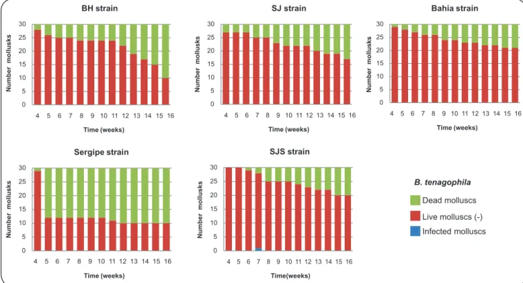
Table 1 displays the number of mollusks that were infected
and the mortality rate. Figure 1 and Figure 2 display the
infectivity of mollusks from the fourth week of infection to
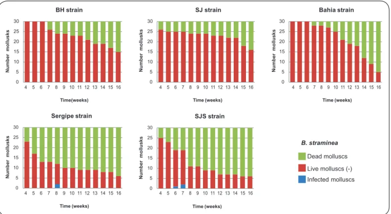
the end of the experiment. Only four of the specimens of B.
straminea that were exposed to infection exhibited elimination of cercariae: two that were exposed to the SJS strain and two that were exposed to the SE strain. The prepatent period was six weeks for the SJS strain and eight weeks for the SE strain. The
specimens of B. straminea that were exposed to the SE strain and
eliminated cercariae died between the eighthand ninth weeks of
infection. The specimens of B. straminea that were exposed to
the SJS strain and eliminated cercariae died between the eighth and ninth weeks of infection: one survived for one week and the
other for two weeks. Only one of the specimens of B. tenagophila
that were exposed to infection eliminated cercariae: the SJS strain of S. mansoni. The prepatent period was seven weeks, and this
mollusk died between the seventhand eighthweeks of infection.
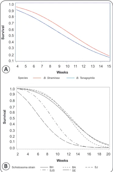
Analysis of the survival curves (Figure 3, Figure 4A, and
Figure 4B), which were softened by the Weibull distribution,
revealed a highly signifi cant difference between the curves
(p<0.0001). The shortest survival times were associated with
the SE strain (Sergipe). No signifi cant difference (p=0.32580)
was observed for the effect of the mollusk species (Figure 4A).
A signifi cant difference (p<0.0001) was observed for the effect
of the strain (Figure 4B), which disappeared (p=0.7018) when
the SE strain was removed from the data set (Sergipe).
The susceptibility of mollusks that are vectors of S. mansoni
TABLE 1 - Infection and mortality rates of Biomphalaria straminea (n=30)and Biomphalaria tenagophila (n=30)from Argentina after exposure to 10 miracidia from fi ve different strains of Schistosoma mansoni.
Strain of Number of mollusks Infection rate Mortality rate
Species of Biomphalaria Schistosoma mansoni that eliminated cercariae (%) n %
Biomphalaria straminea BH 0 0.0 15 50.0
SE 2 6.7 24 80.0
SJS 2 6.7 24 80.0
SJ 0 0.0 14 46.6
BA 0 0.0 25 83.3
Biomphalaria tenagophila BH 0 0.0 20 66.6
SE 0 0.0 20 66.6
SJS 1 3.3 10 33.3
SJ 0 0.0 13 43.3
BA 0 0.0 9 30.0
BH strain (Belo Horizonte, State of Minas Gerais, Brazil); SE strain (Sergipe, Brazil); SJS strain (São José dos Campos, genetically selected, State of São Paulo, Brazil); SJ strain (São José dos Campos, State of São Paulo, Brazil); BA strain (Bahia, Brazil).
0 5 10 15 20 25 30
4 5 6 7 8 9 10 11 12 13 14 15 16 4 5 6 7 8 9 10 11 12 13 14 15 16
4 5 6 7 8 9 10 11 12 13 14 15 16
N
u
m
b
e
r
m
o
ll
u
s
k
s
N
u
m
b
e
r
m
o
ll
u
s
k
s
Time (weeks) SJ strain
0 5 10 15 20 25 30
4 5 6 7 8 9 10 11 12 13 14 15 16
Time(weeks) SJS strain
0 5 10 15 20 25 30
N
m
o
ll
u
s
k
s
u
m
b
e
r
N
m
o
ll
u
s
k
s
u
m
b
e
r
Time (weeks) Sergipe strain
0 5 10 15 20 25 30
Time (weeks) Bahia strain
B. tenagophila
Dead molluscs
Live molluscs (-)
Infected molluscs
0 5 10 15 20 25 30
4 5 6 7 8 9 10 11 12 13 14 15 16
N
u
m
b
e
r
m
o
ll
u
s
k
s
Time (weeks) BH strain
FIGURE 1 - Prepatent period and infectivity of Biomphalaria tenagophila from Argentina after exposure to fi ve different strains of Schistosoma mansoni. BH: Belo Horizonte; SJ: São José dos Campos; SJS: São José dos Campos, genetically selected.
The results of the present study demonstrated that, of the fi ve
strains of S. mansoni used, only the SJS and SE strains were
able to develop in populations of Argentinean mollusks. The SJS strain originated from a population of B. tenagophila that is sympatric to the SJ strain19,which was genetically selected for
N m o ll u s k s u m b e r N m o ll u s k s u m b e r N m o ll u s k s u m b e r N m o ll u s k s u m b e r N m o ll u s k s u m b e r B. straminea Dead molluscs
Live molluscs (-)
Infected molluscs 0 5 10 15 20 25 30
4 5 6 7 8 9 10 11 12 13 14 15 16 4 5 6 7 8 9 10 11 12 13 14 15 16 4 5 6 7 8 9 10 11 12 13 14 15 16
4 5 6 7 8 9 10 11 12 13 14 15 16 4 5 6 7 8 9 10 11 12 13 14 15 16
Time (weeks) Sergipe strain 0 5 10 15 20 25 30 Time (weeks) SJS strain 0 5 10 15 20 25 30 Time (weeks) Bahia strain 0 5 10 15 20 25 30 Time (weeks) SJ strain 0 5 10 15 20 25 30 Time(weeks) BH strain
FIGURE 2 - Prepatent period and infectivity of Biomphalaria straminea from Argentina after exposure to fi ve different strains of Schistosoma mansoni. BH: Belo Horizonte; SJ: São José dos Campos; SJS: São José dos Campos, genetically selected.
1.0 Survival 0.9 0.8 0.7 0.6 0.5 0.4 0.3 0.2 0.1 0.0
B. Straminea BH B. Straminea Bahia B. Straminea SJS B. Straminea SJ
B. Straminea Sergipe
B. Tenagophila Bahia B. Tenagophila BH B. Tenagophila SJS
B. Tenagophila SJ B. Tenagophila Sergipe
Weeks
4 5 6 7 8 9 10 11 12 13 14 15
FIGURE 3 - Survival curves adjusted for the Weibull distribution with respect to the experimental groups. B: Biomphalaria; BH: Belo Horizonte; SJS: São José dos Campos; SJS: São José dos Campos, genetically selected.
Schistosoma mansoni. Among the five Brazilian strains of
S. mansoni used in the susceptibility tests, B. tenagophila
specimens from the province of Buenos Aires (Argentina) were susceptible only to the SJS strain, with liberation of cercariae and
an infection rate of 3.3% (Table 1). This result demonstrates that
B. tenagophila is only susceptible to parasites that originate in mollusks of the same species, corroborating the results of Borda & Rea12,14,16,who found that liberation of cercariae only occurred in
B. tenagophila from Corrientes (Argentina) after exposure to
the SJ strain of S. mansoni. The infection rate confi rmed by the
authors was between 2% and 22%14,with a prepatent period of 31
to 54 days. In the present study, the prepatent period was seven weeks. The longer prepatent periods and lower infection rates indicate that the development of the trematode was hampered in
the mollusk. However, subsequent generations of B. tenagophila
were more susceptible to S. mansoni, as reported by Borda &
Rea14, who found a higher infection rate in the F1 generation of B.
tenagophila. Susceptibility is easier to obtain than an increase in
resistance25. Bernadini & Machado1 confi rmed that B. tenagophila
was the vector species in schistosomiasis foci in Santa Catarina (Brazil). Along the coast of Rio Grande do Sul (Brazil), the
population of B. tenagophila in the Taim Ecological Station has
been shown to be resistant to infection by S. mansoni26. These
Brazilian states share a border with the Argentinean province
of Corrientes. Paraense27 reported morphological differences in
the penile complex of planorbids in Rio Grande do Sul (Brazil)
and Uruguay, suggesting the existence of a subspecies called B.
tenagophila guaibensis, which had previously28 been shown to
be resistant to infection by the BH and SJ strains of S. mansoni.
However, specimens of B. tenagophila tenagophila from
2 4 6 8 10 12 14 16 18 20 1.0
Survival
0.9
0.8
0.7 0.6
0.5
0.4
0.3
0.2 0.1
0.0
BH Schistosoma strain
SJS
BA SE
SJ
Weeks 1.0
0.9
0.8
0.7
0.6
0.5
0.4
0.3
0.2
0.1
Survival
4 5 6 7 8 9 10 11 12 13 14 15
Species B. Straminea B. Tenagophila
Weeks
Weeks
FIGURE 4 - Survival curves adjusted for the Weibull distribution with respect to the two species of mollusks, B. straminea and B. tenagophila (A), and the strains of Schistosoma mansoni used in the experiments, BA, BH, SE, SJ, and SJS (B). B: Biomphalaria; BH: Belo Horizonte; SJS: São José dos Campos, genetically selected; BA: Bahia, SE: Sergipe; SJ: São José dos Campos.
Biomphalaria straminea specimens from the province of Corrientes (Argentina) were infected by the SJS and SE
strains (Table 1), an unprecedented result for this species from
Argentina. Paraense & Corrêa17 observed a morphologically
similar species infection of B. straminea from Uruguay after
exposure to the SJ strain. Biomphalaria straminea specimens
from Argentina were notably susceptible to the Sergipe strain
(SE), which was isolated from naturally infected B. glabrata.
Infection by this strain, which has a high degree of endemicity for schistosomiasis, caused a higher mortality rate in the mollusks (Figure 3, Figure 4A and Figure 4B). This increased mortality was attributed to the parasite; the larval stages of the trematode compromise the tissues and organs of mollusks infected with
S. mansoni29. The prepatent period ranged from six to eight weeks,
indicating restricted S. mansoni development, although one of
the specimens survived for two weeks. Figueiredo30 observed a
natural infection rate of 2.5% for B. straminea from Laranjeiras,
Sergipe (Brazil), which the author considered to be relatively
high. This species is a signifi cant transmitter of S. mansoni in this Brazilian state. The SE strain used in the present study
was obtained from naturally infected B. glabrata and collected
from Ilha das Flores in Sergipe (SE), Brazil. The signifi cance of
B. straminea as a vector of S. mansoni is associated with greater endemicity in northeastern Brazil. Furthermore, an extended
geographical distribution of B. straminea in South America has
been noticed in recent years6,10. Michelson & Dubois31 defi ned
B. straminea as a species of competitive superiority due to its capacity to invade territory occupied by other species. A
number of authors30,32,33 have commented on the displacement of
B. glabrata by B. straminea in northeastern Brazil and the high
capacity of B. straminea to resist periods of drought, which are
characteristic of this region. Naturally infected B. straminea34
have been found in Cruzeiro, in the valley of the Paraíba do
Sul River (SP, Brazil). In experimental tests35, specimens from
this municipality were susceptible to human and wild strains of
S. mansoni. This species was reported6 in a pisciculture aquarium
in Porto Alegre (RS, Brazil). B. straminea and B. tenagophila
have been identifi ed in rice fi elds in Corrientes (Argentina);
B. straminea was more abundant than B. tenagophila10.
The results of the present study confi rm the susceptibility
of B. tenagophila from Argentina to a S. mansoni strain that
originated in sympatric B. tenagophila. The susceptibility of
B. straminea to two Brazilian strains demonstrates the potential of this species to establish schistosomiasis foci in Argentina.
REFERENCES
The authors declare that there is no confl ict of interest.
CONFLICT OF INTEREST
1. Bernardini OJ, Machado MM. Esquistossomose mansoni em Santa Catarina: isolamento do Schistosoma mansoni primeiro foco de transmissão ativa em São Francisco do Sul. Arq Cat Med1981; 10:212. 2. Espíndola KS, Machado MM, Hofmann PR. Natural and experimental
infection of planorbids from the island of Santa Catarina (Brazil). Rev Inst Med Trop São Paulo 1992; 34:289-294.
3. Carvalho OS, Nunes IM, Caldeira, RL. First report of Biomphalaria glabrata in the state of Rio Grande do Sul, Brazil. Mem Inst Oswaldo Cruz 1998; 93:39-40.
4. Graeff-Teixeira C, Anjos CB, Oliveira VC, Velloso CFP, Fonseca MBS,
Valar C, et al. Identifi cation of a transmission focus of Schistosoma mansoni in the southernmost brazilian state, Rio Grande do Sul. Mem Inst Oswaldo Cruz 1999; 94:9-10.
5. Gorodner J, Alonso JM, Zibelman O, Galván M, Merino D, Balbachan
SE, Miranda O. Impacto ambiental de modifi cações ecológicas realizadas
em uma área subtropical. Rev Soc Bras Med Trop 2004; 37:154-157. 6. Teles HMS, Pareira PAC, Richinitti LMZ. Distribuição de Biomphalaria
(Gastropoda, Planorbidae) nos Estados do Rio Grande do Sul e Santa Catarina, Brasil. Rev Saude Publica S Paulo1991; 25:350-352.
7. Schlemper Jr BR, Ferreira Neto JA, Thiago PTS, Bressan C, Amarante
AR, Distribuição geográfi ca de planorbídeos em Santa Catarina, Brasil.
Rev Soc Bras Med Trop 1996; 29:411-418.
8. Paraense WL. Estado atual da sistemática dos planorbídeos brasileiros. Arq Mus Nac Rio de Janeiro1975; 55:105-128.
A
9. Bonetto A, Bechara JA, Tassara M. Los moluscos de La família Planorbidae en el área Del río Paraná Medio. PhysisSec B 1982; 41:1-6. 10. Rumi A, Hamann MI. Potential schistosome-vector snails and associated
trematodes in ricefi elds of Corrientes Province, Argentina. Preliminary
results. Mem Inst Oswaldo Cruz1990; 85:321-328.
11. Borda CE, Rea MJF. Susceptibilidad de Biomphalaria tenagophila de las cuencas de los ríos Paraná y Uruguay a Schistosoma mansoni. Rev Panam Salud Publica 1997; 1:167-173.
12. Borda CE, Rea MJF. Biomphalaria tenagophila potencial vector of Schistosoma mansoni in the Paraná River basin (Argentina and Paraguay). Mem Inst Oswaldo Cruz2007; 102:191-195.
13. Rumi A, Gregoric DEG, Roche A. Tendencias del crecimiento individual en poblaciones naturales de Biomphalaria spp. (Gastropoda, Planorbidae) en La cuenca del Plata, Argentina. Comun Soc Malacol Uruguay2009; 9:185-193. 14. Borda CE, Rea MJF. Susceptibility and compatibility of Biomphalaria
tenagophila from the Río de la Plata basin with Schistosoma mansoni from Brazil. Mem Inst Oswaldo Cruz2010; 105:496-498.
15. Borda CE, Pellegrino J, Susceptibilidad de Biomphalaria tenagophila y B. glabrata a dos cepas de Schistosoma mansoni. Rev Inst Med Trop São Paulo 1976; 18:157-164.
16. Borda CE, Rea MJF. Intermediate and defi nitive hosts of Schistosoma mansoni in Corrientes province, Argentina. Mem Inst Oswaldo Cruz 2006; 101:233-234.
17. Paraense WL, Corrêa LR. A potential vector of Schistosoma mansoni in Uruguay. Mem Inst Oswaldo Cruz1989; 84:281-288.
18. Magalhães LA. Estudo dos dados obtidos de uma população de Biomphalaria glabrata de Belo Horizonte infectada por Schistosoma mansoni da mesma cidade
e de uma população de Biomphalaria tenagophila de Campinas infectada por S. mansoni de São José dos Campos. Rev Soc Bras Med Trop1969; 3:195-196. 19. Paraense WL, Corrêa LR. Variation in susceptibility of populations of
Australorbis glabratus to a strain of Schistosoma mansoni . Rev Inst Med Trop São Paulo1963; 5:15-22.
20. Zanotti-Magalhães EM, Magalhães LA, Carvalho JF. Relação entre a patogenicidade de Schistosoma mansoni em camundongos e a susceptibilidade do molusco vetor. I. Infecciosidade das cercarias e carga de vermes. Rev Saude Publica S Paulo1991; 25:359-366.
21. Newton WL. The inheritance of susceptibility to infection with Schistosoma mansoni in Australorbis glabratus. Exp Parasitol1953; 2:242-257.
22. Richards CS. Genetic of a molluscan vector of schistosomiasis. Nature 1970; 227:806-810.
23. Santana JV, Magalhães LA, Rangel HA. Seleção de linhagens de Biomphalaria glabrata e B. tenagophila visando maior susceptibilidade ao Schistosoma mansoni. Rev Saúde Pública São Paulo, 1978; 12:67-77.
24. Zanotti-Magalhães EM, Magalhães LA, Carvalho JF. Relação entre a patogenicidade do Schistosoma mansoni em camundongos e a susceptibilidade do molusco vetor. IV. Infecciosidade dos miracídios. Rev Saúde Pública Sao Paulo 1997; 31:488-494.
25. Zuim NRB, Zanotti-Magalhães EM, Magalhães LA, Linhares AX.
Seleção genética de Biomphalaria glabrata e Biomphalaria tenagophila
visando a alteração da suscetibilidade e resistência ao Schistosoma mansoni. Rev Soc Bras Med Trop 2005; 38:387-390.
26. Coelho PMZ, Carvalho OS, Andrade ZA, Martins-Sousa RL, Rosa FM, Barbosa L, et al. Biomphalaria tenagophila/Schistosoma mansoni interaction: premises for a new approach to biological control of schistosomiasis. Mem Inst Oswaldo Cruz 2004; 99:109-111.
27. Paraense WL, Biomphalaria tenagophila guaibensis ssp.n. from Southern Brazil and Uruguay (Pulmonata:Planorbidae). I. Morphology. Mem Inst Oswaldo Cruz 1984; 79:465-469.
28. Paraense WL, Corrêa LR. Probable extension of schistosomiasis mansoni to southernmost Brazil. Mem Inst Oswaldo Cruz 1987; 82:577.
29. Coelho MV. O parasito -Schistosoma mansoni. In: Cunha AL, editor. Esquistossomose mansoni, São Paulo: USP Edition; 1970, p. 1-12. 30. Figueiredo CCSB. Dispersão de Biomphalaria straminea no estado de
Sergipe: um estudo comparativo com dezenove anos de intervalo. Mem Inst Oswaldo Cruz 1989; 84:383-387.
31. Michelson EH, Dubois L. Competitive interaction between two snails hosts of Schistosoma mansoni: laboratory studies of Biomphalaria glabrata and B. straminea. Rev Inst Med Trop São Paulo 1979; 21: 246-253.
32. Barbosa FS, Pereira da Costa DP, Arruda F. Competitive interactions between species of freshwater snails. I. Laboratory studies: IB. Comparative studies of the dispersal and the vagility capabilities of Biomphalaria glabrata and B. straminea. Mem Inst Oswaldo Cruz1984; 79:163-167.
33. Barbosa FS, Pereira da Costa DP, Arruda F. Competitive interactions between species of freshwater snails. I. Laboratory studies: IC. Comparative survival of Biomphalaria glabrata and B. straminea kept out of water. Mem Inst Oswaldo Cruz1985; 80:155-157.
34. Santos L, Costa IB, Figueiredo CCSB, Altomani MAG. Primeiro encontro de Biomphalaria straminea (Dunker, 1848) no município de Cruzeiro, vale do Paraíba, Estado de São Paulo, naturalmente infectada por cercarias de Schistosoma mansoni. Rev Inst Adolpho Lutz São Paulo 1980; 40:165-166.