Analysis of heparanase isoforms and cathepsin B in the
plasma of patients with gastrointestinal carcinomas:
analytical cross-sectional study
Análises das isoformas de heparanase e da catepsina B em plasma de pacientes
com carcinomas gastrointestinais: estudo transversal analítico
Carina Mucciolo Melo
I, Clarice Silvia Taemi Origassa
I, Thérèse Rachell Theodoro
II, Leandro Luongo Matos
III, Thaís Aguilar
Miranda
IV, Camila Melo Accardo
V, Rodrigo Ippolito Bouças
VI, Eloah Rabello Suarez
V, Madalena Maria Nunes Silva Pares
VII,
Daniel Reis Waisberg
VIII, Giovanna Canato Toloi
IX, Helena Bonciani Nader
X, Jaques Waisberg
XI, Maria Aparecida Silva Pinhal
XIIUniversidade Federal de São Paulo (Unifesp), Faculdade de Medicina do ABC (FMABC) and Hospital do Servidor Público Estadual
(IAMSPE), São Paulo, Brazil
ABSTRACT
CONTEXT AND OBJECTIVE: Heparanase-1 degrades heparan sulfate and has been correlated with tumor progression. Although the isoform heparanase-2 has no catalytic activity, it seems to be important for modulating heparanase-1 activity. Cathepsin B is a proteinase involved in tumor metastasis. The aim of this study was to analyze heparanase isoform expression and cathepsin B activity in plasma samples from patients with gastrointestinal carcinomas, compared with healthy individuals (control group).
DESIGN AND SETTING: This was an analytical cross-sectional study. Peripheral blood samples were col-lected at a Brazilian public hospital, from 21 patients with histopathological diagnoses of gastrointestinal carcinomas and from 43 healthy individuals. The analyses were performed in two Brazilian medical schools. METHODS: Heparanase isoforms were identiied and quantiied in plasma samples by means of Western blot. The enzymatic activities of heparanase-1 and cathepsin B were also measured.
RESULTS: The results demonstrated that the expression of both heparanase isoforms was signiicantly greater in plasma samples from gastrointestinal carcinoma patients, compared with the control group. Logistic regression analysis showed that increased heparanase-1 and heparanase-2 expression was exclu-sively dependent on the tumor. There was a signiicant increase in heparanase-1 and cathepsin B activity in the patients’ plasma.
CONCLUSION: Overexpression of heparanase-1 and heparanase-2, along with increased heparanase-1 and cathepsin B activity in plasma, is associated with the diagnosis of gastrointestinal carcinoma. These indings provide support for using non-invasive assays (plasma samples) as an auxiliary method for diag-nosing gastrointestinal tumors.
RESUMO
CONTEXTO E OBJETIVO: A heparanase-1 degrada heparam sulfato e está relacionada à progressão de tumor. Apesar de a isoforma heparanase-2 não possuir atividade catalítica, parece ser importante para modular a atividade da heparanase-1. A catepsina B é uma proteinase envolvida na metástase de tumores. O objetivo deste estudo foi analisar a expressão das isoformas de heparanase e atividade da catepsina B em amostras de plasma de pacientes com carcinomas gastrointestinais, comparando-se com indivíduos saudáveis (grupo controle).
TIPO DE ESTUDO E LOCAL: Este é um estudo transversal analítico. Foram coletadas amostras de sangue pe-riférico, em hospital público brasileiro, de 21 pacientes com diagnóstico histopatológico de carcinoma gas-trointestinal e 43 indivíduos saudáveis. As análises foram realizadas em duas faculdades de medicina brasileiras. MÉTODOS: As isoformas da heparanase foram identiicadas e quantiicadas em amostras de plasma por Western blot. As atividades enzimáticas de heparanase-1 e catepsina B foram também mensuradas. RESULTADOS: Os resultados demonstraram que as expressões das isoformas de heparanase foram sig-niicativamente maiores nas amostras de plasma de pacientes com carcinoma gastrointestinal em com-paração com o grupo controle. A análise feita por regressão logística mostra que aumento na expressão de heparanase-1 e heparanase-2 é exclusivamente dependente da presença de tumor. Houve aumento signiicativo na atividade da heparanase-1 e catepsina B no plasma dos pacientes.
CONCLUSÃO: A superexpressão de heparanase-1 e heparanase-2, bem como o aumento da atividade de heparanase-1 e catepsina B no plasma, está associada com o diagnóstico de carcinoma gastrointestinal. Tais achados dão suporte ao uso de ensaios não invasivos (amostras de plasma) como método auxiliar para o diagnóstico de tumores gastrointestinais.
IMSc. Doctoral Student, Department of
Biochemistry, Universidade Federal de São Paulo (Unifesp), São Paulo, Brazil.
IIMSc. Doctoral Student, Faculdade de Medicina
do ABC (FMABC), Santo André, São Paulo, Brazil.
IIIMD, PhD. Associate Professor, Department of
Biochemistry, Faculdade de Medicina do ABC (FMABC), Santo André, São Paulo, Brazil.
IVMSc. Research Collaborator, Department of
Biochemistry, Universidade Federal de São Paulo (Unifesp), São Paulo, Brazil.
VPhD. Postdoctoral Student, Department of
Biochemistry, Universidade Federal de São Paulo (Unifesp), São Paulo, Brazil.
VIPhD. Research Collaborator, Department of
Biochemistry, Universidade Federal de São Paulo (Unifesp), São Paulo, Brazil.
VIIPhD. Research Collaborator, Associação
Beneicente de Coleta de Sangue (Colsan), São Paulo, São Paulo, Brazil.
VIIIMD. Attending Physician. Surgery Department,
Universidade de São Paulo (USP), São Paulo, Brazil.
IXMedical Student. Faculdade de Medicina do
ABC (FMABC), Santo André, São Paulo, Brazil.
XPhD. Titular Professor, Department of
Biochemistry, Universidade Federal de São Paulo (Unifesp), São Paulo, Brazil.
XIMD, PhD. Adjunct Professor, Department
of Surgery, Faculdade de Medicina do ABC (FMABC), Santo André, São Paulo, Brazil.
XIIPhD. Titular Professor, Department of
Biochemistry, Faculdade de Medicina do ABC (FMABC), Santo André, São Paulo, Brazil.
KEY WORDS:
Glucuronidase. Cathepsin B.
Tumor markers, biological. Carcinoma.
Plasma.
PALAVRAS-CHAVE:
Glucuronidase. Catepsina B.
Marcadores biológicos de tumor. Carcinoma.
INTRODUCTION
Gastrointestinal carcinomas remain one of the leading causes of cancer-related death worldwide.1 he major cause of mortality
among patients with diferent gastrointestinal cancers is distant metastasis, rather than the primary carcinoma itself.
he incidence of hepatocarcinoma ranges from 1 to 9.34 cases per 100,000 inhabitants, depending on the region of Brazil.2
his type of cancer presents very fast development and therefore an early diagnosis is important for successful treatment.
In 2008, one million new cases of gastric neoplasms were expected. Gastric cancer has the fourth highest incidence in the world, and a high mortality rate.3
Duodenal papillary carcinoma is an extremely rare type of cancer and it is generally diagnosed by means of biopsy.4 In the
United States, the incidence rate of duodenal papillary carcinoma is less than 5% of all cases of digestive neoplasms.5
In 2013, 102,480 new cases of colon cancer and 40,340 of rec-tal carcinoma were recorded. Colorecrec-tal carcinomas are usually diagnosed by means of biopsy.6
Esophageal cancer is a relatively uncommon malignancy and is an extremely lethal tumor. In Brazil, during 2005, the expected incidence was 8.99 and 2.61 new cases of esophageal cancer per 100,000 men or women, respectively.7
Invasion of malignant cells involves interaction with the extracellular matrix and the basement membranes. Basement membranes consist essentially of collagen type IV, laminin and heparan sulfate proteoglycans. Tumor cells need to degrade base-ment membrane constituents in order to promote metastasis, and this activity of malignant cells involves proteinases and other enzymes such as heparanase.8
Heparanase is an endo-beta-glucuronidase that degrades heparan sulfate chains of proteoglycans and has a multifunctional modulatory efect on cancer cell progression and cell-extracellu-lar matrix interaction.9-11 Research has shown that heparanase
has important roles in relation to the progression of esophageal, stomach and colonic cancer. Heparanase expression has been closely correlated with the diagnosis/prognosis of gastrointesti-nal cancer. Heparanase silencing can be useful as a potential anti-cancer therapy.12
here are two members of the heparanase family: heparan-ase-1 (HPSE1) and heparanase-2 (HPSE2), which are encoded by diferent genes: 4q21.3 and 10q23.24, respectively.13-17
he two isoforms of heparanase present diferent tissue distribu-tions and diferent cell locadistribu-tions. Immunoluorescence and cell fractionation studies have shown that heparanase-1 is present in the nucleus, perinuclear region and plasma membrane.18,19
It is well known that heparanase-1 shows a direct correla-tion with tumor metastasis. Heparanase-1 presents two isoforms: irstly, a precursor with no apparent enzymatic activity (65 kDa),
which then undergoes proteolytic activity to form the mature active enzyme, which consists of a heterodimer containing a 50 kDa subunit in association with an 8 kDa subunit.9,20
Nevertheless, more studies need to be conducted to clarify the relative contribution of heparanase-2 to human health and dis-ease, since heparanase-2 does not present enzymatic activity.15,21
Heparanase-2 has three alternative variant splice transcripts (hep-aranase-2a, heparanase-2b, and heparanase-2c), which encode the putative proteins of 480, 534, and 592 amino acids, respectively. It has been predicted from sequence analysis that all three hepa-ranase-2 proteins are intracellular, membrane-bound proteins.15
Alignment of the predicted coding region of heparanase-2a to heparanase-1 has revealed that the two proteins have overall identicalness of 35%.15 here is some evidence that heparanase-2
attenuates heparanase-1 activity, possibly conferring a favorable outcome upon head and neck cancer patients.21
Cathepsin B is a lysosomal cysteine proteinase, which has been implicated in a variety of diseases, such as inlammation and tumor metastasis. Overexpression of cathepsin B has been observed in malignant tumors, and speciically in the cells at the invasive edge of these tumors. Cathepsin B may facilitate inva-sion directly by degrading extracellular matrix components or indirectly by activating other proteases capable of digesting the extracellular matrix.22
Many studies have been conducted using tissue samples to investigate the expression of cathepsin B and heparanase iso-forms in a variety of cancers. he present study had the advan-tage of using a noninvasive assay, using plasma samples.
OBJECTIVE
he aim of this study was to evaluate isoforms of heparanase and cathepsin B in plasma samples from patients with primary gas-trointestinal carcinoma, in comparison with healthy individuals.
METHODS
Study design and ethics
his was an analytical cross-sectional study that aimed to evalu-ate heparanase isoforms and cathepsin B in plasma samples from patients with diferent gastrointestinal carcinomas, in compari-son with a control group (healthy individuals).
André, Brazil), under number 025/2008, and the Human Ethics Research Committee of the Servidor Público Estadual Hospital (São Paulo, Brazil), under number 021/08.
Settings
he samples were collected in a Brazilian public hospital that provides primary, secondary and tertiary care. his study was conducted in two diferent Brazilian medical schools.
All patients who had been admitted for surgical treatment over the period from January to March 2009 and who fulilled the eligibility criteria were selected. he control group was composed of adult volunteers of both genders without any evidence or sus-picion of malign gastrointestinal neoplasia, pre-neoplasic lesions or any inlammatory intestinal disease, who were recruited between January and March 2009.
Patients and clinical features
he eligibility criteria were as follows: age ≥18 years; histopatho-logically conirmed diagnosis of gastrointestinal cancer; no prior chemotherapy or radiotherapy; no prior treatment for other can-cer; and no associated acute disease. he patient group included 21 individuals who had undergone operative procedures in order to remove primary gastrointestinal cancer, while 43 healthy indi-viduals constituted the control group.
he patient group included 16 men (76.2%) and 5 women (23.8%). he average age in the patient group was 68.7 ± 10.0 years (ranging from 50 to 82 years). All the patients were Caucasian. he control group included 32 men (74.4%) and 11 women (25.6%) with an average age of 52.9 ± 5.0 years (ranging from 46 to 65 years). he carcinomas were located in the stomach in 5 patients (23.8%), colon in 5 (23.8%), esophagus in 4 (19.0%), rectum in 4 (19.0%), duodenal papilla in 2 (9.5%) and liver in 1 (4.8%). Concerning histological diferentiation, 4 lesions (19.0%) were poorly diferentiated, 10 (47.6%) were moderately diferentiated and 5 (23.8%) were well diferentiated. he lymph nodes were compromised by the tumor in 14 patients (66.6%) and were dis-ease-free in 7 (33.3%). Venous invasion occurred in 4 patients (19.0%), lymphatic invasion occurred in 3 (14.3%) and neural iniltration was present in 2 (9.5%). Among these 21 patients, dis-tant metastasis was revealed in 7 (33.3%). Four patients (19.0%) died due to metastasis or local recurrence from carcinoma.
Blood sample collection
Peripheral blood samples were collected using ethylenediamine tetraacetic acid (EDTA), as an anticoagulant. Sodium citrate could not be used as an anticoagulant, because of platelet activa-tion and the release of heparanase-rich platelet-dense granules in plasma samples. Plasma samples were obtained ater peripheral blood centrifugation (1,500 x g, 15 min and 4 °C). he plasma
samples were frozen in small aliquots and were thawed just once for the analysis.23,24
Western blot analysis
For immunoblot analysis, 2 or 5 µg of total plasma protein ali-quots were loaded for electrophoresis on 10% sodium dodecyl sulfate-polyacrylamide gel (SDS-PAGE), for heparanase-1 and heparanase-2 detection, respectively. It is important to note that more than twice the amount of total plasma protein was used to analyze heparanase-2 isoforms in the Western blot assays compared with the amount used for heparanase-1 proteins. he proteins were transferred to an Immobilon-P membrane (Millipore, Bedford, Massachusetts, USA), followed by succes-sive incubations with block solution (1% nonfat milk) contain-ing anti-heparanase polyclonal antibody (anti-HPA) H-80 (Santa Cruz Biotechnology, Santa Cruz, California, USA) and anti-HPA C-17 (Santa Cruz Biotechnology, Santa Cruz, California, USA), for heparanase-1 and heparanase-2 analysis, respectively. he primary anti-heparanase antibodies were diluted 1:100 in 0.1% bovine serum albumin, 10 mmol/L Tris-HCl (pH 7.5), 100 mmol/L NaCl and 0.05% Tween-20. he secondary anti-body was horseradish peroxidase-conjugated anti-rabbit/mouse antibody (Jackson Laboratories, Bar Harbor, Maine, USA). he immunoblot reaction was detected using 0.5% 3,3’-diamino-benzidine (DAB) and 0.01% H2O2. A mixture of standard pro-tein markers (Sigma Chemical Co., St. Louis, Missouri, USA) was used for relative molecular mass determination. he Western blot quantiication was performed by means of densitometry analy-sis using Scion Image sotware, version 4.03 (Scion Corporation, Frederick, Maryland, USA).
Quantiication of the product obtained through heparanase he heparanase activity level was determined in terms of the hep-aran sulfate products generated through enzymatic activity, using 15% biotinylated heparan sulfate as the substrate, immobilized in a poly-L-lysine multi-well. he assay was performed using an Enzyme-Linked Immunosorbent Assay (ELISA)-like method.25
count/s) was processed automatically, using MultiCalc sotware (PerkinElmer Life Sciences-Wallac Oy, Turku, Finland). A stan-dard curve of diferent concentrations of biotinylated heparan sulfate was produced and the result was determined as the per-centage of degraded heparan sulfate.
Cathepsin B assay
he cathepsin B enzymatic action was measured using a spectro-luorometer, using the luorogenic substrate carbobenzoxy-Phe-Arg-7-amide-4-methylcoumarin (Z-FR-MCA, Sigma-Aldrich), as described previously.26 he incubations were carried out on
dark microplates (Nunc, Sigma-Aldrich), at 37 ºC in 50 mM sodium phosphate bufer (pH 6.3), containing 200 mM NaCl (Millipore) and 2 mM EDTA (Sigma-Aldrich). he luorescence intensity was measured in a microplate reader (FLEXStation 3) with the SotMax sotware (Molecular Devices), with the excita-tion and emission wavelengths set as 360 and 465 nm, respectively. he assay was performed initially by pre-incubating the plasma samples with the enzyme activator in 2 mM of dithiothreitol (DTT, Sigma-Aldrich), for 20 minutes at room temperature, and then 5 µM of the irreversible inhibitor E-64 (Sigma-Aldrich) and the substrate (10 µM) were added. hus, the enzymatic assay was performed for 24 hours, at 37 ºC. he level of cathep-sin B action was determined in terms of substrate degradation, expressed as arbitrary units of luorescence (AUF).
Statistical analysis
Statistical analysis was performed using the SPSS 16.0 sotware for Windows (SPSS, Chicago, Illinois, USA). he Kolmogorov-Smirnov test was used to analyze whether the data had normal dis-tribution, and the study variables were considered to be parametric. Subsequently, statistical diferences among the experimental groups were evaluated using Student’s t test and the Mann-Whitney U-test. he Pearson correlation was analyzed to evaluate any possible inter-connection between patients’ ages and the results obtained. Logistic regression analysis was performed to investigate whether the levels of heparanase isoforms and cathepsin B alterations were dependent on the presence of a tumor. he signiicance level was set at P < 0.05.
RESULTS
Protein expression of heparanase isoforms in the plasma
Figure 1 shows heparanase-1 and heparanase-2 protein detection
in the plasma of gastrointestinal patients and healthy individuals (controls), through analysis using Western blot.
Two isoforms of heparanase-1 (50 kDa and 65 kDa) can be observed in Figure 1A. It was also demonstrated that the heparan-ase-1 levels in the plasma of gastrointestinal carcinoma patients are higher than in the plasma of the control group (Figure 1A).
he polyclonal antibody anti-heparanase-2 C-17, which was used in the Western blot analysis, was able to identify three iso-forms containing 480, 534 and 592 amino acids, named respec-tively, heparanase-2a, heparanase-2b and heparanase-2c. he levels of all heparanase-2 isoforms were higher in the plasma of patients with gastrointestinal carcinoma than in the control group, as shown in Figure 1B.
Quantitative analysis from Western blot assay was performed for each heparanase isoform, i.e. for heparanase-1 (50 kDa), heparanase-1 (65 kDa) and heparanase-2, as shown in Table 1. he protein expression levels in the plasma of the gastrointestinal carcinoma patients was signiicantly higher than in the plasma of the healthy individuals for all heparanase isoforms (Table 1).
The active form of heparanase-1 (50 kDa) presented higher protein expression in the gastrointestinal carcinoma patients than in the control group: respectively, 28.87 ± 9.70 pixels/µg of total protein and 12.06 ± 4.85 pixels/µg of total protein. The data showed a significant difference in the expression of plasma heparanase-1 (50 kDa) (P < 0.001; Student’s t test), as demonstrated in Table 1.
Similar results were obtained for pro-enzyme heparan-ase-1 (65 kDa), which demonstrated plasma expression of 17.01 ± 6.80 pixels/µg of total protein in the control group, whereas higher protein expression (23.33 ± 7.99 pixels/µg of total protein) was observed in the plasma of the gastrointestinal car-cinoma patients (P = 0.002; Student’s t test), as shown in Table 1. he average heparanase-2 expression in the plasma of the patients with gastrointestinal carcinoma was signiicantly greater (39.21 ± 13.09 pixels/µg of total protein) than in the control group (25.39 ± 9.95 pixels/µg of total protein), as shown in Table 1 (P < 0.001; Student’s t test).
Figure 1. Heparanase isoform detection by means of Western blot on plasma samples. (A) represents heparanase-1: both isoforms of heparanase-1, i.e. 65 kDa (precursor) and 50 kDa (active fraction). (B) represents heparanase-2: three isoforms of heparanase-2 comprising respectively 65 kDa, 58 kDa and 55 kDa. Patient and Control correspond to plasma samples from patients with gastrointestinal carcinoma and plasma samples from healthy individuals, respectively. MW refers to standard protein markers.
65
Patient
B A
65
50
Molecular Mass
kDa
130
100
70
55
40 MW Control
130
100 70
55
40
Patient MW Control
55 58
Molecular Mass
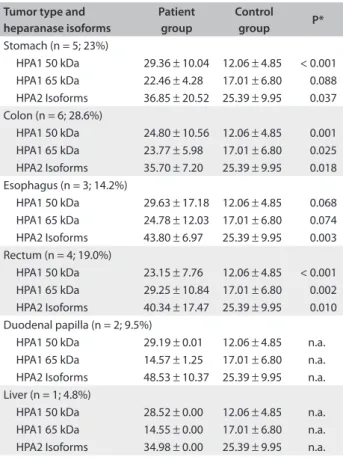
Furthermore, patients were grouped according to their gas-trointestinal tumor types and the heparanase isoforms were quantitatively analyzed in each subgroup, in relation to the con-trol group (Table 2). hese results demonstrated that the level of the active fraction of heparanase-1 (50 kDa) was signiicantly higher in all tumor subtypes, compared with the control group
(Table 2). Moreover, heparanase-2 protein expression was
signif-icantly higher in all subtypes of gastrointestinal carcinomas. Univariable statistical analysis showed a direct correlation between heparanase isoforms and the patient’s age. Since the aver-age aver-age of patients with gastrointestinal carcinoma is normally higher than the average age in the control group (the donors), Pearson’s correlation analysis was performed. he results clearly demonstrated that patients with diferent types of gastrointestinal carcinomas were older and presented higher expression of heparan-ase isoforms (heparanheparan-ase-1 50 kDa: R = 0.658 and P < 0.0001; hepa-ranase-1 65 kDa: R = 0.380 and P = 0.002; heparanase-2: R = 0.345 and P = 0.005), in comparison with the donor group (control).
In order to elucidate possible bias between older age and the presence of gastrointestinal carcinomas, multivariable statisti-cal analysis was performed using logistic regression. he statististatisti-cal parameters obtained through logistic regression proved that each heparanase isoform expression was straightforwardly dependent on the tumor [heparanase-1 (50 kDa): P < 0.0001; heparanase-1 (65 kDa): P = 0.002; and heparanase-2: P < 0.0001] and was inde-pendent of the patient’s age [heparanase-1 (50 kDa): P = 0.270; hepa-ranase-1 (65 kDa): P = 0.218; and heparanase-2: P = 0.717].
Clinical feature correlations demonstrated that there was no signiicant diference between heparanase-1 (50 kDa), heparan-ase-1 (65 kDa) or heparanase-2 expression and tumor diferen-tiation, tumor metastasis, tumor vascular invasion, lymphatic invasion or perineural iniltration, as observed through statisti-cal analysis (data not shown).
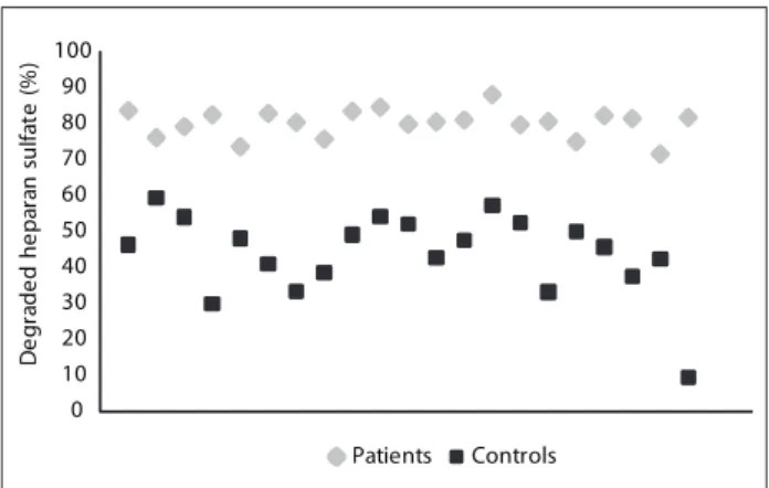
Detection of heparanase-1 enzymatic action on the plasma samples
he percentage of degraded heparan sulfate was used to quantify heparanase-1 enzymatic activity on the plasma samples. Figure 2 shows that there was signiicantly greater heparanase-1 enzymatic activity in the plasma of the gastrointestinal carcinoma patients (80.05%), compared with the control plasma (44.07%). hus, the heparanase-1 enzyme activity in the plasma patients with gastroin-testinal carcinoma was seen to be approximately twice as much as in the plasma of the unafected individuals (Student’s t test, P < 0.0001).
Plasma cathepsin B
he plasma cathepsin B enzymatic assay showed that its level was signiicantly higher in the patient group (1223 ± 147.2 AUF/µl) than in the control group (345 ± 32 AUF/µl), as demonstrated in Figure 3 (Student’s t test, P < 0.0001).
DISCUSSION
In non-neoplastic human tissues, heparanase-1 mRNA is restricted to hematopoietic cells and placental cells, as described in the literature.9,10,16 In contrast, heparanase-2 expression is low
in these tissues, but high in the brain, mammary gland, prostate, small intestine, testis, uterus and bladder tissues.15,17 his
mark-edly diferent pattern of heparanase-2 expression suggests that, at least in normal tissues, the two heparanase isoforms may ful-ill diferent functions.19 he results obtained in the present study
demonstrated that heparanase-1 and heparanase-2 proteins were both present in the plasma.
Table 1. Quantitative analysis of heparanase-1 and heparanase-2 in the plasma of patients with gastrointestinal (GI) tumors and healthy
controls, expressed as mean ± standard deviation of pixels/µg of total
plasma protein
Heparanase isoforms GI tumor group Control group P*
Heparanase-1 (50 kDa) 28.87 ± 9.70 12.06 ± 4.85 < 0.001 Heparanase-1 (65 kDa) 23.33 ± 7.99 17.01 ± 6.80 0.002 Heparanase-2 39.21 ± 13.09 25.39 ± 9.95 < 0.001 *Student’s t test.
Table 2. Quantitative HPA1 and HPA2 analysis on the plasma of
diferent gastrointestinal carcinoma types expressed as mean ±
standard deviation of pixels/µg of total plasma protein
Tumor type and heparanase isoforms
Patient group
Control
group P*
Stomach (n = 5; 23%)
HPA1 50 kDa 29.36 ± 10.04 12.06 ± 4.85 < 0.001 HPA1 65 kDa 22.46 ± 4.28 17.01 ± 6.80 0.088 HPA2 Isoforms 36.85 ± 20.52 25.39 ± 9.95 0.037 Colon (n = 6; 28.6%)
HPA1 50 kDa 24.80 ± 10.56 12.06 ± 4.85 0.001 HPA1 65 kDa 23.77 ± 5.98 17.01 ± 6.80 0.025 HPA2 Isoforms 35.70 ± 7.20 25.39 ± 9.95 0.018 Esophagus (n = 3; 14.2%)
HPA1 50 kDa 29.63 ± 17.18 12.06 ± 4.85 0.068 HPA1 65 kDa 24.78 ± 12.03 17.01 ± 6.80 0.074 HPA2 Isoforms 43.80 ± 6.97 25.39 ± 9.95 0.003 Rectum (n = 4; 19.0%)
HPA1 50 kDa 23.15 ± 7.76 12.06 ± 4.85 < 0.001 HPA1 65 kDa 29.25 ± 10.84 17.01 ± 6.80 0.002 HPA2 Isoforms 40.34 ± 17.47 25.39 ± 9.95 0.010 Duodenal papilla (n = 2; 9.5%)
HPA1 50 kDa 29.19 ± 0.01 12.06 ± 4.85 n.a. HPA1 65 kDa 14.57 ± 1.25 17.01 ± 6.80 n.a. HPA2 Isoforms 48.53 ± 10.37 25.39 ± 9.95 n.a. Liver (n = 1; 4.8%)
HPA1 50 kDa 28.52 ± 0.00 12.06 ± 4.85 n.a. HPA1 65 kDa 14.55 ± 0.00 17.01 ± 6.80 n.a. HPA2 Isoforms 34.98 ± 0.00 25.39 ± 9.95 n.a.
Friedman et al. reported heparanase-1 tissue overexpres-sion in cases of colon cancer progresoverexpres-sion and metastasis, among 16 patients with colon adenocarcinoma. hey also concluded that the most poorly diferentiated carcinoma tissues presented the highest expression of heparanase-1. High expression of heparan-ase-1 was also noted in other tissues like lung, lymph node and liver, as well as in the stromal tumor tissues. However, low hepa-ranase-1 levels were observed in non-neoplastic tissues.27
he results obtained here showed that the level of the active form of heparanase-1 (50 kDa) was signiicantly higher in the plasma of the gastrointestinal carcinoma patients than in the control group. he greater enzymatic action of heparanase-1 in the plasma of the gastrointestinal carcinoma patients corroborates with the higher of protein expression found through Western blot analysis.
Furthermore, statistical analysis on the tumors grouped as diferent subtypes showed that the heparanase-1 (50 kDa) expres-sion was signiicantly higher in all samples analyzed.
Cleavage of heparan sulfate by heparanase-1 (50 kDa) gen-erated active oligosaccharides that were able to modulate cellu-lar processes. Additionally, it was proved that these heparan sul-fate oligosaccharides could increase the biological function of growth factors, angiogenic factors and cytokines, thus mediating cell proliferation, migration, inlammation and angiogenesis.28-30
It had already been observed in previous studies from our laboratory that heparanase-1 and heparanase-2 isoforms were overexpressed in the blood (mononuclear cell fraction) of women with breast cancer, thereby suggesting that this tumor can pos-sibly modulate the expression of both heparanases.31 Similar
results have been described, suggesting that higher heparanase expression in gastric cancer tissues was closely correlated with lower treatment responsiveness and poor prognosis.32
he data obtained in the present study demonstrated that heparanase-2 protein expression was signiicantly greater in plasma samples from patients with gastrointestinal carcino-mas, compared with healthy individuals. Moreover, statistical analysis was also performed on diferent tumor type subgroups. It was observed that heparanase-2 levels were signiicantly higher in all subtypes of gastrointestinal carcinomas, compared with the control group.
Previous results from our group had shown that heparan-ase-2 was upregulated in all stages of colorectal carcinomas.33,34
Heparanase-2 may be involved in the carcinogenesis process through mediating tumor cell adhesion and migration or apop-tosis, as shown by the latent form of heparanase-1 (65 kDa).35
herefore, heparanase-2 may initiate signal transduction that activates proliferation and survival of colorectal carcinoma cells, independent of enzymatic activity.33,34 Nevertheless, further
inves-tigation is necessary, in order to validate this hypothesis concern-ing heparanase-2 function in colorectal carcinoma development. As suggested by Giordano, heparanase-2 upregulation in colorec-tal cancer may constitute a new marker for this neoplasia.33
Cathepsin B enzymatic activity was higher in the plasma samples of patients with gastrointestinal carcinomas than in the control group. Because cathepsin B cellular traic can be modu-lated by the heparan sulfate proteoglycan cell surface, there may have been a correlation between cathepsin B and heparanase activity. In addition, cathepsin B seems to be localized in the perinuclear region of tumor cells and, consequently, the cellular Figure 3. Cathepsin B activity was measured in plasma
samples, using a luorometric assay, as described in the
Methods section. The values represent mean ± standard
deviation. The assays were performed in triplicate and were expressed as arbitrary units of luorescence (AUF) per µl of plasma. The plasma samples were collected from gastrointestinal carcinoma patients (Patients) and healthy individuals (Controls) (P < 0.0001).
0 200 400 600 800 1000 1200 1400 1600
C
at
h
e
p
si
n
B
(A
U
F
/µ
L
)
Patients
Controls Figure 2. Analysis on plasma heparanase-1 activity by
quantiication of degraded heparan sulfate. Heparanase-1 enzymatic activity in plasma samples was measured by means of an ELISA-like method, using 15% biotinylated heparan sulfate immobilized in poly-L-lysine multiwall, as the substrate. The samples were obtained from plasma of gastrointestinal carcinoma patients (Patients) and healthy individuals (Controls). The values represent percentages of degraded heparan sulfate. The assay was performed in triplicate and repeated four times. There was a statistically signiicant diference between patients and controls (P < 0.0001).
0 10 20 30 40 50 60 70 80 90 100
0 5 10 15 20
D
e
g
ra
d
e
d
he
p
ar
an
s
u
lfa
te
(
%
)
distribution of heparan sulfate associated with growth factors may also have been altered. herefore, the cathepsin B and hep-aran sulfate proteoglycan complex may play a role in nuclear functions, thereby becoming part of the transformation process that is observed in carcinogenesis.22,26
It is important to emphasize that the small number of sam-ples for each type of gastrointestinal carcinoma may be a lim-itation to the present study. Nevertheless, the analysis on each subgroup demonstrated that there was a signiicant diference between the control and tumor samples.
he combined results obtained through analysis on heparan-ase and cathepsin B may have future implications for diagnos-ing gastrointestinal cancer. Heparanase and cathepsin B analysis may develop into a safer and less invasive novel diagnostic tool for gastrointestinal carcinomas.
CONCLUSIONS
he analysis on heparanase isoforms and cathepsin B in the plasma of patients with gastrointestinal carcinomas that was pro-posed in the present study revealed that this is a potential new diagnostic tool.
his noninvasive assay for detecting diferent gastrointestinal carcinomas can be performed using plasma samples. he diferences in plasma samples that were observed between the gastrointestinal carcinoma patients and unafected individuals suggest that the protein expression of heparanase-1 (50 kDa) and heparanase-2 and also the enzymatic action of heparanase-1 and cathepsin B can possibly be used to detect the presence of carcinoma.
Further studies need to be undertaken in order to investigate whether the expression of heparanase isoforms and higher lev-els of enzymatic activity of heparanase-1 and cathepsin B in the plasma can be used to improve the diagnosis and also as potential novel therapeutic targets in cases of gastrointestinal carcinoma.
REFERENCES
1. Cancer Research UK. World cancer factsheet A3. Available from:
http://publications.cancerresearchuk.org/downloads/Product/CS_
FS_WORLD_A3.pdf. Accessed in 2013 (Nov 13).
2. Brasil. Ministério da Saúde. Instituto Nacional de Câncer. Câncer de
fígado. Available from: http://www.inca.gov.br/conteudo_view.
asp?id=330. Accessed in 2013 (Nov 13).
3. Arregi MMU, Férrer DPC, Assis ECV, et al. Peril clínico-epidemiológico
das neoplasias de estômago atendidas no Hospital do Câncer
do Instituto do Câncer do Ceará, no período 2000-2004
[Clinical-epidemiological proile of gastric neoplasms in the cancer Hostpital
of Ceara’s Cancer Institute, from the period 2000-2004]. Rev Bras
Cancerol. 2009;55(2):121-8.
4. Wakatsuki T, Irisawa A, Takagi T, et al. Primary adenocarcinoma of the
minor duodenal papilla. Yonsei Med J. 2008;49(2):333-6.
5. Devuni D, Birk JW. Papillary tumors. Medscape. Available from: http://
emedicine.medscape.com/article/187464-overview#a1/. Accessed
in 2014 (Jan 27).
6. National Cancer Institute. What You Need To Know About™
Cancer of the Colon and Rectum. Available from: http://www.
cancer.gov/cancertopics/wyntk/colon-and-rectal/. Accessed in
2014 (Jan 27).
7. Queiroga RC, Pernambuco, AP. Câncer de esôfago: epidemiologia,
diagnóstico e tratamento [Esophageal cancer: epidemiology,
diagnosis and treatment]. Rev Bras Cancerol. 2005;52(2):173-8.
8. Chen JQ, Zhan WH, He YL, et al. Expression of heparanase gene,
CD44v6, MMP-7 and nm23 protein and their relationship with the
invasion and metastasis of gastric carcinomas. World J Gastroenterol.
2004;10(6):776-82.
9. Vlodavsky I, Friedmann Y, Elkin M, et al. Mammalian heparanase:
gene cloning, expression and function in tumor progression and
metastasis. Nat Med. 1999;5(7):793-802.
10. Hulett MD, Freeman C, Hamdorf BJ, et al. Cloning of mammalian
heparanase, an important enzyme in tumor invasion and metastasis.
Nat Med. 1999;5(7):803-9.
11. Naomoto Y, Takaoka M, Okawa T, et al. The role of heparanase in
gastrointestinal cancer (Review). Oncol Rep. 2005;14(1):3-8.
12. Vlodavsky I, Eldor A, Haimovitz-Friedman A, et al. Expression of
heparanase by platelets and circulating cells of the immune system:
possible involvement in diapedesis and extravasation. Invasion
Metastasis. 1992;12(2):112-27.
13. Vlodavsky I, Goldshmidt O, Zcharia E, et al. Molecular properties
and involvement of heparanase in cancer progression and normal
development. Biochimie. 2001;83(8):831-9.
14. McKenzie E, Tyson K, Stamps A, et al. Cloning and expression proiling
of Hpa2, a novel mammalian heparanase family member. Biochem
Biophys Res Commun. 2000;276(3):1170-7.
15. Baker E, Crawford J, Sutherland GR, et al. Human HPA
endoglycosidase heparanase. Map position 4q21.3. Chromosome
Res. 1999;7(4):319.
16. Vlodavsky I, Goldshmidt O. Properties and function of heparanase
in cancer metastasis and angiogenesis. Haemostasis. 2001;31
Suppl 1:60-3.
17. Dempsey LA, Brunn GJ, Platt JL. Heparanase, a potential regulator of
cell-matrix interactions. Trends Biochem Sci. 2000;25(8):349-51.
18. Marchetti D, Li J, Shen R. Astrocytes contribute to the brain-metastatic
speciicity of melanoma cells by producing heparanase. Cancer Res.
2000;60(17):4767-70.
19. Goldshmidt O, Zcharia E, Cohen M, et al. Heparanase mediates
cell adhesion independent of its enzymatic activity. FASEB J.
2003;17(9):1015-25.
21. Nadav L, Eldor A, Yacoby-Zeevi O, et al. Activation, processing and
traicking of extracellular heparanase by primary human ibroblasts.
21. Levy-Adam F, Feld S, Cohen-Kaplan V, et al. Heparanase 2 interacts
with heparan sulfate with high ainity and inhibits heparanase
activity. J Biol Chem. 2010;285(36):28010-9.
22. Yan S, Sameni M, Sloane BF. Cathepsin B and human tumor
progression. Biol Chem. 1998;379(2):113-23.
23. Eldor A, Bar-Ner M, Yahalom J, Fuks Z, Vlodavsky I. Role of heparanase
in platelet and tumor cell interactions with the subendothelial
extracellular matrix. Semin Thromb Hemost. 1987;13(4):475-88.
24. Ishai-Michaeli R, Eldor A, Vlodavsky I. Heparanase activity expressed by
platelets, neutrophils, and lymphoma cells releases active ibroblast
growth factor from extracellular matrix. Cell Regul. 1990;1(11):833-42.
25. Bouças RI, Trindade ES, Tersariol IL, Dietrich CP, Nader HB. Development
of an enzyme-linked immunosorbent assay (ELISA)-like luorescence
assay to investigate the interactions of glycosaminoglycans to cells.
Anal Chim Acta. 2008;618(2):218-26.
26. Almeida PC, Nantes IL, Chagas JR, et al. Cathepsin B activity regulation.
Heparin-like glycosaminoglycans protect human cathepsin B from
alkaline pH-induced inactivation. J Biol Chem. 2001;276(2):944-51.
27. Friedmann Y, Vlodavsky I, Aingorn H, et al. Expression of heparanase
in normal, dysplastic, and neoplastic human colonic mucosa and
stroma. Evidence for its role in colonic tumorigenesis. Am J Pathol.
2000;157(4):1167-75.
28. Dreyfuss JL, Regatieri CV, Jarrouge TR, et al. Heparan sulfate
proteoglycans: structure, protein interactions and cell signaling. An
Acad Bras Cienc. 2009;81(3):409-29.
29. Sanderson RD, Yang Y, Kelly T, et al. Enzymatic remodeling of heparan
sulfate proteoglycans within the tumor microenvironment: growth
regulation and the prospect of new cancer therapies. J Cell Biochem.
2005;96(5):897-905.
30. Vreys V, David G. Mammalian heparanase: what is the message? J Cell
Mol Med. 2007;11(3):427-52.
31. Theodoro TR, de Matos LL, SantAnna AV, et al. Heparanase expression
in circulating lymphocytes of breast cancer patients depends on
the presence of the primary tumor and/or systemic metastasis.
Neoplasia. 2007;9(6):504-10.
32. Takaoka M, Naomoto Y, Ohkawa T, et al. Heparanase expression
correlates with invasion and poor prognosis in gastric cancers. Lab
Invest. 2003;83(5):613-22.
33. Giordano RJ. Heparanase-2 and syndecan-1 in colon cancer: the
ugly ducklings or the beautiful swans? Eur J Gastroenterol Hepatol.
2008;20(8):716-8.
34. Peretti T, Waisberg J, Mader AM, et al. Heparanase-2, syndecan-1,
and extracellular matrix remodeling in colorectal carcinoma. Eur J
Gastroenterol Hepatol. 2008;20(8):756-65.
35. Marques RM, Focchi GR, Theodoro TR, et al. The immunoexpression
of heparanase 2 in normal epithelium, intraepithelial, and
invasive squamous neoplasia of the cervix. J Low Genit Tract Dis.
2012;16(3):256-62.
Acknowledgements: The authors are grateful for the inancial support from Fundação de Amparo à Pesquisa do Estado de São Paulo (Fapesp),
Coordenação de Aperfeiçoamento de Pessoal de Nível Superior (Capes) and
Conselho Nacional de Desenvolvimento Cientíico e Tecnológico (CNPq)
Conlict of interest: None
Sources of funding: Fundação de Amparo à Pesquisa do Estado de
São Paulo (Fapesp; grant numbers 2009/50061-0 and 2011/18688-3),
Coordenação de Aperfeiçoamento de Pessoal de Nível Superior (Capes;
fellowship grant) and Conselho Nacional de Desenvolvimento Cientíico
e Tecnológico (CNPq; fellowship grant)
Date of irst submission: April 29, 2013 Last received: December 18, 2013 Accepted: January 29, 2014
Address for correspondence: Carina Mucciolo Melo
Rua Três de Maio, 100
São Paulo (SP) — Brasil
CEP 04044-020
Tel. (+55 11) 5549-4629