Original Article
Remodeling Pattern and Ventricular Function in Rats Exposed to
Cigarette Smoke
Paula S. Azevedo, Marcos F. Minicucci, Beatriz B. Matsubara, Luiz S. Matsubara, Daniella R. Duarte, Sergio A. R. Paiva,
Leonardo A. M. Zornoff
Faculdade de Medicina de Botucatu – UNESP, Botucatu, SP – Brazil
Abstract
Background: The relevance of the remodeling pattern in the model of rats exposed to cigarette smoke is not known.
Objective: Analisar a presença de diferentes padrões de remodelação nesse modelo e sua relação com a função ventricular. Analyzing the presence of different remodeling patterns in this model and its relation with the ventricular function.
Methods: Smoking rats (n=47) have been divided according to the geometry pattern, analyzed by echocardiogram: normal (normal mass index and normal relative wall thickness), concentric remodeling (normal mass index and increased relative wall thickness), concentric hypertrophy (increased mass index and increased relative wall thickness) and eccentric hypertrophy (increased mass index and normal relative wall thickness).
Results: Smoking rats presented one of the following geometry patterns: normal pattern, 51%; eccentric hypertrophy; 32%; concentric hypertrophy, 13% and concentric remodeling, 4%. The normal and eccentric hypertrophy groups presented smaller ejection fraction values and fractional shortening than the concentric hypertrophy group. Thirteen animals (28%) presented systolic dysfunction detected by the ejection fraction and by fractional shortening.
In the single regression analysis, geometry patterns and mass index could not predict ventricular dysfunction (p<). On the other hand, the increased relative thickness of the wall could predict ventricular dysfunction in the single regression analysis (p<0,001) and in the multiple regression analysis after adjustment to the mass index (p=0,003).
Conclusion: Rats exposed to cigarette smoke presented one of the four different remodeling patterns. Among the geometric variables analyzed, only the increased relative thickness of the left ventricle wall could predict ventricular dysfunction in this model. (Arq Bras Cardiol 2010;94(2): 209-212)
Key Words: Remodelling; geometry ; ventricular function; smoking.
Mailing Address: Leonardo Antônio Mamede Zornoff •
Departamento de Clínica Médica - Rubião Jr - 18618-000 - Botucatu, SP - Brazil
E-mail: [email protected], [email protected]
Manuscript received May 14, 2009; revised manuscript received June 29, 2009; manuscript accepted July 09, 2009.
Introduction
Remodeling refers to gene, molecular, cellular and cardiac interstitial variations, which clinically manifest by changes in size, mass, geometry and heart function,
in response to a given agression1-3. The remodeling
process plays a key role in the ventricular dysfunction physiopathology. By reacting to a given aggression, genetic, structural and biochemical changes result in a progressive
deterioration of heart functions4-6.
A relevant aspect is that in patients with high blood pressure, it was found that secondary remodeling to arterial hypertension may result in different geometry patterns. Using the mass index and the left ventricle (LV) wall relative thickness, the patients were divided into four different geometry patterns: normal (normal mass index and
normal relative thickness), concentric remodeling (normal mass index and increased relative thickness), concentric hypertrophy (increased mass index and increased relative thickness) and eccentric hypertrophy (increased mass index and normal relative thickness). An important data to be considered is that the remodeling pattern predicted
cardiovascular events7.
Recently, various studies reported that the exposure to cigarette smoke causes cardiac remodeling in rats, damaging
their ventricular function8-15. Nevertheless, the importance
of the cardiac remodeling pattern, in this model, is not known. Hence, the purpose of this study was to analyze the presence of different remodeling patterns ventricular functions in the model rats exposed to cigarette smoke.
Methods
The experimental protocol of this paper has been approved by the Ethics Commission on Animal Experimentation (CEEA) of our institution. It complies with the Ethic Principles on Animal Experimentation adopted by the Brazilian Board of Animal Experimentation.
Original Article
Arq Bras Cardiol 2010;94(2): 209-212
Azevedo et al Remodeling pattern and smoking
Protocol and experimental groups
Male Wistar rats weighing 200g to 230g were divided into 2 experimental groups: 1) group C, n=30 - composed of animals not exposed to cigarette smoke; 2) group F, n=47 - composed of animals exposed to cigarette smoke for a period of 2 months.
Later, the animals of group F have been classified according to the remodeling pattern: normal (normal mass index and normal relative thickness), concentric remodeling (normal mass index and increased relative thickness), concentric hypertrophy (increased mass index and increased relative thickness) and eccentric hypertrophy (increased mass index and normal relative thickness).
To expose the animals to cigarette smoke, we used a
method standardized in our laboratory8-13. During the first
week, the smoke was released at a rate of 5 cigarettes, twice a day in the afternoon with resting intervals of 10 minutes. The number of cigarettes was increased to a rate of 10 cigarettes/30 minutes, twice in the morning and twice in the afternoon, until the completion of the study.
Morphologic and functional evaluation by echocardiogram
Twenty-four hours past the end of the observation period, the animals underwent echocardiographic study. A Philips equipment (model TDI 5500) provided with multifrequency transducer was used up to 12 MHz. Cardiac structures were measured according to the recommendations of the American Society of Echocardiography/ European Association
of Echocardiography16. The measurements were made by
the same observer, who did not know which groups the animals belonged to. For the relative wall thickness of the LV, the following formula was used: (RWT= 2 x posterior wall thickness (WT)/diastolic diameter). The relative wall
thickness was considered increased when > 0.4216. For the
mass index (LVMI), the following formula was used: LVMI =
LVM/BW, considering LVM = {[LVDD + (2 x WT)3 – (LVDD)3]
x 1.04}/1000 and BW is the body weight of the animal. The mass index was considered increased when > 1.733. This value corresponds to the values above the percentile 75% found in the control animals. To issue a diagnosis on systolic ventricular dysfunction, we considered the values below 49% for the shortening percentage and below 0.87 for the ejection fraction. These values correspond to the values below the percentile 25% found in the control animals.
Statistic method
The comparisons were made by one-way ANOVA, complemented by Tukey test, when the data presented normal distribution. When the data did not present normal distribution, the comparisons between the groups were made by Kruskal-Wallis test. The data were expressed in mean ± standard deviation (for normal distribution) or average with percentiles 25 and 75 (for non normal distribution). The analysis of ventricular dysfunction prediction factors was made by simple regression analysis. The significance level was 5%. Statistic analysis were made with the program SigmaStat for Windows v3.5 (SPSS Inc, Chicago, IL).
Results
Considering the geometric pattern of control animals, 20 animals presented normal pattern, 5 had eccentric hypertrophy , 2 presented concentric hypertrophy and 3 had concentric remodeling. Concerning the cardiac dimension measures, the results of control animals were: LVDD (7.13 ± 0.68 mm), posterior wall thickness (1.39 ± 0.27 mm) relative wall thickness (0.40 ± 0.14) and mass index (1.5 ± 0.31 g/kg).
Considering our rats exposed to cigarette smoke, the animals presented one of the four geometry patterns, in the following percentages: normal pattern, 51%; eccentric hypertrophy; 32%; concentric hypertrophy, 13% and concentric remodeling, 4%. As only two animals presented the concentric remodeling pattern, these animals were not included in the other analyses.
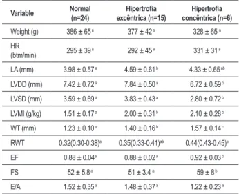
The echocardiogram results, according to the remodeling pattern, are shown in Table 1. We can observe that the left atrium diameter was bigger in the eccentric hypertrophy than in the normal pattern. The normal and eccentric hypertrophy patterns presented higher ventricular cavity values than the concentric hypertrophy group, both in the systole and in the diastole. Concerning the LV mass index, the animals belonging to the hypertrophy group (concentric and eccentric hypertrophy) presented higher values than the normal group. Concerning the LV wall relative thickness, the animals falling under the normal pattern presented lower values than the concentric hypertrophy pattern. Considering the systolic function, the normal and eccentric hypertrophy groups presented smaller ejection fraction values and fractional shortening than the concentric hypertrophy group. There were no differences between the groups related to the diastolic function.
Considering the smoking animals, thirteen (13) animals (13) presented systolic dysfunction detected by the ejection fraction and by fractional shortening. These animals presented only two geometry patterns: 61% presented normal geometry pattern and 39% presented eccentric hypertrophy pattern.
As to the ventricular dysfunction prediction factors, we analyzed the influence of the mass index, the wall relative thickness and the geometry pattern. In the single regression analysis, geometry patterns and mass index could not predict ventricular dysfunction (p<). On the other hand, the increased relative thickness of the wall could predict ventricular dysfunction in the single regression analysis (p<0.001) and in the multiple regression analysis after adjustment to the mass index (p=0,003).
Discussion
Several studies showed that the exposure to cigarette smoke results in cardiac remodeling, in the rat model,
with a drop in the systolic function indexes8-15. Considering
the lack of information on the relevance of remodeling pattern in the model of rats exposed to cigarette smoke, the purpose of this study was to analyze the presence of different remodeling patterns and its relation with ventricular function in this model.
Original Article
Arq Bras Cardiol 2010;94(2): 209-212
Azevedo et al
Remodeling pattern and smoking
Table 1 - Echocardiogram data according to geometry
Variable Normal
(n=24)
Hipertroia
excêntrica (n=15)
Hipertroia
concêntrica (n=6)
Weight (g) 386 ± 65 a 377 ± 42 a 328 ± 65 a
HR
(btm/min) 295 ± 39
a 292 ± 45 a 331 ± 31 a
LA (mm) 3.98 ± 0.57 a 4.59 ± 0.61 b 4.33 ± 0.65 ab
LVDD (mm) 7.42 ± 0.72 a 7.84 ± 0.50 a 6.72 ± 0.59 b
LVSD (mm) 3.59 ± 0.69 a 3.83 ± 0.43 a 2.80 ± 0.72 b
LVMI (g/kg) 1.51 ± 0.17 a 2.00 ± 0.31 b 2.10 ± 0.28 b
WT (mm) 1.23 ± 0.10 a 1.40 ± 0.16 b 1.57 ± 0.14 c
RWT 0.32(0.30-0.38)a 0.35(0.33-0.41)ab 0.44(0.43-0.45)b
EF 0.88 ± 0.04a 0.88 ± 0.02 a 0.92 ± 0.03 b
FS 52 ± 5.8 a 51 ± 3.4 a 59 ± 8 b
E/A 1.52 ± 0.35 a 1.48 ± 0.37 a 1.22 ± 0.23 a
LA - left atrium diameter; LVDD - left ventricle diastolic diameter; LVSD - left ventricle systolic diameter; WT - diastolic thickness of the posterior wall; RTW - left ventricle relative wall thickness; LVMI - left ventricle mass index; FS- fractional shortening; EF – ejection fraction. The data are expressed in mean±standard deviation (for normal distribution) or average with percentiles 25 and 75 (for non normal distribution).The presence of different letters indicates statistically signiicant differences.
The first relevant information of our study was that animals exposed to cigarette smoke presented one of the four geometry patterns described: normal pattern, eccentric hypertrophy, concentric hypertrophy, and concentric remodeling. Nevertheless, we should consider that the mechanisms involved in the geometry pattern variability are not known.
Considering the presence of one of the four different remodeling patterns, an aspect that is worthy of note is that the same phenomenon was observed in clinical studies,
in arterial hypertension models7,17. In the case of arterial
hypertension, although the mechanisms are not completely clear, the different geometry patterns are explained by the different physiopathological mechanisms for hypertension, with different hemodynamic patterns. Hence, the geometry would depend on the vasoconstriction degree, the intensity of activation of neurohumoral factors and the presence of
volume overcharge17. In our study, however, the animals
exposed to cigarette smoke present the same characteristics. Then, our results suggest that in the model of exposure to cigarette smoke, animals with the same characteristics may present different morphological adaptations even if submitted to the same aggression.
Concerning the relevance of the ventricular remodeling patterns, in the Jackson Cohort of the Atherosclerosis Risk in Communities (ARIC) Study, the type of hypertrophy was associated to the type of ventricular dysfunction. Eccentric
hypertrophy was associated to a systolic dysfunction, while concentric hypertrophy was associated to diastolic dysfunction. On the other hand, concentric remodeling
was not associated to ventricular dysfunction18. In the
MESA study, however, patients with concentric remodeling presented systolic dysfunction, assessed by means of magnetic
resonance19. Therefore, the association between the geometry
and the ventricular dysfunction is still controversial. In our study, only the animals with normal geometry patterns and eccentric hypertrophy presented systolic dysfunction. Another aspect to be considered is that different authors showed that the remodeling poattern may have prognostic implications. This way, in patients with hypertension, the concentric hypertrophy pattern was associated with increased risk of
cardiovascular events compared to other geometry patterns20.
Additionally, studies suggest that the concentric remodeling is
associated to an increased risk of cardiovascular events21-26.
On the other hand, other studies found that geometry was
not associated to a worse prognosis27-29, suggesting that the
relevance of the remodeling pattern in patients with high blood pressure remains undetermined. In our study, the geometry patterns were not factors of prediction of systolic ventricular dysfunction. Therefore, in the model of rats exposed to cigarette smoke, the remodeling pattern did not present relevance in relation to cardiac function.
Another aspect to be considered in our study refers to the importance of the LV relative thickness. In clinical trials with patients with high blood pressure, the mass index was more important than the relative thickness of wall as a prognostic
factor29. In patients with acute myocardial infarction, the
geometry pattern, the mass index and the relative thickness
of the wall were independent prediction factors of death30.
In our study, among the geometric variables analyzed, the relative thickness of the wall was the only variable that could predict ventricular dysfunction, emphasizing the importance of such variable in the model of smoking rats.
The conclusion we have drawn is that rats exposed to cigarette smoke presented one of the four different remodeling patterns. Among the geometric variables analyzed, only the increased relative wall thickness of the left ventricle could predict ventricular dysfunction in this model.
Potential Conflict of Interest
No potential conflict of interest relevant to this article was reported.
Sources of Funding
There were no external funding sources for this study.
Study Association
This study is not associated with any post-graduation program.
Original Article
Arq Bras Cardiol 2010;94(2): 209-212
Azevedo et al Remodeling pattern and smoking
References
1. Pfeffer MA, Braunwald E. Ventricular remodeling after myocardial infarction: experimental observations and clinical implications. Circulation. 1990; 81: 1161-72.
2. Pfeffer JM, Pfeffer MA, Braunwald E. Influence of chronic captopril therapy on the infarcted left ventricle of the rat. Circ Res. 1985; 57: 84-95.
3. Cohn JN, Ferrari R, Sharpe N. Cardiac remodeling- concepts and clinical implications: a consensus paper from an international forum on cardiac remodeling. J Am Coll Cardiol. 2000; 35: 569-82.
4. Zornoff LAM, Paiva SAR, Duarte DR, Spadaro J. Ventricular remodeling after myocardial infarction: concepts and clinical implications. Arq Bras Cardiol. 2009; 92: 150-6.
5. Francis GS. Pathophysiology of chronic heart failure. Am J Med. 2001; 110: 37S-46S.
6 Swynghedauw B. Molecular mechanisms of myocardial remodeling. Physiol Rev. 1999; 79: 215-62.
7. Ganau A, Devereux RB, Roman MJ, de Simone G, Pickering TG, Saba PS, et al. Patterns of left ventricular hypertrophy and geometric remodeling in essential hypertension. J Am Coll Cardiol. 1992; 19: 1550-8.
8. Paiva SAR, Zornoff LAM, Okoshi MP, Okoshi K, Cicogna AC, Campana AO. Behavior of cardiac variables in animals exposed to cigarette smoke. Arq Bras Cardiol. 2003; 81: 221-8.
9. Castardeli E, Paiva SAR, Matsubara BB, Matsubara LS, Minicucci MF, Azevedo PS, et al. Chronic cigarette smoke exposure results in cardiac remodeling and impaired ventricular function in rats. Arq Bras Cardiol. 2005; 84: 320-4.
10. Zornoff LAM, Matsubara LS, Matsubara BB, Okoshi MP, Okoshi K, Dal Pai-Silva, et al. Beta-carotene supplementation attenuates cardiac remodelling induced by one-month tobacco-exposure in rats. Tox Sci. 2006; 90: 259-66.
11. Zornoff LAM, Matsubara BB, Matsubara LS, Minicucci MF, Azevedo PS, Campana AO, et al. Cigarette smoke exposure intensifies ventricular remodelling process following myocardial infarction. Arq Bras Cardiol. 2006; 86: 276-82.
12. Castardeli E, Duarte DR, Minicucci MF, Azevedo PS, Matsubara BB, Matsubara LS, et al. Tobacco smoke-induced left ventricular remodelling is not associated with metalloproteinase-2 or -9 activation. Eur J Heart Fail. 2007; 9: 1081-5.
13. Castardeli E, Duarte DR, Minicucci MF, Azevedo PS, Matsubara BB, Matsubara LS, et al. Exposure time and ventricular remodeling induced by tobacco smoke exposure in rats. Med Sci Monit. 2008; 14: BR62-66.
14. Houdi AA, Dowell RT, Diana JN. Cardiovascular responses to cigarette smoke exposure in restrained conscious rats. J Pharmacol Exp Ther. 1995; 275: 646-53.
15. Gu L, Pandey V, Geenen DL, Chowdhury SAK, Piano MR. Cigarette smoke-induced left ventricular remodelling is associated with activation of mitogen-activated protein kinases. Eur J Heart Fail. 2008; 10: 1057-64.
16. Lang RM, Bierig M, Devereaux RB, Flachskampf FA, Foster E, Pellikka PA, et al. Recommendations for chamber quantification: a report from the American Society of Echocardiography’s Guidelines and Standards Committee and the Chamber Quantification Writing Group, developed in conjunction with the
European Association of Echocardiography, a branch of the European Society of Cardiology. J Am Soc Echocardiogr. 2005; 18: 1440-63.
17. Dávila DF, Donis JH, Odreman R, Gonzalez M, Landaeta. Patterns of left ventricular hypertrophy in essential hypertension: should echocardiography guide the pharmacological treatment? Int J Cardiol. 2008; 124: 134-8.
18. Fox E, Taylor J, Taylor H, Han H, Samdarshi T, Arnett D, et al. Left ventricular geometric pattern in the Jackson Cohort of the Atherosclerosis Risk in Communities (ARIC) Study: clinical correlates and influences on systolic and diastolic dysfunction. Am Heart J. 2007; 153: 238-44.
19. Rosen BD, Edvardsen T, Lai S, Castillo E, Pan L, Jerosch-Herold M, et al. Left ventricular concentric remodeling is associated with decreased global and regional systolic function: the Multi-Ethinic Study of Atherosclerosis. Circulation. 2005; 112: 936-8.
20. Koren MJ, Devereux RB, Casale PN, Savage DD, Laragh JH. Relation of left ventricular mass and geometry to morbidity and mortality in uncomplicated essential hypertension. Ann Intern Med. 1991; 114: 345-52.
21. de Simone G, Kitzman DW, Chinali M, Oberman A, Hopkins PN, Rao DC, et al. Left ventricular concentric geometry is associated with impaired relaxation in hypertension: the HyperGEN study. Eur Heart J. 2005; 26: 1039-45.
22. Gardin JM, McClellan R, Kitzman D, Lima JA, Bommer W, Klopfenstein HS, et al. M-mode echocardiographic predictors of six-to-seven year incidence of coronary heart disease, stroke, congestive heart failure, and mortality in an elderly cohort. Am J Cardiol. 2001; 87: 1051-7.
23. Verdecchia P, Schillaci G, Borgioni C, Ciucci A, Battistelli M, Bartoccini C, et al. Adverse prognostic significance of concentric remodeling of the left ventricle in hypertensive patients with normal left ventricular mass. J Am Coll Cardiol. 1995; 25: 871-8.
24. Muiesan ML, Salvetti M, Monteduro C, Bonzi B, Paini A, Viola S, et al. Left ventricular concentric geometry during treatment adversely affects cardiovascular prognosis in hypertensive patients. Hypertension. 2004; 43: 731-8.
25. Pierdomenico SD, Lapenna D, Bucci A, Manente BM, Cuccurullo F, Mezzetti A. Prognostic value of left ventricular concentric remodeling in uncomplicated mild hypertension. Am J Hypertens. 2004; 17: 1035-9.
26. Di Tullio MR, Zwas DR, Sacco RL, Sciacca RR, Homma S. Left ventricular mass and geometry and the risk of ischemic stroke. Stroke. 2003; 34: 2380-4.
27. Verdecchia P, Schillaci G, Borgioni C, Ciucci A, Gattobigio R, Zampi I, et al. Prognostic value of left ventricular mass and geometry in systemic hypertension with left ventricular hypertrophy. Am J Cardiol. 1996; 78: 197-202.
28. Krumholz HM, Larson M, Levy D. Prognosis of left ventricular geometric patterns in the Framingham Heart Study. J Am Coll Cardiol. 1995; 25: 879-84.
29. Ghali JK, Liao Y, Cooper RS. Influence of left ventricular geometric patterns on prognosis in patients with or without coronary artery disease. J Am Coll Cardiol. 1998; 31: 1635-40.
30. Verma A, Meris A, Skali H, Ghali JK, Arnold JM, Bourgoun M, et al. Prognostic implications of left ventricular mass and geometry following myocardial infarction: the VALIANT (VALsartan In Acute myocardial iNfarcTion) Echocardiographic Study. JACC Cardiovasc Imaging. 2008; 1: 582-91.