PROPOSAL FOR AN EVALUATING PROTOCOL
FOR THE ELECTRICAL ACTIVITY OF MASSETER
AND SUPRAHYOID MUSCLES IN NEWBORN
PRETERM INFANTS DURING THE FEEDING
Proposta de um protocolo de avaliação da atividade elétrica
dos músculos masseter e supra-hióideos em recém-nascidos
pré-termo durante a alimentação
Rebeca Domingues Raposo (1), Hilton Justino da Silva (2)
(1) Speech Therapist, Professor of Postgraduate course in
Orofacial Motricity at the Instituto de Medicina Integral Pro-fessor Fernando Figueira – IMIP; PhD in Child and Ado-lescent Health at the Federal University of Pernambuco,
activation of skeletal muscles. Through it can be studied the function of striated muscle, by analyzing the signal captured during rest and/or during muscle contraction, recording the voltage variations produced by the membrane of muscle fibers1.
In recent decades, the surface EMG has been used in studies of various dysfunctions and in evalu-ating methods of infant feeding, especially to verify muscle the activity during lactation and its possible implications on the growth of oral structures and ABSTRACT
Purpose: present a protocol for evaluating the electrical activity of the masseter and supra-hyoid in newborn preterm infants during feeding. Method: the information was gathered from papers catalogued in Lilacs and MEDLINE, and printed literature in order to identify protocols used in national and international studies on electromyographic evaluation in infants during feeding, prioritizing the last fifteen years. Information was also collected from protocols used in research with other populations that could be used as a theoretical basis for this study. From the reading and analyzing the found materials, an initial electromyographic evaluation protocol was drawn up and was applied in six newborn preterm infants, to verify its applicability.Results: the literature search and testing in the population led to a protocol composed by sub-definition of muscles evaluated, recommendations for skin preparation, electrode placement, positioning for evaluation, normalization of the signal for the evaluation activities, and suggestions analysis and interpretation of the signal. Conclusion: the study shows the possibility of applying this protocol in the evaluation of muscle electromyography in preterm infants, during feeding. The masseter and supra-hyoid are recommended as a good option for studying the electrical activity of muscles activated during feeding in preterm infants. The protocol also recommended procedures for standardizing the signal for better interpretation of data.
KEYWORDS: Evaluation; Electromyography; Masseter Muscle; Infant, Premature; Protocols; Infant, Newborn
INTRODUCTION
protruding, elevating and retracting the jaw, the same movements that will be used in mastication in the future. A large part of this muscle has been evaluated during the feeding function: masseter, temporalis, orbicularis, suprahyoid (milohyoid, geniohyoid, stilohyoid and digastric) and buccinator. The masseter proved to be a muscle that actively participates in the function of suction, protrusion by rising and retracting the jaw, as well as the supra-hyoid, which participate in the movement and stabi-lization of the jaw and tongue movements3.
Studies using4-10 electromyography to evaluate the feeding in infants born at term and preterm do not mention the use of a protocol, thus not allowing comparisons between them due to the lack of similarity in the use of methods, muscles evaluated or feeding method2.
Dorland11 defines as protocol “an explicit plan, detailed from experience, procedure, examination or test” (p.1432). The need to use protocols was gradually becoming clear in all areas of knowledge and this was not different in Speech Pathology. Overtime, practitioners realized the importance of using specific protocols so they could get more reliable records. The protocols provide the ability to make relationships between facts; adopt attitudes thought from episodes that recur; allow further determine the best procedure, besides guiding the assessment and ensuring the quality of data collected, the use of protocols also allows their reapplication12.
Thus, the aim of this study was to develop a proposal for a protocol to assess the electrical activity of the masseter and suprahyoid muscles in preterm newborn, which can be performed in the different feeding methods and verified their applicability.
METHOD
This research was submitted to the Ethics Committee in Human Research of the Instituto de Medicina Integral Professor Fernando Figueira – IMIP and approved under the number 2172-11 for being in accordance with the Resolution 196/96 of the National Health Council (CNS). The babies were assessed after reading and had the consent form signed for their parents.
Literature searches were conducted in order to identify the protocols used in national and interna-tional studies on electromyographic evaluation in infants during feeding. The databases and printed literature between 1995 and 2011 were consulted. For such were used the indexers: Evaluation, Electromyography, Masseter Muscle, Premature, Protocol, and Newborn. It also collected information
of protocols used in research with other populations that could be used as theoretical basis for this study.
Articles of interest were identified from conducting a search by title and abstract. Articles whose subject was not relevant to the study were excluded, along with letters to the editor and editorials. In a second step, the selected articles were analyzed with regard to their objectives, method of the study and results obtained.
From then, it was created an initial protocol following the steps described in the studies on electromyographic evaluation in infants (born on preterm or term) and the assumptions of existing protocols applied in children and adults. With the initial proposal, it was applied as a pilot project in six preterm newborn aiming at testing and determining the size and location of the electrodes, the standard-ization procedures of the electromyographic signal and verifying its applicability to final design of the protocol.
In this phase were evaluated preterm newborn from the Kangaroo Unit of the Instituto de Medicina Integral Professor Fernando Figueira – IMIP. Babies participant in this study were those clinically stable (no respiratory support, without the presence of neurological complications – intracranial hemor-rhage grades III and IV and hydrocephalus, absence of serious underlying diseases – congenital heart diseases and genetic syndromes, absence of severe craniofacial malformations – tracheo-esophageal changes), tolerating enteral feeding; suction coordination, swallowing and breathing. They should also be children of healthy mothers who could breastfeed (no nipple problems that hindered the handle and without diseases or use of medications against breastfeeding). At the time of evaluation, the average days of life was 30 days (SD = 18 days), the corrected gestational age was 35.5 weeks (SD = 2 weeks) and weight 1.677g (SD = 196g).
We also used two SDS500 sensors with connection by claws; reference cable (ground) and calibrator were also used.
For data analysis, a survey of each method used in the application of the electromyography technique described in studies was conducted, besides details of each step of application of the technique in infants evaluated, thus comprising the final proposal of evaluation protocol for electrical activity of the masseter and suprahyoid muscles in newborn preterm infants during feeding.
RESULTS
In the searches carried out in the literature were found six articles5-10 on the use of electromyography in the evaluation of infants during feeding (Figure 1). There was a wide variety of muscles evaluated, with different goals. Most articles did not mention the environment, positioning of the subjects and prepa-ration for the exam (skin cleaning and description and placement of electrodes). None of the articles reviewed referred to the use of evaluation protocols, some described in more detail the exam, others only cited the material used. All analyzes were performed without normalization of the EMG signal.
Studies on electromyography in the evaluation of the stomatognathic system revealed that for better definition of protocols used in the musculature of the head and neck and oral function are recommended in the precise definition of muscles and the definition of normalization procedures of electromyographic signal13,14.
From the literature review and the application of the initial proposal of the protocol were defined: – Regarding the electrodes, studies on infants
do not describe which models and sizes of electrodes used. To set the size electrodes were tested cut pediatric electrodes, once neonatal electrodes were not found. These were cut into round and square shapes with sizes of 1.5 centi-meters to prevent the cross-talk (signal capture by neighboring muscles). The cut in square shape showed better adhesion to the skin and therefore it was chosen.
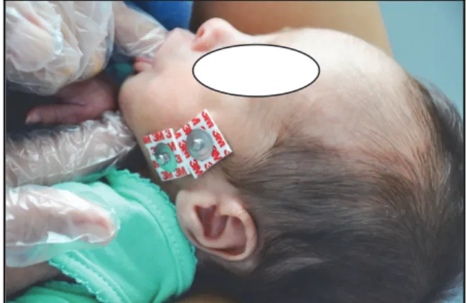
– For electrode placement in the direction of the fibers were stimulated phasic bite reflex (by stimulating the lateral alveolar region) for palpation and visualization of the most robust region of the masseter and suction reflex to palpation of milohyoid muscle and electrode placement of the suprahyoid region (Figures 2, 3, 4 and 5).
– Among the normalization procedures described in the literature, it was verified the possibility of use some procedures described below in the electromyography assessment of this population. Standards from the rest (in which the value found during rest is used as a reference), standardi-zation related to the maximum peak (in which is used in the highest value of electromyographic signal found in the movement or studied cycle) and normalization with respect to the maximum reflex activity (being used as the reference the highest value found in a maximal isometric contraction).
The results of implementing the initial proposal of the protocol are described in Figure 6. The mean muscle electrical activity (AEM) captured during electromyographic evaluation in µV were normalized (transformed into percentage of the reference value) by the different procedures for each subject and for each muscle.
The results (described in figure 7) show that the normalized values from the peak for the masseter muscle range from 0.20% to 38.69%, compared to the peak range between 16.56% and 94.60%, and compared to SEA between 28.61% and 100.74%. Regarding the suprahyoid muscles, the normalized values from the peak vary between 0.65% and 13.16%, relative to the peak range from 65.59% to 94.65% and relative to the MAR between 7.72 % and 102.84%.
AUTHOR GOALS METHOD RESULTS
Inoue et al., 1995
Analyze the masseter activity in infants during bottle-feeding and breastfeeding.
- Studied the masseter for being the main muscle of mastication in 24 term infants, aged two to six months,
- The babies were divided into two groups: breast- and bottle-feeding.
The masseter activity was much lower in bottle-fed babies.
Tamura et al., 1996
Analyze differences in muscle activity to classify the contribution of different muscles
- Studied the temporalis, masseter, orbicularis and suprahyoid muscles unilaterally in 25 term infants, mean age of three months, divided into three groups: breastfed, bottle-fed, breastfed + bottle-fed.
Masseter and temporalis (mandible elevators) are activated in the opening and closing and/or protrusion and retrusion of the mandible. The phase of expression - dominated by the orbicularis, and the suction phase dominated by suprahyoid. Sakashita et al.,
1996
Analyze differences in muscle activity to classify the contribution of the different muscles
- Studied the masseter of 36 term infants, aged between one and five months, divided into three groups: breastfed, bottle with nipple and bottle with nipple and valves.
Regarding the masticatory system development, it is preferable to bottle nipple and valves (mastication-type nipples) when it is necessary to interrupt or abandon the breastfeeding. Tamura et al.,
1998
Investigate the suction development in infants born in term using electromyography during breastfeeding
- Studied the temporalis, masseter, orbicularis and suprahyoid unilaterally. 48 term infants, aged between one and five months, divided into five groups according to age.
There was no significant difference in the activities of the masseter, temporalis and orbicularis between groups. The activity of the suprahyoid musculature was intensified with age.
The active movements of lowering the tongue/ mandible have a key role in increasing the suction force on breastfeeding. Nyqvist et al.,
2001
Describe the oral behavior of preterm newborn infants during breastfeeding and determine the validity of the direct observation of suction during breastfeeding.
- Studied the orbicularis and suprahyoid muscles in the pharyngeal region of 26 healthy preterm infants with a mean gestational age of 32.5 weeks and the mean 18 days of age.
Early suction capacity in preterm infants during breastfeeding, with large individual variations and the surface EMG and direct
observation can be
recommended as valid methods to evaluate the breastfeeding behavior in this population. Gomes et al.,
2006
Measure and compare the activity in different feeding methods.
- Evaluated the masseter, temporalis and buccinators muscles of 60 infants, aged between two and three months, divided into three groups: breastfeeding, breastfeeding and + bottle and breastfeeding + cup during the evaluation
Amplitude and mean contraction in the masseter and contraction amplitude in the temporalis in the AMEs, and in the mean
contraction in the cup. Results in buccinator contraction amplitude n the bottle group.
There are similarities between muscle activity in the AME group and cup suggesting the use of glass as an alternative and temporary feeding method for infants
Figure 2 – Fixing the electrode in the olecranon of the ulna – elbow
Figure 3 – Stimulation of phasic bite for palpation and visualization of the most robust region of the masseter muscle
Figure 4 – Stimulation of non-nutritive suction
for palpation of the mylohyoid muscle
NEONATES (N)
ELECTRODE SIZE
ELECTRODE LOCATION
NORMALIZATION
PROCEDURE APPLICABILITY
6 preterm Pediatric
electrode - cut or neonatal.
Masseter muscle: Phasic bite stimulation for palpation and visualization of the most robust region of the muscle.
Suprahyoid muscles: Stimulation of non-nutritive suction for palpation of the mylohyoid musculature
Regarding Maximum reflex activity:
- Masseter: stimulates phasic bite reflex for maintaining occlusion in contraction. - Suprahyoid: stimulates the suction reflex during
stimulation of non-nutritive suction (NNS).
Compared to the peak - Peak of electromyographic signal found during feeding activity evaluated.
From rest
- A single record and usual position, with lips together without activity.
In all cases there was no difficulty in applying the protocol.
Figure 6 – Table showing the results of applying the initial proposal of the Assessment Protocol of the electrical activity of the masseter and suprahyoid muscles in preterm newborn infants during feeding
RN
ELECTRICAL ACTIVITY OF MUSCLES DURING BREASTFEEDING (%)
MASSETER SUPRAHYOID
From rest Compared to the peak
Compared
to the MAR From rest
Compared to the peak
Compared to the MAR
1 4.30 74.89 74.89 13.16 87.95 7.72
2 11.83 18.94 80.62 2.80 82.92 100.53
3 38.69 16.56 28.61 10.85 94.65 102.84
4 2.29 91.37 92.36 9.19 72.20 95.90
5 2.04 94.26 100.74 9.87 65.59 98.57
6 0.20 92.74 94.60 0.65 81.49 98.01
Figure 7 – Electrical activity of the masseter and suprahyoid muscles in preterm newborn infants, during breastfeeding, normalized in percentage from the rest and compared to the peak and maximum
Musculature evaluated
- The definition of muscles or muscle groups to be analyzed is the first step of the evaluation.
These should be selected according to the purpose of the study and movements to be performed.
Skin preparation - Before placing the electrodes is necessary to perform skin cleansing (removal of grease and
dirt). It is recommended to clean the baby’s skin, the use of cotton or gauze soaked in alcohol 70°.
Placement of Electrodes
- Reference electrode or “ground” is placed at a point distant from the place of registration of the
muscles evaluated, and here agreed the olecranon of the ulna of the baby’s arm.
- The electrode is positioned in a bipolar configuration in the venter of the chosen muscle, arranged longitudinally to the muscle fibers.
- For the location of the region in which the masseter electrode will be fixed, the phasic bite reflex
is stimulated (by stimulating the lateral alveolar region) for palpation and visualization of the most robust region of the masseter that is the midline of the muscle venter.
- The electrode of the suprahyoid region is fixed by palpation of the mylohyoid musculature.
- After attaching the electrodes on the baby’s skin, it is made the placement of sensors with claws, obeying the order of electrode placement.
- The distance between the electrodes is approximately 1 cm.
Positioning for evaluation
- The progenitor and evaluator remain comfortably seated in chairs with back support and no support for head and arms.
- The progenitor should stand with feet flat on the floor to facilitate the baby positioning.
- The baby should be well positioned:
• On breastfeeding: the infant must remain in front of the mother and near (baby’s tummy
facing the mother’s body). The head and spine straight, on the same axis with the mouth
facing the nipple. The motheshould support the baby’s body with her arm and hand, bring the baby’s mouth right in front of the chest, so he can grab most of the areola (the darker area and rounded in the breast) into the mouth.
• In the little cup: the baby should be on alert, remaining sit or semi-sit on the lap. • Translactation: the baby should remain in the same position as breastfeeding.
Signal normalization
Normalization regarding the Maximum reflex activity-MAR
- MAR of the masseter (MARM): stimulates phasic bite reflex for maintaining occlusion in contraction for five seconds. It is considered for the analysis the mean in µV of three seconds intermediate to the test. The stimulation of the reflex is repeated three times 10-second interval
between each contraction.
- MAR of the suprahyoid (MARSH): is used the suction reflex during stimulation of non-nutritive suction (NNS). The gloved finger is inserted into the oral cavity. It is considered for the analysis the mean in µV of the three seconds of suction intermediate to the five seconds in total (the 1st and 5th second are excluded).
Normalization regarding the peak
- Peak of electromyographic signal found during feeding activity evaluated.
Normalization from the rest
- A single record and usual position, with lips together, without performing any activity for five
seconds. It is considered for the analysis the mean in µV of three seconds intermediate to the test.
Activities for evaluation
- Must be performed after the activities for signal normalization. - On the position previously described for each evaluation method.
- Breast-feeding: the baby is placed in the breast to nurse. the baby must mouthful most of the areola (the darker area and rounded breast) . The baby’s chin should touch the breast of the mother. The mouth should be wide open, lips turned outward (everted); areola more visible at the
top to the bottom; cheek round (“full”) and the baby’s tongue should involve the nipple. Let the
baby suck as long as necessary.
- Little cup: touching the edge of the cup on the baby’s lower lip and the little cup should be tilted until the milk touches his lower lip. One should allow the baby to remove milk sucking it, and wait he swallows it. Do not pour the milk into the baby’s mouth.
- Translactation: A 20 ml syringe without plunger is attached to the maternal cervix, coupled with
a gavage number 4, with the end holes placed at the nipple. When putting the baby to breast,
the baby grasps the areola and the probe. The previously expressed breast milk is placed in the
Figure 8 – Protocol for evaluating the electrical activity of the masseter and suprahyoid muscles in preterm newborn infants during feeding
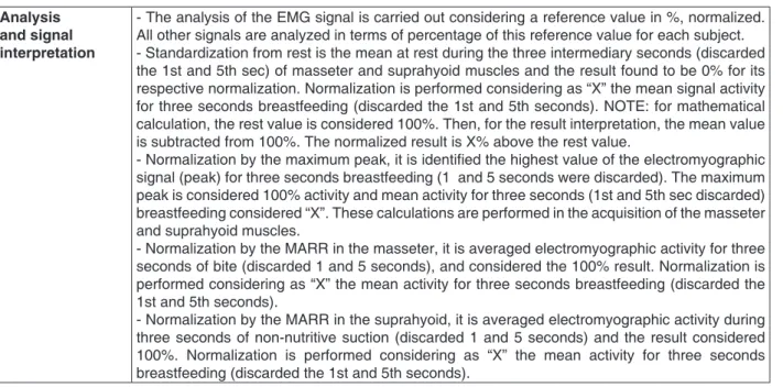
Analysis and signal interpretation
- The analysis of the EMG signal is carried out considering a reference value in %, normalized. All other signals are analyzed in terms of percentage of this reference value for each subject. - Standardization from rest is the mean at rest during the three intermediary seconds (discarded the 1st and 5th sec) of masseter and suprahyoid muscles and the result found to be 0% for its
respective normalization. Normalization is performed considering as “X” the mean signal activity
for three seconds breastfeeding (discarded the 1st and 5th seconds). NOTE: for mathematical calculation, the rest value is considered 100%. Then, for the result interpretation, the mean value is subtracted from 100%. The normalized result is X% above the rest value.
- Normalization by the maximum peak, it is identified the highest value of the electromyographic signal (peak) for three seconds breastfeeding (1 and 5 seconds were discarded). The maximum
peak is considered 100% activity and mean activity for three seconds (1st and 5th sec discarded)
breastfeeding considered “X”. These calculations are performed in the acquisition of the masseter
and suprahyoid muscles.
- Normalization by the MARR in the masseter, it is averaged electromyographic activity for three seconds of bite (discarded 1 and 5 seconds), and considered the 100% result. Normalization is
performed considering as “X” the mean activity for three seconds breastfeeding (discarded the
1st and 5th seconds).
- Normalization by the MARR in the suprahyoid, it is averaged electromyographic activity during three seconds of non-nutritive suction (discarded 1 and 5 seconds) and the result considered
100%. Normalization is performed considering as “X” the mean activity for three seconds
breastfeeding (discarded the 1st and 5th seconds).
COMPUTER DATA SHEET
Newborn___________________________________ Birthdate:___________ IG at birth:___________ Weight at birth:____________
Date of evaluation:___________ Age:____________IGC:______ Feeding:______________
µV Masseter rest
Suprahyoid rest MAR of Masseter MAR of Suprahyoid AEM in feeding Masseter peak Suprahyoid peak
NORMALIZATION (%)
Rest Peak MAR Masseter Supra Masseter Supra Masseter Supra
AEM feeding
DISCUSSION
To evaluate the electrical activity by means of surface EMG are necessary: a system of signal capture (electrodes), record conditioning circuits (amplifiers and filters), log output media (a simple high-speaker – sound output, output on thermal paper, and output on the video monitor screen) notebook with an operational system and software for signal processing. For being captured as analogical signal (continuous in time), the electro-myographic signal needs to be converted into digital signal (defined only for certain time intervals), to get to be recognized by the computer, therefore it is still necessary a converter15.
According to Basmajian and DeLuca16 running electromyography evaluation must be preceded by standardization, in which will be pre-established: muscles evaluated, used equipment, preparation for exam, electrodes placement, patient’s posture and activities performed (sequence of movements). Moraes13 and Pernambuco14 complement affirming the importance of the precise definition of muscles and standardization procedures of the electromyo-graphic signal. The use of a protocol is important for the record of electromyographic activity represents faithfully the electrical signal of the muscle under study.
The first step before the assessment is the identi -fication of muscles or muscle groups to be analyzed. The muscles should be selected according to the purpose of the study and movements to be performed. The studies referenced5-10 evaluated a wide range of muscles activated during feeding function: masseter, temporalis, orbicularis, supra-hyoid (mylosupra-hyoid, geniosupra-hyoid, stylosupra-hyoid and digastric) and buccinator, with different goals.
During milking, four separate movements of the jaw are performed: opening, protrusion, closing and retrusion. Five pairs of mandibular muscles control these movements: depressors of mandible (lateral pterygoid and digastric); protrusors of mandible (masseter and medial pterygoid); elevators (masseter, temporalis and medial pterygoid), and retrusors (masseter and temporalis)17.
In this protocol, the selected muscles to evaluate the suction (mother’s breast milking) in preterm were the masseter and suprahyoid. The masseter for being a muscle that actively participates in the function of suction, protruding, raising and retracting the mandible. There is the advantage of being less deep, allowing easier access, unlike the lateral
After preparation of the equipment and muscle definition, the next step is the electromyographic examination. Several factors can influence the EMG signal: those intrinsic related to anatomical and physiological characteristics of the muscles and those extrinsic, related to the electrode and fixation. Intrinsic factors cannot be modified, but the extrinsic ones so. Therefore, caution is needed in attempting to minimize and standardize the issues that may influence EMG data collection18.
Regarding the electrodes, it must be taken into account: the size, shape, and distance between electrodes (center to center distance between the conductive areas). The size for being directly related to the amplitude of the detected signal, the larger the size, the greater the signal and the lesser electrical noise generated at the interface between the skin and the electrode sensing surface. However, it should be small enough to avoid cross-talk (signal capture of neighboring muscles). The distance between the electrodes affects the frequency and signal amplitude19,20. Thus, this protocol proposed using disposable and cut pediatric surface electrodes with approximate size of 1.5 cm.
The Surface EMG members for the non-invasive assessment of muscles (SENIAM) recommend that some care must be taken to decrease the influence of skin impedance19,21, the skin preparation. Before placing the electrodes is necessary to perform the skin cleansing (removal of grease and dirt), and/ or remove the hair (shaved) and mild abrasion for removal of dead skin cells19,21. In the protocol proposed here, it is recommended the use of cotton or gauze soaked in alcohol 70 for cleaning the baby’s skin, without however, rubbing the baby’s sensitive skin.
masseter. The electrode of the suprahyoid region is determined by palpation of the mylohyoid muscle.
The placement of electrodes must meet standardization, starting with the reference electrode or “ground”, which is used to minimize interferences from external electrical noise. The same is placed at a point distant from the record place of the muscles evaluated22 being here agreed the olecranon of the ulna (elbow). Then, the others electrodes are fixed. In some situations, it is necessary to use tape for better fixation of the electrodes. It is important that it is hypoallergenic to reduce the risk of irritation on the baby’s skin.
The placement of sensors with claws obeys the same order of electrodes placement. Completed this process, the configuration is checked and the activation of channels in the software is performed following a predetermined arrangement that in the current protocol is: Channel 1 – masseter muscle and Channel 2 – suprahyoid muscles. Unused channels should be properly disabled.
Once finalized all the procedures, it is initiated the evaluation of electrical activity of muscles during feeding. The electrical muscle activity (MSA), defined as the mean of the action potentials of the motor units of a muscle group, obtained from the EMG signal expressed in microvolts (uV)19 is captured during feeding, being considered for analysis the mean in µV of the time with greater signal stabilization. Once captured, the EMG signal is amplified, filtered and converted from analogical to digital18.
In order the electromyographic signal can be analyzed and compared in different individuals, muscles and studies, and over time, it is necessary to use normalization techniques in which the µV values of the activity are expressed as a percentage of muscle activity during muscle contraction, obtained under standardized and reproducible conditions. Standardization is a prerequisite for any comparative analysis of the EMG signals18,23.
The analysis of the EMG signal is carried out considering a reference value in percentage (%), normalized. All other signals are analyzed in terms of percentage of this reference value for each subject. The normalization of the EMG signal follows the recommendations of the International Society of Electrophysiology and Kinesiology (Isek)24. In the literature are found some ways to make the normal-ization of the electromyographic signal13, 23. One is the standardization regarding the maximum peak of the signal that uses the highest value electro-myographic signal found in the movement or cycle studied23.
Another way of performing the normalization is through the mean value of the electromyographic
signal as reference, where the mean value of the contraction signal is used. A fixed value of the signal can also be used to perform the normalization, where the benchmark is a submaximal contraction or submaximal isometric contraction24.
Normalization can also be performed by Maximum Resisted Voluntary Activity (MAVR), the highest value found in a maximal isometric contraction for the muscle in question, or even by the mean signal obtained during a dynamic activity13,23,24. The rest was used in some research as normalization procedure; however, there are no reports in the literature about studies that have used it as reference.
The current protocol proposes three normal-ization procedures: the maximum peak, the maximum resisted voluntary activity (MAVR) with some adaptations, and rest. At the maximum peak, the highest value of the electromyographic signal in µV found during the feeding period it is used as a reference for normalization.
The MAVR concept is proposed as a normal-ization procedure; however certain changes are necessary since the population studied did not perform any voluntary activity, just reflex ones. Thus, voluntary activities are replaced by reflex activities, becoming Maximum Reflex Activity (SEA) of the muscles, which must be elicited the reflex of the muscle activity or muscle group being evaluated.
In this protocol, in the MAR of masseter is used the reflex of phasic bite (through stimulation of the gingival lateral region and in response, the baby has a bite) to maintain the occlusion in contraction. For MAR of suprahyoid muscles is used suction reflex (through stimulation of non-nutritive suction with a gloved finger). The intermediate means of the test in microvolts are considered in the analysis, moment when there is greater signal stabilization. The reflex stimulation is repeated at intervals between each contraction14,22.
At rest (Rp), the protocol proposes using a single record in the usual position as a benchmark for the normalization, with lips together, without conducting any activity during the signal capture. It is also considered for analysis the intermediate mean test of the in µV.
The results of electromyographic evaluation of six babies evaluated in this study show a wide range of numerical values of the signals when normalized by the different procedures suggested by the protocol. There are no references in the literature on standardization in the electromiographic signal in studies with this population. The studies analyzed the signal in µV. This indicates the need for further study to understand the cause or causes of this variation and if there influence of the characteristics of the studied population or if there is a more appro-priate procedure for normalization of the EMG signal in this population.
CONCLUSION
The study shows the possibility of applying the proposed protocol in the electromyographic
evaluation of these muscles in preterm newborn infants during feeding. The masseter and suprahyoid muscles are recommended as a good choice for studying the electrical activity of muscles activated during feeding in preterm infants.
The proposed standardization in this protocol attempts to assist the evaluation of electromyo-graphic recordings in preterm newborn, providing studies on this topic, similar methods, thus allowing the comparison between the results achieved. At the same time, it enables the collected data may be further analyzed and from such analyses, more efficient conducts can be imposed. The second phase of this work will include experimental research and statistical analysis.
The protocol also recommends procedures for signal normalization for better data interpretation.
RESUMO
Objetivo: apresentar uma proposta de protocolo de avaliação da atividade elétrica dos músculos masseter e supra-hióideos em recém-nascidos pré-termo durante a alimentação. Método: inicial-mente foi realizada uma revisão da literatura nas bases de dados Lilacs e MEDLINE e literatura
impressa com o objetivo de identificar protocolos utilizados em estudos nacionais e internacionais sobre a avaliação eletromiográfica em bebês durante a alimentação, priorizando os últimos quinze anos. Foram coletadas informações dos protocolos utilizados em pesquisas com outras populações que pudessem ser usados como base teórica para este estudo. A partir da leitura e análise do mate-rial encontrado, foi elaborado um protocolo inicial de avaliação eletromiográfica e este foi aplicado em seis recém-nascidos pré-termo, para verificação da sua viabilidade. Resultados: a busca na lite-ratura e testagem na população resultaram em um protocolo composto por subitens com definição de musculatura avaliada, recomendações de preparação da pele, colocação dos eletrodos, posicio-namento para a avaliação, normalização do sinal, atividades para a avaliação, além de sugestões de análise e interpretação do sinal. Conclusão: o estudomostra a possibilidade de aplicação deste protocolo da eletromiografia na avaliação destes músculos em recém-nascidos pré-termo durante a alimentação. Os músculos masseter e supra-hióideos são recomendados como uma boa opção para o estudo da atividade elétrica de músculos ativados durante a alimentação em pré-termos. O protocolo ainda recomenda procedimentos de normalização do sinal para melhor interpretação dos dados.
DESCRITORES: Avaliação; Eletromiografia; Músculo Masseter; Prematuro; Protocolos; Recém-Nascido
REFERENCES
1. Fialho RA, Anzorandia CS, Herrera EM. Dessarollo histórico y fundamentos teóricos de la
term and preterm infants: a literature rewiew. Dev Med Child Neurol. 2009;51:936-42.
5. Inoue N, Sakashita R, Kamegai T. Reduction of masseter muscle activity in bottle-fed babies. Early Hum Dev. 1995;18:185-93.
6. Tamura Y, Horikawa Y, Yoshida S. Co-ordination of tongue movements and peri-oral muscle activities during nutritive sucking. Dev Med Child Neurol. 1996;38:503-10.
7. Sakashita R, Kamegai T, Inou N. Masseter muscle activity in bottle feeding with the chewing type bottle teat: evidence from electromyographs. Early Hum Dev. 1996;45:83-92.
8. Tamura Y, Matsushita S, Shinoda K, Yoshida S. Development of perioral muscle activity during suckling in infants: a crosssectional and follow-up study. Dev Med Child Neurol. 1998;40:344-8. 9. Nyqvist KH, Farnstrand C, Edebol Eeg-Olofsson K, Ewald U. Early oral behaviour in preterm infants during breastfeeding: an EMG study. Acta Paediatr. 2001;90:658-63.
10. Gomes CF, Trezza EMC, Murade ECM, Padovani CR. Surface electromyography of facial muscles during natural and artificial feeding of infants. J Peditar. 2006;82:103-9.
11. Dicionário Médico Ilustrado Dorland. 28ª ed. São Paulo: Manole; 1999; p. 1432.
12. Genaro KF, Berretin-Felix G, Rehder MIB, Marchesan IQ. Avaliação miofuncional orofacial: protocolo MBGR. Rev CEFAC. 2009;11(2):237-55. 13. Moraes KJR, Cunha RA, Lins OG, Cunha DA, Silva HJ. Eletromiografia de Superfície: padronização da técnica. Neurobiologia. 2010;73(3):151-8. 14. Pernambuco LA, Silva HJ, Nascimento GKBO, Silva EGF, Balata PMM, Santos VS et al. Electrical activity of the masseter during swallowing after total laryngectomy. Braz J Otorhinolaryngol. 2011;77(5):645-50.
15. Button VLSN. Eletromiógrafo. Depto. Engenharia Biomédica, FEEC/UNICAMP, 2002. 24p.
16. Basmajian JV, DeLuca CJ. Muscle alive: their functions revealed by electromyography. Baltimore: Williams & Wilkins; 1985.
17. Casagrande L, Ferreira FV, Hahn D, Unfer DT, Praetzel JR. Aleitamento natural e artificial e o desenvolvimento do sistema estomatognático. Rev Fac Odontol Porto Alegre. 2008;49(2):11-7.
18. DeLuca CJ. The use of electromyography in biomechanics. J Appl Biomech. 1997;13:135-63. 19. Hermens HJ, Freriks B, Disselhorst-Klug C, Rau G. Development of recommendations for SEMG sensors and sensor placement procedures. J Electromyogr Kinesiol. 2000;10:361-74.
20. Neuromuscular research Center. Boston University. [acesso em: 02 jan. 2011] Disponível em: www.delsys.com/library/papers
21. Konrad P. The ABC of EMG. A Practical Introduction to Kinesiological Electromyography. Versão 1.0, 2005 [acesso em: 12 nov. 2010]. Disponível em: www.noraxon.com/emg/php3 22. Cunha DA, Silva HJ, Nascimento GKB, Silva EGF, Cunha RA, Régis RMFL et al. Analysis of the masticatory processo of asthmatic children: clinical and electromyografic research. Int Arch. Otorhinolaryngol. 2012;16(3):358-64.
23. Ball N, Scurr J. An assessment of the reliability and standardisation of test, used to elicit reference muscular actions for electromyographical normalization. J Electromyogr Kinesiol. 2010;20:81-8.
24. Moraes KJR, Cunha DA, Bezerra LA, Cunha RA, Silva HJ. Surface electromyography: proposal of a protocol for cervical muscles. Rev CEFAC. ahead of print, pp. 0-0. Epub 05-Dez-2011.
Received on: May 14, 2012 Accepted on: April 07, 2013
Mailing address:
Rebeca Domingues Raposo
Rua Jornalista Guerra de Holanda, 161/102 – Monteiro – Recife-PE
CEP: 52061-010