Arq Neuropsiquiatr 2009;67(2-A):308-310
308
Clinical / Scientiic note
Subdural Haematoma in a
patient witH meningioma
Paulo Valdeci Worm
1, Marcelo Paglioli Ferreira
2, Nelson Pires Ferreira
3, Fernanda Cechetti
4Hematoma Subdural em paCiente Com meningioma
Hospital São José, Santa Casa de Misericórdia de Porto Alegre, Porto Alegre RS, Brazil: 1MD; 2MD; 3MD, PhD, Professor of Neurological Surgery,
Depart-ment of Neurosurgery, Hospital São José - Santa Casa de Misericórdia de Porto Alegre, Porto Alegre RS, Brazil; 4MD, Programa de Pós Graduação em
Ciências Biológicas-Neurociências, Universidade Federal do Rio Grande do Sul, RS, Brazil. Received 17 September 2008, received in inal form 19 December 2008. Accepted 13 March 2009.
Dr. Paulo Valdeci Worm – Rua Carlos Trein Filho 550 / 403 - 90450-120 Porto Alegre RS - Brasil. E-mail: [email protected]
Non-traumatic subdural haematomas are very rare es-pecially those associated with intracranial meningiomas1.
Intracranial tumors hemorrhage occurs in 39% of all cases. They are a malignant pathology such as metastatic tumors or gliomas. They usually present with seizures hemiparesis visual ield loss aphasia or other focal sumptoms.
We report a case of meningioma of the skull base associated with subdural haematoma after a Valsava maneuver.
CaSe
A 64-year old male patient, smoker, had a loss of conscious-ness with mild headache during a spirometric exam. He was med-icated with analgesics and remained with headache for 12 hours and was discharged from hospital. He persisted with chronic headache and developed a gradual confusional state and left
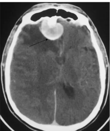
hemiparesia. After the initial episode, ive days after the spiro-metric test, he returned to the hospital for a computed tomog-raphy (CT) of the head (Figs 1 and 2). The CT showed a right sub-dural haematoma with a fresh clot in the anterior skull base with bone thickness of the anterior fossa besides meningioma of the falx with adjacent edema.
The patient had no prior history of trauma or alcoholism. He was using (AAS) 200 mg/day. Coagulation tests were normal.
A right frontal osteoplastic craniotomy was performed draining the haematoma, and during the same surgical proce-dure the tumor was removed using microscopy.
A dark, thick, red blood clot was drained. During the surgi-cal drainage of the subdural haematoma two indings were ob-served by the surgeon: 1) the homolateral side of the subdural haematoma presented with greater vascular density. 2) the clot surface on this side showed a variable aspect, suggesting
ac-Fig 1. CT of skull showing meningioma of falx with adjacent ede-ma and subdural collection in homolateral side. CT shows the brain shift and edema.
Arq Neuropsiquiatr 2009;67(2-A)
309
Subdural haematoma with meningioma Worm et al.
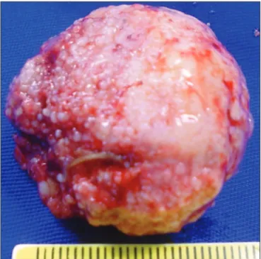
tive bleeding (Fig 3). An en bloc surgical excision of the clot and the tumor were performed. The histopathological diagnosis was meningeoma. Right after drainage his motor state improved but he remained confused. He remained in hospital for seven days. When he was discharged from hospital he was partially orient-ed and had no focal neurological deicit.
diSCuSSion
Benign intracranial tumors like meningiomas rarely present with hemorrhage and the reported incidence is 1.3% of all meningiomas2. The higher incidence of this
as-sociation was found by Niiro et al.2 who described six
cas-es of hemorrhage in a sericas-es of 298 patients with intrac-ranial meningiomas (2%).
The physiopatological mechanisms of this association are not yet well understood, but several hypotheses have been proposed to explain the formation of a spontane-ous hemorrhage in patients with intracranial meningioma: 1) distension of the feeding vessels and weakening of the thin walls with tumor growth and consecutive rupture; 2) stretching of subdural bridging veins caused by tumor ex-pansion; 3) rupture of abnormal tumor vessels; 4) direct invasion of the tumor and disruption of blood vessels; 5) formation of neo-membranes, like chronic subdural hae-matoma caused by a dural reaction to the tumor; 6) vaso-active substances released by the meningioma.
Another hypothesis proposed by Gruskiewicz et al. is that rapid tumor growth and venous thrombosis leading
to tumor necrosis have been implicated. Tumor necrosis can cause direct breakdown of the tumor vessels and sub-sequent hemorrhage; tortuous feeding arteries that are less resistant to blood pressure changes and susceptible to rupture are implicated3.
However, none of the proposed mechanisms alone can explain the phenomenon. The combination of sever-al physiopathogenic factors should be seen as the most common event.
Other factors, such as tumor histological subtype, lo-cation, age and sex of the patient do not show signiicant relationship with the hemorrhagic event2. Subdural
hae-matoma is not related to the presence of systemic arte-rial hypertension or tumor location. Some authors have emphasized a high risk of hemorrage in angioblastic and malignant meningiomas1.
It is conceivable that the localization of the tumor in the cerebral convexity increases the risk of hemorrhage. Chaskis et al.4 described 20 cases of hemorrhagic
menin-giomas. Eleven were situated in the convexity. Other au-thors have hypothesized that the development of a men-ingioma could be secondary to chronic irritation of the meninges exerted by chronic subdural haematoma5.
Intracranial hemorrhage has less associations with be-nign tumors than with malignant ones5. Malignant
meta-static melanoma, choriocarcinoma and bronchogenic car-cinoma are associated with intracranial hemorrhage and are widely discussed in the literature. Chronic subdural haematoma is rarely associated with meningioma.
Itoyama et al.6 described a patient with bilateral
sub-dural haematoma with a left sphenoid wing meningioma. Sinha and Dharker7 presented a case of a meningioma of
the right occipital convexity associated with a left fron-toparietal subdural haematoma. Pozzi et al.8 reported a
right hemisphere convexity subdural haematoma associ-ated left parietal meningioma. This contralateral injury is highly controversial. It could be due to small traumas caused by convulsions in clinical seizures of meningiomas.
Tanaka et al.9 correlated the localization and the
histo-logical subtype of meningiomas and a higher probability of the association with chronic subdural haematoma.
Some CT findings can help correlate a meningioma with subdural haematoma, such as: 1) bone alteration 2) focal cerebral distortion and cerebral perifocal ede-ma due to the presence of meningioede-ma inside the sub-dural clot collection 3) potential differences in the sig-nal density betwen the different areas measured for the Hounsield scale.
In the case reported we found bone alterations at the meningioma implantation site, evidence of a rich and vas-cular density in the homolateral side far from the haema-toma; moreover, we found more macrocospic character-istic changes on this side.
Arq Neuropsiquiatr 2009;67(2-A)
310
Subdural haematoma with meningioma Worm et al.
These indings call attention to the fact that the pos-sible etiology of the hemorrhage had its genesis in a Val-salva maneuver that ruptured during the manipulation of forced expiration triggered for the spirometric examina-tion. The patient probably already had the intracranial haematoma, and the use of AAS may have been one of the facilitating factors. This has not yet been described in the literature. The mechanisms of haematoma formation associated with the Valsalva maneuver are poorly under-stood but Shojima et al.10 mentioned that events such as
seizures or a rapid change in blood pressure may also act as etiologic agents for hemorrhage. In the CT the homo-lateral area near the haematoma was more clearly shown by the contrast beyond the presence of surrounding ce-rebral edema (Fig 3).
Although several hypotheses have been described to explain the bleeding in meningiomas, so far this event has been rare. We do not have a satisfactory explanation for this phenomenon. Besides observations of the histolog-ical properties and radiologhistolog-ical change, possibly macro-scopic observation of the tumor and the adjacent vascu-lature during surgery could, in future, help us reveal the histopathogenesis of the phenomenon.
referenCeS
1. Helle TL, Conley FK. Haemorrhage associated with meningio-ma: a case report and review of the literature. J Neurol Neuro-surg Psychiatry 1980;43:725-729.
2. Niiro M, Ishimaru K, Hirano H, et al. Clinico-pathological study of meningiomas with haemorrhagic onset. Acta Neu-rochir (Wien ) 2003;145:767-772.
3. Bloomgarden GM, Byrne TN, Spencer DD, et al. Meningioma associated with aneurysm and subarachnoid hemorrhage: case report and review of the literature. Neurosurgery 1987;20:24-26. 4. Chaskis C, Raftopoulos C, Noterman J, et al. Meningioma as-sociated with subdural haematoma: report of two cases and re-view of the literature. Clin Neurol Neurosurg 1992;94:269-274. 5. Baskinis N, Grotenhuis A, Wandt H. Chronic subdural hemato-ma associated with an intracapsular meningiohemato-ma: case report and short review of the literature. J Neurosurg Sci 1984;28:17-23. 6. Itoyama Y, Fukumura A, Itoh Y, et al. [Primary brain tumor com-plicating subdural hematoma]. No To Shinkei 1987;39:1157-1161. 7. Sinha VD, Dharker SR. Meningioma associated with contral-ateral chronic subdural haematoma: a short report. Neurol In-dia 2001;49:204-206.
8. Pozzi M, Dario A, Marra A, et al. Associated chronic subdural haematoma and meningeal neoplasm: two case reports. J Neu-rosurg Sci 1993;37:113-117.
9. Tanaka N, Yamamoto M, Jimbo M, et al. Meningioma associat-ed with chronic subdural hematoma and meningothelial cell cluster within the hematoma capsule: case report. Neurol Med Chir (Tokyo) 1994;34:176-179.