Instituto de Cardiologia do Rio Grande do Sul/Fundação Universitária de Car-diologia e Instituto de Geriatria da Pontifícia Universidade Católica do RS. Wake Forest University School of Medicine, Lipoprotein Laboratory. Winston Salem, North - Caroline, USA.
Mailing address: Vera Lúcia Portal – IC/FUC -Serviço de Epidemiologia - Av. Princesa Isabel, 395 90.620001 Porto Alegre, RS Brasil
-E-mail: [email protected]
Objective - To study the differences between fluvas-tatin and pravasfluvas-tatin regarding LDL susceptibility to oxi-dation, plasma levels of total cholesterol (TC), HDL–C, LDL–C and triglycerides (TG) in hypercholesterolemic patients with established coronary heart disease (CHD).
Methods - A double-blind randomized parallel study was conducted that included 41 hypercholesterolemic outpatients with CHD treated at the Instituto de Cardiolo-gia do Rio Grande do Sul. The inclusion criteria were LDL–C above 100 mg/dL and triglycerides below 400 mg/ dL based on 2 measures. After 4 weeks on a low cholesterol diet, those patients that fullfilled the inclusion criteria were randomized into 2 groups: the fluvastatin group (fluvastatin 40 mg/day) and the pravastatin group (pra-vastatin 20 mg/day), for 24 weeks of treatment. LDL susceptibility to oxidation was analyzed with copper-induced production of conjugated dienes (Cu2+) and
wa-ter-soluble free radical initiator azo-bis (2’-2’amidino-propanil) HCl (AAPH). Spectroscopy nuclear magnetic resonance was used for determination of lipids.
Results - After 24 weeks of drug therapy, fluvastatin and pravastatin significantly reduced LDL susceptibility to oxidation as demonstrated by the reduced rate of oxida-tion (azo and Cu) and by prolonged azo-induced lag time (azo lag). The TC, LDL-C, and TG reduced significantly and HDL-C increased significantly. No differences bet-ween the drugs were observed.
Conclusion - In hypercholesterolemic patients with CHD, both fluvastatin and pravastatin reduced LDL susce-ptibility to oxidation.
Key words: LDL oxidation, statins, hypercholesterolemia
Arq Bras Cardiol, volume 80 (nº 2), 156-61, 2003
Vera Lúcia Portal, Emílio H. Moriguchi, José Luiz da Costa Vieira, Sadi Schio, Eduardo T. Mastalir, Fabiana Buffé, Eleni Borges Bortolini, Ricardo Santalucia Brüch, Rubem Rodrigues
Porto Alegre, RS - Brazil
Comparison of the Effect of Two HMG CoA Reductase
Inhibitors on LDL Susceptibility to Oxidation
Atherosclerosis is a progressive and multifactorial in-flammatory process involving a series of highly specific cel-lular and molecular responses that may lead to clinical coro-nary events, such as acute myocardial infarction, unstable angina, and sudden cardiac death 1,2. A causal relationship
between high cholesterol levels, especially high low-density lipoprotein cholesterol (LDL-C) levels, and coro-nary heart disease (CHD) is well established 3-7. During the
last years, a developing set of evidences demonstrated that low-density lipoproteins (LDL) particle oxidation has an im-portant role in the pathogenesis of atherosclerosis 1,8-12 .
Ex-perimental in vitro and in vivo studies show that an isolated increase in LDL-C levels by itself would not totally explain all processes associated with atherogenesis 13-17. Oxidative
modification undergone by LDL particles in the vessel inti-ma would prepare the particle for uptake by inti-macrophages, leading to the formation of foam cells 18-20, the first step in the
formation of early lesions in the pathogenesis of atheros-clerosis 21,22.
It is believed that part of the beneficial effect obtained through the use of statins in the reduction of cardiovas-cular events 3-7, in addition to LDL-C reduction effects,
could be due to its antioxidant action adding an antiathero-genic effect 23-39.
For that reason, we conducted a randomized study to evaluate the antioxidant action of 2 statins, fluvastatin and pravastatin, used in clinical practice as lipid-lowering drugs to test the hypothesis that these drugs reduce LDL suscep-tibility to oxidation.
Methods
The study was undertaken between July 1998 and June 1999 at the Instituto de Cardiologia do Rio Grande do Sul. The protocol was approved by the institutional review board. All patients gave written informed consent.
po-pulation, 45 elegible consecutive patients were included in the study according to inclusion and exclusion criteria des-cribed below.
According to the established inclusion criteria, only patients fulfilling the following requirements could be inclu-ded in the study: LDL-cholesterol (LDL-C) above 100 mg/dL (more than 2 measurements); triglycerides below 400 mg/dL; diabetic patients needed to be well-controlled with stable plasma glucose levels, eg, a fasting glucose level below 110 mg/dL during at least 3 consecutive months.
All 45 patients received orientation to proceed with the American Heart Association Step II (NCEP - ATP II) diet to be followed throughout the study. After the first 4 weeks on the diet, the 41 patients who still had LDL-C above 100 mg/dL were randomized into 2 groups in a double-blind mode: 1 group received 40 mg/day of fluvastatin (fluvasta-tin group) and the other 20 mg/day of pravasta(fluvasta-tin (pravasta-tin group). The follow-up period was 24 weeks, with patient examination at intervals of 4 weeks. If after a 4-week period from the start of drug treatment the LDL-C was still above 100 mg/dL, the drug dosage was doubled at the next visit. At each follow-up visit, study staff provided counseling regar-ding adherence to the study regimen and using a standard questionnaire asked about the occurrence of any relevant events and side-effects after the previous visit.
Exclusion criteria were secondary hyperlipidemia; a history of acute myocardial infarction or stroke occurring within the previous 3 months; severe cardiac failure (NYHA class III or IV); acute atrial fibrillation; alcohol dependance; hepatic disease (transaminases or bilirubins levels more than 2 times the normal level); renal disease (creatinine > 1.5 mg/dL); chronic pancreatitis; systemic lupus erythema-tosus; porphyria; severe gastrointestinal disease; morbid obesity (>140% ideal body weight); use of drugs, such as hormones other than postmenopausal hormone reposition, immunosuppressants, statins, nicotinic acid, resins, or both, in the last 8 weeks and probucol, fibrates, or both, in the last 12 weeks.
Arterial blood pressure was measured with patients in the sitting position after at least 5 minutes of rest, with a pre-viously calibrated Tycos anaeroid sphygmomanometer. Patients with hypertension were considered those taking antihypertensive medication or having mean pressure levels above 140/90 mmHg in at least 2 pressure monitorings.
Patients considered diabetic were those who identi-fied themselves as such, were using hypoglycemic medica-tion, or had fasting serum glucose concentrations above 126 mg/dL in 2 readings.
Body mass index (BMI) was calculated with the ratio body weight (in kilograms)/square height (in meters). Pa-tients were classified as current smokers, past smokers, and nonsmokers. In the statistical analysis of baseline characte-ristics, the only percentage considered was that of current smokers. Patients were asked about alcohol abuse. Seden-tary patients were those who practiced some sort of physi-cal activity less than 3 times a week and for less than 30 minu-tes per session 41.
Blood samples were collected at 4-weeks intervals, after the patient fasted overnight for 12 hours, and were immediately centrifuged for 15 minutes at 1,600 G for plasma collection. Levels of glucose, alanine aminotransferase (ALT), urea (BUN), creatinine and total proteins, and thyro-tropin hormone were measured, and prothrombin time esti-mation, activated partial thromboplastin time (APTT), fibri-nogen, and blood sample analysis and blood platelet count were determined immediately after blood was collected at the laboratory of the Instituto de Cardiologia do Rio Grande do Sul, in Porto Alegre. Glucose, ALT, creatine kinase (CK), urea, creatinine, and total proteins were measured with commercially available kits (Merck Diagnostics). Thyrotro-pin hormone was evaluated by polarized fluorescence with Opus of Dade and Boehringer Equipment. Blood sample and platelets were analyzed with an automatic counter, model 818 by AVL. Fibrinogen, prothrombin time, and APTT were evaluated on Dade Boehringer’s Fibrintimer II. The remaining plasma was placed in a polypropylene vial, which was placed under nitrogen flow before being closed with a silicone screw cap to avoid plasma contact with oxygen, to determine LDL susceptibility to oxidation. Samples were stored in boxes containing dry ice (between 0° and 4°C) and sent to the Lipoproteins Laboratory of Wake Forest University School of Medicine, Winston-Salem, North Carolina, United States less than 24 hours after collection. Additional plasma samples were frozen at -75°C for later lipid analysis at the same laboratory.
Lipid determination was accomplished with a Bruker WM 250 spectrometer, with a recently validated spectros-copy method that uses proton magnetic nuclear resonance (MNR), which has become a more precise, rapid, and less costly alternative in relation to previously existing methods for the dosage of lipoprotein subfractions 42-44. The basis of
analysis with this method rests on the fact that each lipo-protein particle, within a diameter band, “irradiates” a distinct MNR sign that is proportional to the total lipid mass concentration.
LDL susceptibility to oxidation was evaluated with 2 methods. One was by the formation of conjugated dienes induced by copper (Cu+2). In short, the heparinized fresh
depending on the LDL particle concentration and on the amount of polyunsaturated fatty acids (PUFA) carried by LDL. A longer lag phase demonstrates a reduced suscepti-bility of LDL particles to oxidation, caused by a higher an-tioxidant concentration, whereas a shorter lag phase means that LDL particles take less time to oxidize, due to the lower presence of antioxidants 45-47.
The other method of LDL oxidation evaluation was by particle incubation with 5 mM of AAPH (2’-2’ amidinopro-panil hydrochloride- azo), for 4 h, at 37°C. AAPH is a free radical generator system that decomposes thermically and generates water-soluble peroxyl radicals at a constant rate. Plasma samples were analyzed as for their oxidation condi-tions, at the end of the incubation period, by using thiobar-bituric acid reactive substances (TBARS) to which equiva-lent amounts of malondialdehyde were measured. Lipidic peroxidation was calculated subtracting the obtained va-lues in the absence of AAPH from those obtained in the pre-sence of AAPH. Results were also presented in the form of phase lag and rate 45-47.
Values of p<0.05 in the 2-tailed test were considered significant. Baseline differences between the groups in rela-tion to demographic and biochemical variables were analy-zed with the Student t test for independent samples, in the case of continuous variables, and by the chi-square test for categoric variables. Variations throughout the period of treatment of lipid levels, ie, TC, LDL-C, HDL-C, and TG, as well as measure evaluations of LDL susceptibility to oxida-tion, ie, Cu rate, Azo rate, Cu lag, and Azo lag, were evalua-ted within each group and between both groups by the fac-torial analysis of variance (ANOVA), with the Tukey-Kramer localization test being used, when necessary.
Results
The study was concluded with 39 patients. Approxi-mately 60% of them needed a dosage of 40 mg/day of pra-vastatin or 80 mg/day of flupra-vastatin to reach the target level of the study of LDL < 100 mg/dL. Of the 41 randomized patients, one was excluded because of adverse gastrointes-tinal effects (fluvastatin group), and the other patient withdrew for no specific reason (pravastatin group). Adherence to treatment measured by counting the remai-ning tablets was 97% in the fluvastatin group and 96% in the pravastatin group.
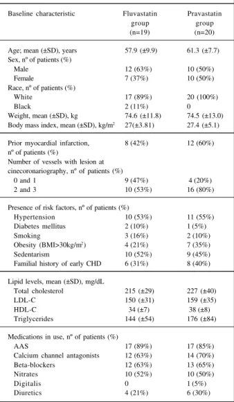
The baseline characteristics of the 2 randomized groups (fluvastatin and pravastatin) are summarized in table I. No significant differences occurred between the groups in relation to demographic characteristics, previous myocardial infarction history, number of coronary vessels with lesions, the presence of coronary risk factors, lipid levels, and use of medications (tab. I).
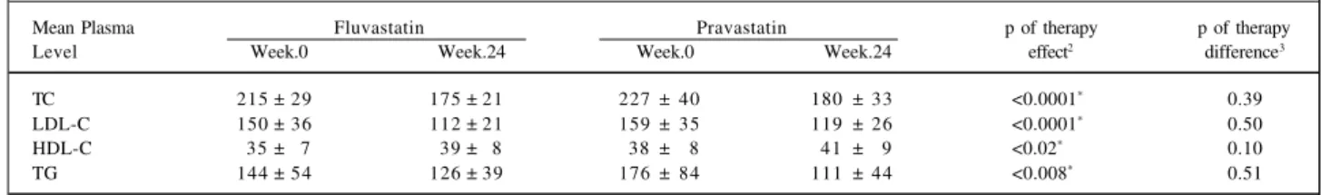
Results of mean plasma levels of TC, LDL-C, HDL-C, and TG at baseline and the 24th week are presented in table II. A significant decrease was seen in TC, LDL-C, and TG levels and a significant increase in HDL-C by the end of treatment period. The fluvastatin group had 18% lower TC,
25% lower LDL-C, and 13% lower triglycerides plasma levels and an 11% increase in HDL-C levels, whereas the pravastatin group had reductions of 21% in TC, 25% in LDL-C, and 37% in triglycerides levels, and a 10% increase in HDL-C (tab. II).
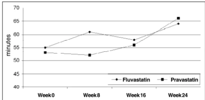
The results of LDL susceptibility to oxidation throu-ghout the study are presented in table III and figures 1 through 4. The LDL susceptibility to oxidation decreased after the use of either drug. When the susceptibility to oxi-dation was evaluated by the rate of oxioxi-dation catalyzed by Cu+2 (Cu rate), a 24% reduction was observed in the
fluvas-tatin group and a 26% reduction in the pravasfluvas-tatin group, and using the oxidation initiator AAPH (Azo rate), a 48% reduction occurred in both groups.
When the LDL susceptibility to oxidation was evalua-ted with the lag time duration using initiator AAPH as the catalyzer (Azo lag), an increase of 17% and 24% was demonstrated respectively in fluvastatin and pravastatin groups. No difference was seen in either group when Cu+2
was used (Cu lag). In all these measurements, no difference was seen between the 2 drugs (tab. III) (fig. 1-4).
Table I – Baseline characteristics of patients according to treatment group at randomization
Baseline characteristic Fluvastatin Pravastatin
group group
(n=19) (n=20)
Age; mean (±SD), years 57.9 (±9.9) 61.3 (±7.7) Sex, nº of patients (%)
Male 12 (63%) 10 (50%)
Female 7 (37%) 10 (50%)
Race, nº of patients (%)
White 17 (89%) 20 (100%)
Black 2 (11%) 0
Weight, mean (±SD), kg 74.6 (±11.8) 74.5 (±13.0) Body mass index, mean (±SD), kg/m2 27(±3.81) 27.4 (±5.1)
Prior myocardial infarction, 8 (42%) 12 (60%) nº of patients (%)
Number of vessels with lesion at cinecoronariography, nº of patients (%)
0 and 1 9 (47%) 4 (20%)
2 and 3 10 (53%) 16 (80%)
Presence of risk factors, nº of patients (%)
Hypertension 10 (53%) 11 (55%)
Diabetes mellitus 2 (10%) 1 (5%)
Smoking 3 (16%) 2 (10%)
Obesity (BMI>30kg/m2) 4 (21%) 7 (35%)
Sedentarism 10 (52%) 9 (45%)
Familial history of early CHD 6 (31%) 8 (40%)
Lipid levels, mean (±SD), mg/dL
Total cholesterol 215 (±29) 227 (±40)
LDL-C 150 (±31) 159 (±35)
HDL-C 34 (±7) 38 (±8)
Triglycerides 144 (±54) 176 (±84)
Medications in use, nº of patients (%)
AAS 17 (89%) 17 (85%)
Calcium channel antagonists 12 (63%) 14 (70%) Beta-blockers 12 (63%) 13 (65%)
Nitrates 10 (52%) 10 (50%)
Digitalis 0 1 (5%)
Both fluvastatin and pravastatin demonstrated good tolerability and safety. The most frequently observed adver-se events were thoadver-se related to the gastrointestinal tract
(2.5% ), sim ilar to those reported in the literature 48-51.
Discussion
A body of randomized clinical studies has consisten-tly demonstrated that reduction in LDL-C levels, through the use of statins, decreases coronary events 3-7,
cardiovas-cular mortality 3-7, and total mortality 3-6. On the other hand,
in vitro 13-15 and in vivo 16-17 experimental studies show that
the isolated increase in LDL levels by itself would not explain all processes related to atherogenesis. Possibly, oxi-dative modifications undergone by the particle inside the vessel intima would target it for uptake by the macrophages that will result in foam cells 18-20, the first step in the formation
of early lesions in the pathogenesis of atherosclerosis 21-22. It
is considered that part of the favorable effect obtained with statins in the reduction of cardiovascular events, besides LDL-C reduction effects, could be due to its antioxidant action, adding an antiatherogenic effect 37-40,48.
In this randomized, double-blind clinical trial, 2 statins were used and although both of them have the same mecha-nism of action for reducing cholesterol levels through the inhibition of HMG CoA reductase, they have distinct chemi-cal structures and different metabolites 52, which could
make them different regarding their antioxidant effects 24.
This study evaluated LDL susceptibility to oxidation through the formation of copper-induced conjugated dienes (Cu+2), with an increase in absorbancy at 234 nm, and of
AAPH (azo) induced conjugated dienes, a generator system of peroxyl radicals. They are simple, provide reproducible
results within a short period, do not require sophisticated equi-pment, and can be semiautomated for routine clinical use 53.
The results are presented as lag time and rate of oxida-tion (fig. 1-4). After 24 weeks of active therapy, LDL parti-cles’ tendency toward oxidation was reduced by both drugs as demonstrated by the increase in Azo lag and by the decrease in Azo rate and Cu rate. The lag phase, when LDL autoxidation was catalyzed by copper (Cu lag), did not show significant differences regarding the 2 study drugs.
The lag phase for both drugs, when autoxidation was copper catalyzed, did not show statistically significant an-tioxidant effects. However, a few studies, including that pu-blished by Thomas et al 46 pointed out that although the lag
phase should be considered the most relevant parameter of LDL susceptibility to oxidation, when the autoxidation was copper catalyzed, the results can be misleading. These studies showed that the correlation between the lag phase and the amount of α-tocopherol carried by individuals’ LDLs 46, 47 was more evident with the azo system.
Table II - Lipid plasma levels (mg/dL) estimated by MNR1 in both groups
Mean Plasma Fluvastatin Pravastatin p of therapy p of therapy
Level Week.0 Week.24 Week.0 Week.24 effect2 difference3
TC 215 ± 29 175 ± 21 227 ± 40 180 ± 33 <0.0001* 0.39
LDL-C 150 ± 36 112 ± 21 159 ± 35 119 ± 26 <0.0001* 0.50
HDL-C 35 ± 7 39 ± 8 38 ± 8 41 ± 9 <0.02* 0.10
TG 144 ± 54 126 ± 39 176 ± 84 111 ± 44 <0.008* 0.51
1 Nuclear magnetic resonance; 2 ANOVA for evaluation of therapy effect throughout the study; 3 ANOVA for evaluation of differences between therapies; * -p<0.05 (Tukey-Kramer).
Fig. 1 – LDL oxidation rate catalyzed by Cu+2 (Cu rate) in both groups throughout the study.
Fluvastatin Pravastatin
Week 0 Week 8 Week 16 Week 24
Table III - LDL susceptibility to oxidation catalyzed by Cu+2 and by AAPH1 (Azo) in both groups
Oxidation Test Fluvastatin Pravastatin *p of treatment p of difference
Week 0 Week 24 Week 0 Week 24 effect 2 between therapies 3
Cu rate 4 2422 (±752) 1836 (±425) 2522 (±650) 1857 (±309) <0.0001* 0.93 Azo rate 4 438 (±185) 228 (±113) 412 (±162) 214 (±105) <0.0001* 0.69
Cu lag 5 48 (±18) 50 (±15) 42 (±13) 46 (±17) <0.02* 0.18
Azo lag 5 54 (±14) 63 (±13) 53 (±13) 66 (±16) <0.008* 0.22
We did not find any statistically significant difference between the 2 drugs in relation to their antioxidant effects. In a literature review, we noted that in studies of Suzumura et al 29 and in Yasuhara et al 31, it was demonstrated that
fluvas-tatin was superior to pravasfluvas-tatin. On the other hand, Klein-veld et al 35 compared pravastatin’s effects with those of
simvastatin in in vitro LDL oxidation and did not find any significant difference between the drugs.
It is well demonstrated that the PUFA content of LDL par-ticles influences their susceptibility to oxidation. The PUFA content of lipoproteins depends basically on the diet 45,47.
Although we had not measured the PUFA content of LDL, the standard dietary treatment given to all patients, by the same nutritionist, since weeks before and during all the pharmacolo-gical treatment, would allow us to deduce that the PUFA content of LDL had not changed during statins treatment.
The lag phase elevation and the rate of reduction after fluvastatin and pravastatin treatment were probably due to antioxidant properties of the 2 drugs and their metabolites 33
and not secondary to modifications in the fatty acid compo-sition of particles 28.
Our results showed that fluvastatin and pravastatin significantly reduced LDL-C levels and LDL susceptibility to oxidation, with no significant difference between drugs in relation to such effects.
Both drugs reduced LDL-C by 25% and increased serum levels of HDL-C by 10% and 11%, with no difference between them. In relation to triglycerides, we found a de-crease of 13% in the fluvastatin group and of 37% in the pravastatin group. We believe that this difference in trigly-ceride level changes, although not significant, might be a consequence of no homogeneity between the groups be-cause a major values dispersion has occurred in the pravastatin group. Another possibility to be considered in the explanation of our results is that baseline levels of triglycerides in the pravastatin group were 22% above that in the fluvastatin group.
Concluding, our results showed that fluvastatin and pravastatin significantly reduced LDL-C levels and LDL susceptibility to oxidation, with no significant difference between drugs in relation to such effects. Because they are drugs with different chemical structures and metabo-lites, one may conclude that in addition to the effects on plasma levels of LDL-C, these 2 HMG CoA reductase inhibitors have antioxidant effects that may be linked to coronary protection.
References
1. Ross R. The pathogenesis of atherosclerosis: a perspective for the 1990s. Nature 1993; 362: 801-09.
2. Ross R. Mechanisms of disease: atherosclerosis – an inflammatory disease. N Engl J Med 1999; 340: 1-10.
3. Pedersen TR, Kjekshus J, Berg K, et al. Randomised trial of cholesterol lowering in 4444 pacients with coronary heart disease: the Scandinavian Simvastatin Sur-vival Study (4S). Lancet 1994; 344: 1383-9.
4. Sacks FM, Pfeffer MA, Move LA, et al. The effect of pravastatin on coronary events after myocardial infarction in patients with average cholesterol levels: the Cho-lesterol and Recurrent Events Trial (CARE). N Engl J Med 1996; 335: 1001-9.
5. The Long-Term Intervention with Pravastatin in Ischaemic Disease (LIPID) StudyGroup. Prevention of cardiovascular events and death with pravastatin in patients with coronary heart disease and a broad range of initial cholesterol levels. N Engl J Med 1998; 339: 1349-57.
6. Shepherd J, Cobbe SM, Ford I, et al. Prevention of coronary heart disease with pra-vastatin in men with hypercholesterolemia: the West Of Scotland Coronary Pre-vention Study (WOSCOPS). N Engl J Med 1995; 333: 1301-07.
7. Downs JR, Clearfield M, Weis S, et al. Primary prevention of acute coronary events with lovastatin in men and women with average cholesterol levels: results of AFCAPS/TexCAPS. JAMA 1998;279:1615-22.
Fig. 2 - LDL oxidation rate catalyzed by AAPH (Azo rate) in both groups throug-hout the study.
Fluvastatin Pravastatin
Week 0 Week 8 Week 16 Week 24
Fig. 3 - Lag time for LDL oxidation catalyzed by Cu+2 (Cu lag) in both groups throug-hout the study.
Fluvastatin Pravastatin
Week 0 Week 8 Week 16 Week 24
m
in
u
te
s
Fig. 4 – Lag time for LDL oxidation catalyzed by AAPH (Azo lag) in both groups throughout the study.
Fluvastatin Pravastatin
Week 0 Week 8 Week 16 Week 24
m
in
u
te
8. Libby P. Molecular bases of the acute coronary syndromes. Circulation 1995; 91: 2844-50.
9. Gofman JW, Dellala O, Glazier F, et al. The serum lipoprotein transport system in health, metabolic disorders, atherosclerosis and coronary artery disease. Plasma 1954; 2: 414-84. Apud Steinberg D, Gotto AM. Preventing coronary artery di-sease by lowering cholesterol levels: fifty years from bench to bedside. JAMA 1999; 282: 2-9.
10. Rhoads G, Gulbrandsen C, Kagan A. Serum lipoproteins and coronary heart disea-se in a population study of Hawaii Japonedisea-se men. N Eng J Med 1976; 294: 293-8. 11. Prevention of coronary heart disease in clinical practice: recommendations of the Second Joint Task Force of European and Other Societies on Coronary Preven-tion. Eur Heart J 1998; 19: 1434-503.
12. Ross R. The Pathogenesis of Atherosclerosis. In: Braunwald E (Ed.). Heart Di-sease. A Textbook of Cardiovascular Medicine. New York: W.B. Saunders, 1997: 1105-25.
13. Buja LM, Kita T, Goldstein JL, Watanabe Y, Brown MS. Cellular pathology of progressive atherosclerosis in the WHHL rabbit: an animal model of hypercho-lesterolemia. Arteriosclerosis 1983; 3: 87-101.
14. Heinecke JW, Rosen H, Chait A. Iron and copper promote modification of low density lipoprotein by human arterial smooth muscle cells in culture. J Clin Invest 1984; 74: 1890-94.
15. Morel DW, DiCorleto PE, Chisolm GM. Endothelial and smooth muscle cells alter low density lipoprotein in vitro by free radical oxidation. Atherosclerosis 1984; 4: 357-64.
16. Haberland ME, Fong D, Cheng L. Malondialdehyde-altered protein occurs in athe-roma of Watanabe heritable hyperlipidemic rabbits. Science 1988; 241: 215-8. 17. Palinski W, Rosenfeld ME, Yla-Herttuala S, et al. Low density lipoprotein
un-dergoes oxidative modification in vivo. Proc Natl Acad Sci USA 1989; 86: 1372-6.
18. Cathcart MK, Morel DW, Chisolm GM III. Monocytes and neutrophils oxidize low-density lipoproteins making it cytotoxic. J Leukoc Biol 1985; 38: 341-50. 19. Parthasarathy S, Printz DJ, Boyd D, Joy L, Steinberg D. Macrophage oxidation of low dendity lipoprotein generates a modified form recognized by the scavenger receptor. Arteriosclerosis 1986; 6: 505-10.
20. Hiramatsu K, Rosen H, Heinecke, JW, Wolfbauer G, Chait A. Superoxide initiates oxidation of low-density lipoprotein by human monocytes. Arteriosclerosis 1987; 7: 55-60.
21. Gerrity RG. The role of the monocyte in atherogenesis. II. Migration of foam cells from atherosclerotic lesions. Am J Pathol 1981; 103: 191-200.
22. Faggiotto A, Ross R. Studies of hypercholesterolemia in the nonhuman primate. I. Changes that lead to fatty streak formation. Arteriosclerosis 1984; 4: 323-56. 23. Chen L, Haught WH, Yang B, et al. Preservation of endogenous antioxidant activity and inhibition of lipid peroxidation as common mechanisms of antiathe-rosclerotic effects of vitamin E, lovastatin and amlodipine. J Am Coll Cardiol 1997; 30: 569-75.
24. Giroux LM, Davignon J, Naruszewicz M. Simvastatin inhibits the oxidation of low density lipoproteins by activated human monocyte-derived macrophages. Biochim Biophys Acta Lipids Lipid Metab 1993; 1165: 335-8.
25. Aviram M, Dankner C, Cogan U, et al. Lovastatin inhibits LDL oxidation and alters its fluidity and uptake by macrophages: in vitro and in vivo studies. Meta-bolism 1992; 41: 229-35.
26. Kimura M, Kurose I, Russell J, Granger DN. Effects of fluvastatin on leukocyte-endothelial cell adhesion in hypercholesterolemic rats. Arterioscler Thromb Vasc Biol 1997; 17: 11521-6.
27. Weber C, Erl W, Weber KSC, Weber PC. HMG CoA reductase inhibitors decrease CD11b expression and CD11b-dependent adhesion of monocytes to endothe-lium and reduce increased adhesiveness of monocytes isolated from patients with hypercholesterolemia. J Am Coll Cardiol 1997; 30: 1212-17.
28. Hussein O, Schlezinger S, Rosenblat M, Keidar S, Aviram M. Reduced suscepti-bility of low density lipoprotein (LDL) to lipid peroxidation after fluvastatin therapy is associated with the hypocholesterolemic effect of the drug and its bin-ding to the LDL. Atherosclerosis 1997; 128: 11-8.
29. Suzumura K, Yasuhara M, Tanaka K, Odawara A, Narita H, Suzuki T. Na in vitro of the hydroxyl radical scavenging property of fluvastatin, and HMG CoA reduc-tase inhibitor. Chem Pharm Bull 1999; 47: 1010-2.
30. Suzumura K, Yasuhara M, Tanaka K, Suzuki T. Protective effect of fluvastatin so-dium (XU-62-320), a 3-hydroxy-3-methylglutaryl coenzyme a (HMG CoA) re-ductase inhibitor, on oxidative modification of human low-density lipoprotein in vitro. Biochem Pharmacol 1999; 57: 697-703.
31. Yasuhara M, Suzumura K, Tanaka K, et al. Fluvastatin, na HMG CoA reductase inhibitor, protects LDL from oxidative modification in hypercholesterolemic rabbits. Biol Pharm Bull 2000; 23: 570-4.
32. Blaha V, Zadak Z, Solichova D, Bratova M, Havel E. Hypocholesterolemic effect of pravastatin is associated with increased content of antioxidant vitamin-E in cholesterol fractions. Acta Medica (Hradec Kralove) 1998; 41: 87-90. 33. Nakashima A, Ohtawa M, Masuda N, Morikawa H, Bando T. Antioxidative
effects of fluvastatin, and its major metabolites. Yakugaku Zasshi 1999; 19: 93-9. 34. Aviram M, Rosenblat M, Bisgaier CL, Newton RS. Atorvastatin and gemfibrozil metabolites, but not the parent drugs, are potent antioxidants against lipopro-tein oxidation. Atherosclerosis 1998; 138: 271-80.
35. Kleinveld HA, Demacker PNM, De Haan AFJ, Stalenhoef AFH. Decreased in vitro oxidizability of low-density lipoprotein in hypercholesterolemic patients treated with 3-hydroxy-3-methylglutaryl-CoA reductase inhibitors. Eur J Clin Invest 1993; 23: 289-95.
36. Leonhardt W, Kurktschiev D, Meissner P, et al. Effects of fluvastatin therapy on lipids, antioxidants, oxidation of low density lipoproteins and trace metals. Eur Clin Pharmacol 1997; 53: 65-9.
37. Steinberg D, Lewis A. Conner Memorial Lecture. Oxidative Modification of LDL and Atherogenesis. Circulation 1997; 95: 1062-71.
38. Steinberg D, Gotto AM. Preventing coronary artery disease by lowering choles-terol levels: fifty years from bench to bedside. JAMA 1999; 282: 2-9. 39. Blumenthal RS. Statins: Effective antiatherosclerotic therapy. Am Heart J 2000;
139: 577-83.
40. Vaughan CJ, Gotto Jr AM, Basson CT. O papel cada vez maior das estatinas no tratamento da aterosclerose. J Am Col Cardiol 2000; 35: 1-10.
41. American College Sports Medicine. ACSM’s Guidelines for Exercise Testing and Prescription. Baltimore: Williams & Wilkins, 1995.
42. Otvos JD. Measurement of lipoprotein subclass profiles by NMR spectroscopy. In: Rifai N, Warnick R, Dominiczak M, eds. Handbook of Lipoprotein Testing. Washington DC: AACC Press, 1997: 497-508.
43. Otvos J. Measurement of triglyceride-rich lipoprotein by NMR spectroscopy. Clin Cardiol 1999; 22: 21-27.
44. Otvos JD, Jeyarajah EJ, Bennett D. A spectroscopic approach to lipoprotein subclass analysis. J Clin Ligand Assay 1996; 19: 184-9.
45. Thomas MJ, Thornburg T, Manning J, et al. Fatty acid composition of lowd-den-sity lipoprotein inflences its suscetibility to autoxidation. Biochemistry 1994; 33: 1828-34.
46. Thomas MJ, Chen Q, Franklin C, Rudel LL. A comparison of the kinetics of low-density lipoprotein oxidation initiated by copper or by azobis (2-amidinopro-pane). Free Radical Biology & Medicine 1997; 23: 927-35.
47. Thomas MJ, Rudel LL. Dietary fatty acids, low density lipoprotein composition and oxidation and primate atherosclerosis. J Nutr 1996; 126: 1058S-62S. 48. Maron DJ, Fazio S, Linton MF. Current perspectives on statins. Circulation 2000;
101: 207-13.
49. Jones P, Kafonek S, Laurora I, et al. Comparative dose efficacy study of atorvasta-tin versus simvastaatorvasta-tin, pravastaatorvasta-tin, lovastaatorvasta-tin, and fluvastaatorvasta-tin in patients with hypwercholesterolemia (the CURVES study). Am J Cardiol 1998; 81: 582-7. 50. McTavish D, Sorkin EM. Pravastatin: a review of its pharmacological properties
and therapeutic potential in hypercholesterolaemia. Drugs 1991; 42: 65-9. 51. Plosker GL, Wagstaff AJ. Fluvastatin: a review of its pharmacology and use in
the management of hypercholesterolaemia. Drugs 1996; 51: 433-59. 52. Endo A. The discovery and development of HMG CoA reductase inhibitors. J
Lipid Res 1992; 33: 1569-82.